Bladder stone (animal)

Last updated
X-ray of a single, large bladder stone in a dog with a bladder located more to the rear than is usual A large bladder stone (urolith) in a small dog, as shown in an x-ray.JPEG
X-ray of a single, large bladder stone in a dog with a bladder located more to the rear than is usual
X-ray of bladder stones in a dog Bladder stones x-ray.JPG
X-ray of bladder stones in a dog
X-ray of a struvite bladder stone in a cat BladderStone.jpg
X-ray of a struvite bladder stone in a cat

Bladder stones or uroliths are a common occurrence in animals, especially in domestic animals such as dogs and cats. [1] Occurrence in other species, including tortoises, [2] has been reported as well. The stones form in the urinary bladder in varying size and numbers secondary to infection, dietary influences, and genetics. Stones can form in any part of the urinary tract in dogs and cats, but unlike in humans, stones of the kidney are less common and do not often cause significant disease, although they can contribute to pyelonephritis and chronic kidney disease. Types of stones include struvite, calcium oxalate, urate, cystine, calcium phosphate, and silicate. Struvite and calcium oxalate stones are by far the most common. Bladder stones are not the same as bladder crystals but if the crystals coalesce unchecked in the bladder they can become stones.

Contents

Signs and symptoms

Bladder stones may cause blood in the urine (animal)in the (hematuria) but sometimes there may be no signs at all. Painful urination or straining to urinate are other signs. Urinary tract infections are commonly associated with bladder stones. Smaller stones may become lodged in the urethra, especially in male animals, causing urinary tract obstruction and the inability to urinate. This condition causes acute kidney failure, hyperkalemia, sepsis, and death within a few days.

Mechanism

Oversaturation of urine with crystals is by far the biggest factor in stone formation in dogs and cats. [3] This oversaturation can be caused by increased excretion of crystals by the kidneys, water reabsorption by the renal tubules resulting in concentration of the urine, and changes in urine pH that influence crystallization. Other contributing factors include diet, frequency of urination, genetics, current medications, and the presence of a urinary tract infection.

The stones form around a nidus , which can consist of white blood cells, bacteria, and organic matrix mixed with crystals, or crystals alone. The nidus makes up about two to ten percent of the mass of the stone. [4] It is possible for the nidus to be made of a different type of crystal than the rest of the stone, also known as epitaxial growth.

Nutrition often plays a major role in the development of bladder stones. Sodium, calcium, phosphorus and potassium ratios and quantities play a large role in urinary health of animals. Research indicates that low dietary inclusion of potassium is associated with increased renal calcium excretion, which lowers urinary pH. By lowering urinary pH, the risk for development of calcium oxalate uroliths increases. By feeding proper amounts of calcium and potassium we avoid this issue, which is especially common in male cats. [5]

Diagnosis

When symptoms indicate bladder stones, the first step is usually to take an x-ray. Most types of stones will appear readily in an x-ray, urate and occasionally cystine stones being the most common exceptions. Stones smaller than three millimeters may not be visible. [4] Ultrasonography is also useful for identifying bladder stones. Crystals identified in a urinalysis may help identify the stones, but analysis of the stones is necessary for identification of the complete chemical composition.

Struvite stones

Struvite stones Struvite stones.JPG
Struvite stones

Struvite stones are also known as magnesium ammonium phosphate stones due to their chemical composition - MgNH4PO4·6H2O. Often there is a small amount of calcium phosphate present. [6] They form at a neutral to alkaline pH of the urine. Bacterial infections contribute to their formation by increasing the pH of the urine through the urease enzyme in dogs. More than 90 percent of dogs with struvite stones have an associated urease-producing bacterial infection in the urinary tract, but in cats struvite stones usually form in sterile urine. [7] The appearance of the stones vary from large solitary stones to multiple smaller stones. They can assume the shape of the bladder or urethra.

Struvite crystals Struvite crystals dog with scale 1.JPG
Struvite crystals

Dissolution of the struvite stones depends on acidification of the urine through diet or urinary acidifiers. Special diets for dissolution also have reduced protein, phosphorus, and magnesium, as well as increased salt to increase water consumption and dilute the urine. The diet needs to be fed exclusively, but it can only be fed for a few months total due to potential side effects. Contraindications to this diet include heart failure, liver failure, kidney failure, pancreatitis, hypertension (high blood pressure), and hypoalbuminemia (low serum albumin). [8] Prevention of struvite stones is with a similar diet with milder restrictions.

Certain dog breeds are predisposed to struvite stones, including Miniature Schnauzers, Bichon Frises, and Cocker Spaniels. [3] They are the most commonly reported bladder stone in female dogs and in ferrets (pregnant ferrets may be especially predisposed). [9] For frequency in cats, see below.

Calcium oxalate stones

Calcium oxalate stones Calcium oxalate stones.JPG
Calcium oxalate stones

Calcium oxalate stones form in an acidic to neutral urine. Two types naturally occur, calcium oxalate monohydrate, or whewellite (CaC2O4·H2O), and calcium oxalate dihydrate, or weddellite (CaC2O4·2H2O). Their appearance can be rough, smooth, spiculated (needle-like), or jackstone. Calcium oxalate stones form more readily in animals with hypercalcaemia, which can caused by Addison's disease or certain types of cancer. Hypercalcaemia results in hypercalciuria, which can also be caused by Cushing's syndrome or hyperparathyroidism.

There is no recommended diet to dissolve calcium oxalate stones. For prevention a diet low in protein and oxalates and high in magnesium, phosphorus, and calcium is recommended. Increased dietary magnesium and phosphorus decreases the amount of calcium in the urine, and increased dietary calcium reduces absorption of oxalates from the intestines. [10] Potassium citrate has been recommended to prevent calcium oxalate stone formation because it forms a soluble complex with oxalates and promotes the formation of alkaline urine. [3]

Dog breeds possibly prone to calcium oxalate stones include Miniature Schnauzers, Lhasa Apsos, Yorkshire Terriers, Miniature Poodles, Shih Tzus, and Bichon Frises. [8] They are the most common stone in male dogs. [10] Calcium oxalate stones are also common in domestic rabbits. Rabbits are prone to hypercalciuria due to intestinal absorption of calcium not being dependent on vitamin D and a high fractional urinary excretion of calcium. [9] The urine will appear thick and creamy or sometimes sandy. Small stones and sand can be removed using urohydropropulsion. Prevention is through reducing calcium intake by feeding more hay and less commercial rabbit pellets, and by not using mineral supplements.

Frequency of struvite and calcium oxalate stones in cats

The Minnesota Urolith Center at the University of Minnesota College of Veterinary Medicine has done detailed analysis of uroliths from animals since 1981 and has noted changing trends in feline uroliths. In 1981, struvite stones were the most common type in cats, making up 78 percent of submitted samples, with only 2 percent comprising calcium oxalate stones. In the mid 1980s there was a substantial increase in the number of calcium oxalate samples, and between 1994 and 2002, 55 percent of feline stones were calcium oxalate and 33 percent were struvite. This may have been caused by the use of dissolution diets for struvite stones in cats and modification of other diets to prevent struvite crystal formation. These modifications predisposed to calcium oxalate crystal formation. However, in 2004, struvite stones once again surpassed calcium oxalate stones 44.9 percent to 44.3 percent, and in 2006, 50 percent of stones were struvite and 39 percent were calcium oxalate. This may have been due to the increased use of diets designed to prevent calcium oxalate crystal formation, which because of increased magnesium in the diet and decreased acidity of the urine help promote struvite crystal formation. [11]

Urethral plugs in cats are usually composed of struvite crystals and organic matter. [12]

Urate stones

Urate stones Urate stones.JPG
Urate stones

Urate (C5H4N4O3) stones, usually ammonium urate (NH4·C5H4N4O3) or sodium urate monohydrate (Na·C5H4N4O3×H2O), form in an acidic to neutral urine. They are usually small, yellow-brown, smooth stones. Urate stones form due to an increased excretion of uric acid in the urine. Dalmatians (especially males) [13] and to a lesser extent Bulldogs are genetically predisposed to the formation of urate stones because of an altered metabolism of purines. Dalmatians have a decreased rate of urate hepatic transport, leading to only about 30 to 40 percent conversion of urate to allantoin, compared with greater than 90 percent conversion in other breeds. [8] Dogs with portosystemic shunts or endstage liver disease also have increased uric acid excretion in the urine due to reduced conversion of uric acid to allantoin and ammonia to urea. Urate stones make up about six percent of all stones in the cat. [14]

Urate stones can be dissolved using a diet with reduced purines that alkalinizes and dilutes the urine. Allopurinol is used in dogs with altered purine metabolism to prevent the formation of uric acid. Feeding a diet high in purines while simultaneously administering allopurinol can result in the formation of xanthine (C5H4N4O2) stones.

Cystine stones

Cystine ((SCH2CHNH2COOH)2) stones form in an acidic to neutral urine. They are usually smooth and round. They are caused by increased urine excretion of cystine (a relatively insoluble amino acid) in dogs with a defect in renal tubule reabsorption of cystine. Dietary reduction of protein and alkalinization of the urine may help prevent formation. Medications such as D-penicillamine and 2-MPG contain thiol, which forms a soluble complex with cystine in the urine. [10] Dog breeds possibly predisposed to formation of cystine stones include Bulldogs, Dachshunds, Basset Hounds, Chihuahuas, Yorkshire Terriers, Irish Terriers, and Newfoundlands. In Newfoundlands, cystinuria is inherited as an autosomal recessive trait, but in the other breeds it is a sex linked trait and found primarily in male dogs. [8]

Calcium phosphate stones

Calcium phosphate, also known as hydroxyapatite (Ca10(PO4)6(OH)2), stones form in neutral to alkaline urine. They are usually smooth and round. Calcium phosphate is usually a component of struvite or calcium oxalate stones and is infrequently a pure stone. They form more readily with hypercalcaemia. Dog breeds possibly predisposed to calcium phosphate stone formation include Yorkshire Terriers, Miniature Schnauzers, and Cocker Spaniels. [3]

Silicate stones

Silicate (SiO2) stones form in acidic to neutral urine. They are usually jackstone in appearance. There is possibly an increased incidence associated with dogs on diets that have a large amount of corn gluten or soybean hulls. Dog breeds possibly predisposed include German Shepherd Dogs, Golden Retrievers, Labrador Retrievers, and Miniature Schnauzers. [3]

Treatment

Reasons for treatment of bladder stones include recurring symptoms and risk of urinary tract obstruction. Some stones, including struvite and urate stones, can be dissolved using dietary modifications and/or medications. Calcium oxalate stones are not able to be dissolved and must be surgically removed. [15] Small stones in female dogs may possibly be removed by urohydropropulsion, a nonsurgical procedure. Urohydropropulsion is performed under sedation by filling the bladder with saline through a catheter, holding the dog vertically, and squeezing the bladder to expel the stones through the urethra. Bladder stones can be removed surgically by a cystotomy, which is a small incision into the bladder to scoop out the stones. Stones lodged in the urethra can often be flushed into the bladder and removed, but sometimes a urethrotomy is necessary. In male dogs with recurrent urinary tract obstruction a scrotal urethrostomy creates a permanent opening in the urethra proximal to the area where most stones lodge, behind the os penis. In male cats, stones lodge where the urethra narrows in the penis. Recurrent cases can be treated surgically with a perineal urethrostomy, which removes the penis and creates a new opening for the urethra. [16]

To prevent recurrence of stones, special diets can be used for each type of stone. Increasing water consumption by the animal dilutes the urine, which prevents oversaturation of the urine with crystals.

Related Research Articles

<span class="mw-page-title-main">Kidney stone disease</span> Formation of mineral stones in the urinary tract

Kidney stone disease, also known as nephrolithiasis or urolithiasis, is a crystallopathy where a solid piece of material develops in the urinary tract. Kidney stones typically form in the kidney and leave the body in the urine stream. A small stone may pass without causing symptoms. If a stone grows to more than 5 millimeters, it can cause blockage of the ureter, resulting in sharp and severe pain in the lower back or abdomen. A stone may also result in blood in the urine, vomiting, or painful urination. About half of people who have had a kidney stone are likely to have another within ten years.

<span class="mw-page-title-main">Urinary system</span> Human anatomical system consisting of the kidneys, ureters, urinary bladder, and the urethra

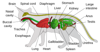
The urinary system, also known as the urinary tract or renal system, consists of the kidneys, ureters, bladder, and the urethra. The purpose of the urinary system is to eliminate waste from the body, regulate blood volume and blood pressure, control levels of electrolytes and metabolites, and regulate blood pH. The urinary tract is the body's drainage system for the eventual removal of urine. The kidneys have an extensive blood supply via the renal arteries which leave the kidneys via the renal vein. Each kidney consists of functional units called nephrons. Following filtration of blood and further processing, wastes exit the kidney via the ureters, tubes made of smooth muscle fibres that propel urine towards the urinary bladder, where it is stored and subsequently expelled from the body by urination (voiding). The female and male urinary system are very similar, differing only in the length of the urethra.

The excretory system is a passive biological system that removes excess, unnecessary materials from the body fluids of an organism, so as to help maintain internal chemical homeostasis and prevent damage to the body. The dual function of excretory systems is the elimination of the waste products of metabolism and to drain the body of used up and broken down components in a liquid and gaseous state. In humans and other amniotes most of these substances leave the body as urine and to some degree exhalation, mammals also expel them through sweating.

<span class="mw-page-title-main">Calcium oxalate</span> Calcium salt of oxalic acid

Calcium oxalate (in archaic terminology, oxalate of lime) is a calcium salt of oxalic acid with the chemical formula CaC2O4. It forms hydrates CaC2O4·nH2O, where n varies from 1 to 3. Anhydrous and all hydrated forms are colorless or white. The monohydrate CaC2O4·H2O occurs naturally as the mineral whewellite, forming envelope-shaped crystals, known in plants as raphides. The two rarer hydrates are dihydrate CaC2O4·2H2O, which occurs naturally as the mineral weddellite, and trihydrate CaC2O4·3H2O, which occurs naturally as the mineral caoxite, are also recognized. Some foods have high quantities of calcium oxalates and can produce sores and numbing on ingestion and may even be fatal. Cultural groups with diets that depend highly on fruits and vegetables high in calcium oxalate, such as those in Micronesia, reduce the level of it by boiling and cooking them. They are a constituent in 76% of human kidney stones. Calcium oxalate is also found in beerstone, a scale that forms on containers used in breweries.

<span class="mw-page-title-main">Bladder stone</span> Concretion of material in the urinary bladder

A bladder stone is a stone found in the urinary bladder.

<span class="mw-page-title-main">Hyperuricemia</span> Medical condition

Hyperuricaemia or hyperuricemia is an abnormally high level of uric acid in the blood. In the pH conditions of body fluid, uric acid exists largely as urate, the ion form. Serum uric acid concentrations greater than 6 mg/dL for females, 7 mg/dL for men, and 5.5 mg/dL for youth are defined as hyperuricemia. The amount of urate in the body depends on the balance between the amount of purines eaten in food, the amount of urate synthesised within the body, and the amount of urate that is excreted in urine or through the gastrointestinal tract. Hyperuricemia may be the result of increased production of uric acid, decreased excretion of uric acid, or both increased production and reduced excretion.

<span class="mw-page-title-main">Oxalate</span> Any derivative of oxalic acid; chemical compound containing oxalate moiety

Oxalate (IUPAC: ethanedioate) is an anion with the formula C2O42−. This dianion is colorless. It occurs naturally, including in some foods. It forms a variety of salts, for example sodium oxalate (Na2C2O4), and several esters such as dimethyl oxalate (C2O4(CH3)2). It is a conjugate base of oxalic acid. At neutral pH in aqueous solution, oxalic acid converts completely to oxalate.

<span class="mw-page-title-main">Cystinuria</span> Amino acid metabolic disorder involving cystine stones forming in the kidneys, ureter, and bladder

Cystinuria is an inherited autosomal recessive disease characterized by high concentrations of the amino acid cystine in the urine, leading to the formation of cystine stones in the kidneys, ureters, and bladder. It is a type of aminoaciduria. "Cystine", not "cysteine," is implicated in this disease; the former is a dimer of the latter.

<span class="mw-page-title-main">Urinary retention</span> Inability to completely empty the bladder

Urinary retention is an inability to completely empty the bladder. Onset can be sudden or gradual. When of sudden onset, symptoms include an inability to urinate and lower abdominal pain. When of gradual onset, symptoms may include loss of bladder control, mild lower abdominal pain, and a weak urine stream. Those with long-term problems are at risk of urinary tract infections.

<span class="mw-page-title-main">Cat food</span> Food for consumption by cats

Cat food is food specifically designed for consumption by cats. As obligate carnivores, cats have specific requirements for their dietary nutrients, namely nutrients found only in meat, such as taurine, arginine, and Vitamin B6. Certain nutrients, including many vitamins and amino acids, are degraded by the temperatures, pressures and chemical treatments used during manufacture, and hence must be added after manufacture to avoid nutritional deficiency.

<span class="mw-page-title-main">Calculus (medicine)</span> Medical condition

A calculus, often called a stone, is a concretion of material, usually mineral salts, that forms in an organ or duct of the body. Formation of calculi is known as lithiasis. Stones can cause a number of medical conditions.

<span class="mw-page-title-main">Struvite</span> Phosphate mineral

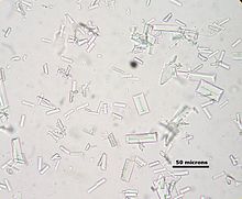
Struvite (magnesium ammonium phosphate) is a phosphate mineral with formula: NH4MgPO4·6H2O. Struvite crystallizes in the orthorhombic system as white to yellowish or brownish-white pyramidal crystals or in platy mica-like forms. It is a soft mineral with Mohs hardness of 1.5 to 2 and has a low specific gravity of 1.7. It is sparingly soluble in neutral and alkaline conditions, but readily soluble in acid.

<span class="mw-page-title-main">Feline lower urinary tract disease</span> Any disorder affecting the bladder or urethra of cats

Feline lower urinary tract disease (FLUTD) is a generic category term to describe any disorder affecting the bladder or urethra of cats.

<span class="mw-page-title-main">Nephrocalcinosis</span> Medical condition

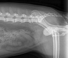
Nephrocalcinosis, once known as Albright's calcinosis after Fuller Albright, is a term originally used to describe the deposition of calcium salts in the renal parenchyma due to hyperparathyroidism. The term nephrocalcinosis is used to describe the deposition of both calcium oxalate and calcium phosphate. It may cause acute kidney injury. It is now more commonly used to describe diffuse, fine, renal parenchymal calcification in radiology. It is caused by multiple different conditions and is determined by progressive kidney dysfunction. These outlines eventually come together to form a dense mass. During its early stages, nephrocalcinosis is visible on x-ray, and appears as a fine granular mottling over the renal outlines. It is most commonly seen as an incidental finding with medullary sponge kidney on an abdominal x-ray. It may be severe enough to cause renal tubular acidosis or even end stage kidney disease, due to disruption of the kidney tissue by the deposited calcium.

Feline idiopathic cystitis (FIC) or feline interstitial cystitis or cystitis in cats, is one of the most frequently observed forms of feline lower urinary tract disease (FLUTD). Feline cystitis means "inflammation of the bladder in cats". The term idiopathic means unknown cause; however, certain behaviours have been known to aggravate the illness once it has been initiated. It can affect both males and females of any breed of cat. It is more commonly found in female cats; however, when males do exhibit cystitis, it is usually more dangerous.

<span class="mw-page-title-main">Renal stone formation in space</span>

Renal stone formation and passage during space flight can potentially pose a severe risk to crew member health and safety and could affect mission outcome. Although renal stones are routinely and successfully treated on Earth, the occurrence of these during space flight can prove to be problematic.

Equil 2 is a computer program used to estimate the risk of nephrolithiasis. The input data includes excretion, concentration, and the saturation of trace elements or other substances which are involved in the creation of kidney stones and the output will be provided in terms of PSF score or other equivalent formats. In some studies SUPERSAT, another program, provided more accurate measurements in some of the parameters such as relative supersaturation (RSS).

Crystallopathy is a harmful state or disease associated with the formation and aggregation of crystals in tissues or cavities, or in other words, a heterogeneous group of diseases caused by intrinsic or environmental microparticles or crystals, promoting tissue inflammation and scarring.

<span class="mw-page-title-main">Idiopathic hypercalcinuria</span>

Idiopathic hypercalcinuria (IH) is a condition including an excessive urinary calcium level with a normal blood calcium level resulting from no underlying cause. IH has become the most common cause of hypercalciuria and is the most serious metabolic risk factor for developing nephrolithiasis. IH can predispose individuals to osteopenia or osteoporosis, and affects the entire body. IH arises due to faulty calcium homeostasis, a closely monitored process, where slight deviations in calcium transport in the intestines, blood, and bone can lead to excessive calcium excretion, bone mineral density loss, or kidney stone formation. 50%-60% of nephrolithiasis patients suffer from IH and have 5%-15% lower bone density than those who do not.

Alkali citrate is an inhibitor of kidney stones. It is used to increase urine citrate levels - this prevents calcium oxalate stones by binding to calcium and inhibiting its binding to oxalate. It is also used to increase urine pH - this prevents uric acid stones and cystine stones.

References

  1. "Dietary treatment of bladder stones". Clinical Nutrition Service at Cummings School. 2017-07-26. Retrieved 2020-02-08.
  2. Giant tortoise cheats death Archived July 26, 2011, at the Wayback Machine ("Evening Express", Aberdeen, 31/01/2009)
  3. 1 2 3 4 5 Ettinger, Stephen J.; Feldman, Edward C. (1995). Textbook of Veterinary Internal Medicine (4th ed.). W.B. Saunders Company. ISBN   0-7216-6795-3.
  4. 1 2 "Urolithiasis: Overview". The Merck Veterinary Manual. 2006. Retrieved 2007-04-14.
  5. PaBlack, N., Brenten, T., Neumann, K., & Zentek, J. (2014). Effects of potassium chloride and potassium bicarbonate in the diet on urinary pH and mineral excretion of adult cats. British Journal of Nutrition, 111(5), 785-797.
  6. Buffington, Tony (2004). "Nutrition and Urolithiasis". Proceedings of the 29th World Congress of the World Small Animal Veterinary Association. Retrieved 2006-07-16.
  7. Hoskins, Johnny D. (November 2006). "Feline Urolithiasis". DVM. Advanstar Communications: 6S–7S.
  8. 1 2 3 4 "Canine Urolithiasis". The Merck Veterinary Manual. 2006. Retrieved 2007-04-14.
  9. 1 2 Hillyer, Elizabeth V.; Quesenberry, Katherin E. (1997). Ferrets, Rabbits, and Rodents: Clinical Medicine and Surgery (1st ed.). W.B. Saunders Company. ISBN   0-7216-4023-0.
  10. 1 2 3 Zoran, Debra (2006). Role of Diet in Feline and Canine Urolithiasis.
  11. Osborne, Carl A.; Lulich, Jody P. (February 2007). "Changing trends in composition of feline uroliths and urethral plugs". DVM. Advanstar Communications: 38–40.
  12. Escolar E, Bellanato J (2003). "Analysis of feline urinary calculi and urethral plugs by infrared spectroscopy and scanning electron microscopy". Vet Rec. 152 (20): 625–8. doi:10.1136/vr.152.20.625. PMID   12790167. S2CID   33483919.
  13. Albasan H, Lulich J, Osborne C, Lekcharoensuk C (2005). "Evaluation of the association between sex and risk of forming urate uroliths in Dalmatians". J Am Vet Med Assoc. 227 (4): 565–9. doi: 10.2460/javma.2005.227.565 . PMID   16117063.
  14. "Feline Urolithiasis and Feline Lower Urinary Tract Disease". The Merck Veterinary Manual. 2006. Retrieved 2007-04-14.
  15. Team, Clinical Nutrition (2017-07-26). "Dietary treatment of bladder stones". Clinical Nutrition Service at Cummings School. Retrieved 2019-12-20.
  16. "Struvite Bladder Stones in Dogs". vca_corporate. Retrieved 2019-12-20.