Calcific tendinitis

Last updated
Calcific tendinitis
Other namescalcified/calcareous tendinitis/tendinopathy, tendinosis calcarea, hydroxyapatite deposition disease, calcific periarthritis
Calcific tendinitis marked.jpg
A plain X ray of the shoulder showing calcific tendinitis
Specialty Rheumatology   OOjs UI icon edit-ltr-progressive.svg
Symptoms Chronic shoulder pain during activities; acute shoulder pain
Duration Self-limiting, typically resolves in 6-9 months
Risk factors Diabetes, hypothyroidism
Diagnostic method X-ray
Treatment Physiotherapy, extracorporeal shockwave therapy, surgical excision
Medication NSAIDs

Calcific tendinitis is a common condition where deposits of calcium phosphate form in a tendon, sometimes causing pain at the affected site. Deposits can occur in several places in the body, but are by far most common in the rotator cuff of the shoulder. Around 80% of those with deposits experience symptoms, typically chronic pain during certain shoulder movements, or sharp acute pain that worsens at night. Calcific tendinitis is typically diagnosed by physical exam and X-ray imaging. The disease often resolves completely on its own, but is typically treated with non-steroidal anti-inflammatory drugs to relieve pain, rest and physical therapy to promote healing, and in some cases various procedures to breakdown and/or remove the calcium deposits.

Contents

Adults aged 30–50 are most commonly affected by calcific tendinitis. It is twice as common in women as men, and is not associated with exercise. Calcifications in the rotator cuff were first described by Ernest Codman in 1934. The name, "calcifying tendinitis" was coined by Henry Plenk in 1952.

Signs and symptoms

Up to 20% of those with calcification in the tendons have no symptoms because it is an integral part of the tendinopathy. [1] For those with symptoms, the symptoms vary based on the phase of the disease. In the initial "formative phase" when the calcium deposits are being formed, people rarely experience any symptoms. [1] Those that do have symptoms tend to have intermittent shoulder pain, particularly during forward shoulder flexion (i.e. lifting the arm in front of the body). [1] In the "resorptive phase" when the calcium deposit is breaking down, many experience severe acute pain that worsens at night. [1] Those affected tend to hold the shoulder rotated inwards to alleviate pain, and have difficulty lying on the affected shoulder. [1] Some people experience heat and redness at the affected shoulder, as well as a limited range of motion. [1]

Cause

Calcific tendinitis is caused by deposits of calcium phosphate crystals in the tendons of the shoulder. [1] These deposits are most frequently found in the supraspinatus tendon (63% of the time), and less frequently in the infraspinatus tendon (7%), subacromial bursa (7%), subscapularis tendon (3%), or in both the supraspinatus and subscapularis tendons at the same time (20%). [1]

The development of calcific tendinitis is often divided into three stages. First, in the "precalcific stage", something causes tendon cells to transform into other cells that can act as sites for calcium deposition. [2] This is followed by the two-part "calcific stage"; first calcium is deposited (the formative phase), then the body begins to break down the calcium deposit (the resorptive phase). [2] Finally, in the "postcalcific stage" the calcium deposits are replaced with new tissue and the tendon completely heals. [2]

The cause of the calcium deposits remains unclear, although several theories have been put forward. [2] Some theories involve the differentiation of tendon cells into other cells, namely cartilages or bone cells. [2] Others associate the condition with cell death due to aging, wear, or lack of oxygen in the tissue; however, the disease is uncommon in the very old, not associated with exercise, and tends to resolve completely even if untreated. [2]

Diagnosis

An x-ray showing calcific deposits in the area of the tendons of the rotator cuff muscles CalcificTendonitisMark.png
An x-ray showing calcific deposits in the area of the tendons of the rotator cuff muscles

Calcific tendinitis is typically diagnosed by physical examination and X-ray imaging. [1] During the formative phase, X-ray images typically reveal calcium deposits with uniform density and a clear margin. [1] In the more painful resorptive phase, deposits instead appear cloudy and with unclear margins. [1] By arthroscopy, formative stage deposits appear crystalline and chalk-like, while resorptive stage deposits appear smooth resembling toothpaste. [1] Ultrasound is also used to locate and assess calcium deposits. In the formative stage, deposits are hyperechoic and arc-shaped; in the resorptive stage deposits are less echogenic and appear fragmented. [1] [3]

Treatment

The first line of treatment for calcific tendinitis is typically nonsteroidal anti-inflammatory drugs to relieve pain, rest for the affected joint, and sometimes physical therapy to avoid joint stiffness. [3] [1] For those with severe pain direct injections of steroids to the affected site are often effective for pain relief, [1] but may interfere with reabsorption of the calcium deposit. [3] For those whose pain doesn't improve with medication and rest, the deposit can be dissolved and removed with techniques called "ultrasound-guided needling", "barbotage", and "US-PICT" (for "ultrasound percutaneous injection in calcific tenditis"). In each, ultrasound is used to locate the deposit and guide a needle to the affected site. There saline and lidocaine are injected to dissolve the deposit, then removed to wash it away. [1] [3] [2] Another common treatment is extracorporeal shockwave therapy, where pulses of sound are used to break up the deposit and promote healing. [1] There is little standardization of energy levels, duration, and time interval of treatment; though most studies report positive outcomes with low- to medium-energy waves (below 0.28 mJ/mm2). [3]

Surgery

Surgery is only recommended once 6 months of conservative, non-operative treatment has failed to reduce symptoms. Surgery is arthroscopic and involves calcification removal with or without acromioplasty of the shoulder. [4] Additionally, debate remains over whether a complete removal of the deposits is necessary, or if equal pain relief can be obtained from a partial removal of calcium deposits. [5]

Removing the deposits either with open shoulder surgery or arthroscopic surgery are both difficult operations, but with high success rates (around 90%). About 10% require re-operation. If the deposit is large, then frequently the patient will require a rotator cuff repair to fix the defect left in the tendon when the deposit is removed or to reattach the tendon to the bone if the deposit was at the tendon insertion into the bone.

Outcomes

Nearly all people with calcific tendinitis recover completely with time or treatment. [6] Treatment helps alleviate pain, but long-term follow-up studies have shown that people recover with or without treatment. [6]

Epidemiology

Calcific tendinitis typically occurs in adults aged 30 to 50, and is rare in those older than 70. It is twice as common in women as men. [1]

Risk factors that increase the chance of developing calcific tendinitis include; hormonal disorders, like diabetes and hypothyroidism, autoimmune disorders, like rheumatoid arthritis, and metabolic disorders that also cause kidney stones, gallstones, and gout. Occupations that consist of repetitive overhead lifting, such as athletes or construction workers, do not seem to significantly increase the likelihood of developing calcific tendinitis. [5]

History

Calcifications in the rotator cuff tendon were first described by Ernest Codman in his 1934 book The Shoulder. [3] In 1952, in his study on x-ray therapy for people with such calcifications, Henry Plenk coined the term "calcifying tendinitis". [3] [7]

Related Research Articles

<span class="mw-page-title-main">Rotator cuff</span> Group of muscles

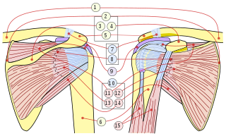
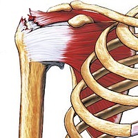
The rotator cuff is a group of muscles and their tendons that act to stabilize the human shoulder and allow for its extensive range of motion. Of the seven scapulohumeral muscles, four make up the rotator cuff. The four muscles are:

<span class="mw-page-title-main">Tendinopathy</span> Inflammation of the tendon

Tendinopathy is a type of tendon disorder that results in pain, swelling, and impaired function. The pain is typically worse with movement. It most commonly occurs around the shoulder, elbow, wrist, hip, knee, or ankle.

<span class="mw-page-title-main">Shoulder problem</span> Medical condition

Shoulder problems including pain, are one of the more common reasons for physician visits for musculoskeletal symptoms. The shoulder is the most movable joint in the body. However, it is an unstable joint because of the range of motion allowed. This instability increases the likelihood of joint injury, often leading to a degenerative process in which tissues break down and no longer function well.

<span class="mw-page-title-main">Shoulder</span> Part of the body

The human shoulder is made up of three bones: the clavicle (collarbone), the scapula, and the humerus as well as associated muscles, ligaments and tendons.

<span class="mw-page-title-main">Achilles tendon</span> Tendon at the back of the lower leg

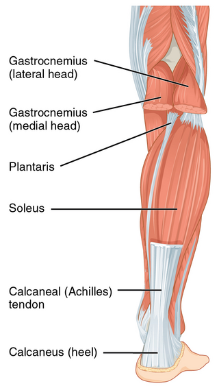
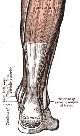
The Achilles tendon or heel cord, also known as the calcaneal tendon, is a tendon at the back of the lower leg, and is the thickest in the human body. It serves to attach the plantaris, gastrocnemius (calf) and soleus muscles to the calcaneus (heel) bone. These muscles, acting via the tendon, cause plantar flexion of the foot at the ankle joint, and flexion at the knee.

<span class="mw-page-title-main">Tennis elbow</span> Condition in which the outer part of the elbow becomes sore and tender

Tennis elbow, also known as lateral epicondylitis or enthesopathy of the extensor carpi radialis origin, is an enthesopathy of the origin of the extensor carpi radialis brevis on the lateral epicondyle. The outer part of the elbow becomes painful and tender. The pain may also extend into the back of the forearm. Onset of symptoms is generally gradual, although they can seem sudden and be misinterpreted as an injury. Golfer's elbow is a similar condition that affects the inside of the elbow.

<span class="mw-page-title-main">Achilles tendinitis</span> Medical condition of the ankle and heel

Achilles tendinitis, also known as achilles tendinopathy, occurs when the Achilles tendon, found at the back of the ankle, becomes sore. Achilles tendinopathy is accompanied by alterations in the tendon's structure and mechanical properties. The most common symptoms are pain and swelling around the affected tendon. The pain is typically worse at the start of exercise and decreases thereafter. Stiffness of the ankle may also be present. Onset is generally gradual.

<span class="mw-page-title-main">Rotator cuff tear</span> Shoulder injury

Rotator cuff tendinopathy is a process of senescence. The pathophysiology is mucoid degeneration. Most people develop rotator cuff tendinopathy within their lifetime.

<span class="mw-page-title-main">Adhesive capsulitis of the shoulder</span> Painful disease restricting movement

Adhesive capsulitis, also known as frozen shoulder, is a condition associated with shoulder pain and stiffness. It is a common shoulder ailment that is marked by pain and a loss of range of motion, particularly in external rotation. There is a loss of the ability to move the shoulder, both voluntarily and by others, in multiple directions. The shoulder itself, however, does not generally hurt significantly when touched. Muscle loss around the shoulder may also occur. Onset is gradual over weeks to months. Complications can include fracture of the humerus or biceps tendon rupture.

<span class="mw-page-title-main">Supraspinatus muscle</span> Muscle of the upper back

The supraspinatus is a relatively small muscle of the upper back that runs from the supraspinous fossa superior portion of the scapula to the greater tubercle of the humerus. It is one of the four rotator cuff muscles and also abducts the arm at the shoulder. The spine of the scapula separates the supraspinatus muscle from the infraspinatus muscle, which originates below the spine.

<span class="mw-page-title-main">Calcium pyrophosphate dihydrate crystal deposition disease</span> Medical condition

Calcium pyrophosphate dihydrate (CPPD) crystal deposition disease, also known as pseudogout and pyrophosphate arthropathy, is a rheumatologic disease which is thought to be secondary to abnormal accumulation of calcium pyrophosphate dihydrate crystals within joint soft tissues. The knee joint is most commonly affected. The disease is metabolic in origin and its treatment remains symptomatic.

<span class="mw-page-title-main">Patellar tendinitis</span> Human disease

Patellar tendinitis, also known as jumper's knee, is an overuse injury of the tendon that straightens the knee. Symptoms include pain in the front of the knee. Typically the pain and tenderness is at the lower part of the kneecap, though the upper part may also be affected. Generally there is no pain when the person is at rest. Complications may include patellar tendon rupture.

<span class="mw-page-title-main">Myositis ossificans</span> Medical condition

Myositis ossificans comprises two syndromes characterized by heterotopic ossification (calcification) of muscle. The World Health Organization, 2020, has grouped myositis ossificans together with fibro-osseous pseudotumor of digits as a single specific entity in the category of fibroblastic and myofibroblastic tumors.

<span class="mw-page-title-main">Enthesopathy</span> Medical condition

An enthesopathy refers to a disorder involving the attachment of a tendon or ligament to a bone. This site of attachment is known as the enthesis . If the condition is known to be inflammatory, it can more precisely be called an enthesitis.

<span class="mw-page-title-main">Subacromial bursitis</span> Medical condition

Subacromial bursitis is a condition caused by inflammation of the bursa that separates the superior surface of the supraspinatus tendon from the overlying coraco-acromial ligament, acromion, and coracoid and from the deep surface of the deltoid muscle. The subacromial bursa helps the motion of the supraspinatus tendon of the rotator cuff in activities such as overhead work.

<span class="mw-page-title-main">Extracorporeal shockwave therapy</span> Ultrasonic, non-invasive, outpatient treatment

Extracorporeal shockwave therapy (ESWT) is a treatment using powerful acoustic pulses which is mostly used to treat kidney stones and in physical therapy and orthopedics.

Therapeutic ultrasound refers generally to any type of ultrasonic procedure that uses ultrasound for therapeutic benefit. Physiotherapeutic ultrasound was introduced into clinical practice in the 1950s, with lithotripsy introduced in the 1980s. Others are at various stages in transitioning from research to clinical use: HIFU, targeted ultrasound drug delivery, trans-dermal ultrasound drug delivery, ultrasound hemostasis, cancer therapy, and ultrasound assisted thrombolysis It may use focused ultrasound or unfocused ultrasound.

<span class="mw-page-title-main">Capsule of the glenohumeral joint</span> Articular capsule of the shoulder joint

The capsule of the glenohumeral (shoulder) joint is the articular capsule of the shoulder. It completely surrounds the joint. It is attached above to the circumference of the glenoid cavity beyond the glenoidal labrum, and below to the anatomical neck of the humerus, approaching nearer to the articular cartilage above than in the rest of its extent.

<span class="mw-page-title-main">Shoulder impingement syndrome</span> Medical condition

Shoulder impingement syndrome is a syndrome involving tendonitis of the rotator cuff muscles as they pass through the subacromial space, the passage beneath the acromion. It is particularly associated with tendonitis of the supraspinatus muscle. This can result in pain, weakness, and loss of movement at the shoulder.

<span class="mw-page-title-main">Calcific bursitis</span> Medical condition

Calcific bursitis refers to calcium deposits within the bursae. This most occurs in the shoulder area. The most common bursa for calcific bursitis to occur is the subacromial bursa. A bursa is a small, fluid-filled sac that reduces friction, and facilitates movements between its adjacent tissues. Inflammation of the bursae is called bursitis.

References

  1. 1 2 3 4 5 6 7 8 9 10 11 12 13 14 15 16 17 18 Kim MS, Kim IW, Lee S, Shin SJ (December 2020). "Diagnosis and treatment of calcific tendinitis of the shoulder". Clin Shoulder Elb. 23 (4): 210–216. doi:10.5397/cise.2020.00318. PMC   7726362 . PMID   33330261.
  2. 1 2 3 4 5 6 7 Chianca V, Albano D, Messina C, Midiri F, Mauri G, Aliprandi A, Catapano M, Pescatori LC, Monaco CG, Gitto S, Pisani Mainini A, Corazza A, Rapisarda S, Pozzi G, Barile A, Masciocchi C, Sconfienza LM (January 2018). "Rotator cuff calcific tendinopathy: from diagnosis to treatment". Acta Biomedica: Atenei Parmensis. 89 (1–S): 186–196. doi:10.23750/abm.v89i1-S.7022. PMC   6179075 . PMID   29350647.
  3. 1 2 3 4 5 6 7 Merolla G, Singh S, Paladini P, Porcellini G (March 2016). "Calcific tendinitis of the rotator cuff: state of the art in diagnosis and treatment". J Orthop Traumatol. 17 (1): 7–14. doi:10.1007/s10195-015-0367-6. PMC   4805635 . PMID   26163832.
  4. (2020). .In: Achar, Suraj A. & Taylor, Kenneth S. (eds.), 5-Minute Sports Medicine Consult, The. 3rd Edition. Two Commerce Square, 2001 Market Street, Philadelphia, PA 19103 USA:Lippincott Williams & Wilkins; Retrieved from: Books@Ovid database.
  5. 1 2 Bechay, Joseph; Lawrence, Cassandra; Namdari, Surena (2020-01-01). "Calcific tendinopathy of the rotator cuff: a review of operative versus nonoperative management". The Physician and Sportsmedicine. 48 (3): 241–246. doi:10.1080/00913847.2019.1710617. ISSN   0091-3847. PMID   31893972. S2CID   209539198.
  6. 1 2 Miller RH, Azar FM, Throckmorton TW (2021). "Chapter 46: Shoulder and Elbow Injuries". In Azar FM, Beaty JH (eds.). Campbell's Operative Orthopaedics. Elsevier. pp. 2374–2425. ISBN   978-0-323-67217-7.
  7. PLENK HP (September 1952). "Calcifying tendinitis of the shoulder; a critical study of the value of x-ray therapy". Radiology. 59 (3): 384–9. doi:10.1148/59.3.384. PMID   14958017.