Shoulder impingement syndrome

Last updated
Shoulder impingement syndrome
Other namesSubacromial impingement, painful arc syndrome, supraspinatus syndrome, swimmer's shoulder, thrower's shoulder
Shoulder joint.svg
Shoulder joint
Specialty Orthopedics, sports medicine

Shoulder impingement syndrome is a syndrome involving tendonitis (inflammation of tendons) of the rotator cuff muscles as they pass through the subacromial space, the passage beneath the acromion. It is particularly associated with tendonitis of the supraspinatus muscle. [1] This can result in pain, weakness, and loss of movement at the shoulder. [2] [3]

Contents

Signs and symptoms

The most common symptoms in impingement syndrome are pain, weakness and a loss of movement at the affected shoulder. [2] The pain is often worsened by shoulder overhead movement and may occur at night, especially when lying on the affected shoulder. The onset of the pain may be acute if due to an injury or insidious if due to a gradual process such as an osteoarthritic spur. The pain has been described as dull rather than sharp, and lingers for long periods of time, making it hard to fall asleep. [2] Other symptoms can include a grinding or popping sensation during movement of the shoulder. [4]

The range of motion at the shoulder may be limited by pain. A painful arc of movement may be present during forward elevation of the arm from 60° to 120°. [4] Passive movement at the shoulder will appear painful when a downward force is applied at the acromion but the pain will ease once the force is removed. [2]

Causes

Acromion types
TypeAppearancePrevalence [5] Angle of
anterior slope [5]
Flat Flat acromion.jpg 17.1%13.18
Curved Curved acromion.jpg 42.9%29.98
Hooked Hooked acromion.jpg 39.3%26.98

When the arm is raised, the subacromial space (gap between the anterior edge of the acromion and the head of the humerus) narrows; the supraspinatus muscle tendon passes through this space. [6] Anything that causes further narrowing has the tendency to impinge the tendon and cause an inflammatory response, resulting in impingement syndrome. Such causes can be bony structures such as subacromial spurs (bony projections from the acromion), osteoarthritic spurs on the acromioclavicular joint, and variations in the shape of the acromion. Thickening or calcification of the coracoacromial ligament can also cause impingement. Loss of function of the rotator cuff muscles, due to injury or loss of strength, may cause the humerus to move superiorly, resulting in impingement. Inflammation and subsequent thickening of the subacromial bursa may also cause impingement. [2]

Weight training exercises where the arms are elevated above shoulder height but in an internally rotated position such as the upright row have been suggested as a cause of subacromial impingement. [7] Another common cause of Impingement syndrome is restrictions in the range movement of the scapulo-thoracic surfaces. Commonly, one or more ribs between rib 2 and rib 7/8 on the side of the impingement may jut out slightly and/or feel hard when the person springs on it or them. When this occurs, the scapula is raised and anteverted (angled forwards). This in turn pushes the acromion and the humeral head out of its usual anatomical position placing pressure downwards at the head of the humerus at the position of the nerve thus causing the impingement syndrome. This is visibly demonstrated by a slightly raised and protracted shoulder girdle. Note: the humerus anteverts in this position causing a more protrusive section of the humerus to press upwards towards the acromion.

Mechanism

The scapula plays an important role in shoulder impingement syndrome. [8] It is a wide, flat bone lying on the posterior thoracic wall that provides an attachment for three different groups of muscles. The intrinsic muscles of the scapula include the muscles of the rotator cuff- the subscapularis, infraspinatus, teres minor and supraspinatus. [9] These muscles attach to the surface of the scapula and are responsible for the internal and external rotation of the glenohumeral joint, along with humeral abduction. The extrinsic muscles include the biceps, triceps, and deltoid muscles and attach to the coracoid process and supraglenoid tubercle of the scapula, infraglenoid tubercle of the scapula, and spine of the scapula. These muscles are responsible for several actions of the glenohumeral joint. The third group, which is mainly responsible for stabilization and rotation of the scapula, consists of the trapezius, serratus anterior, levator scapulae, and rhomboid muscles and attach to the medial, superior, and inferior borders of the scapula. Each of these muscles has its own role in shoulder function and must be in balance with the others in order to avoid shoulder pathology.

Abnormal scapular function is called scapular dyskinesis. One action the scapula performs during a throwing or serving motion is elevation of the acromion process in order to avoid impingement of the rotator cuff tendons. [8] If the scapula fails to properly elevate the acromion, impingement may occur during the cocking and acceleration phase of an overhead activity. The two muscles most commonly inhibited during this first part of an overhead motion are the serratus anterior and the lower trapezius. [10] These two muscles act as a force couple within the glenohumeral joint to properly elevate the acromion process, and if a muscle imbalance exists, shoulder impingement may develop.

The scapula may also be misplaced if a rib deep to it is not moving correctly. Often in the case of Shoulder impingement syndrome, the scapula may be anteverted such that the shoulder on the affected side appears protracted. The ribs that may cause such an anteversion of the scapula include ribs 2–8.

Diagnosis

MRI showing subacromial impingement with partial rupture of the supraspinatus tendon, but no retraction or fatty degeneration of the supraspinatus muscle. MRI. Subacromial impingement..jpg
MRI showing subacromial impingement with partial rupture of the supraspinatus tendon, but no retraction or fatty degeneration of the supraspinatus muscle.

Impingement syndrome can be diagnosed by a targeted medical history and physical examination, [11] [12] but it has also been argued that at least medical imaging [13] (generally X-ray initially) and/or response to local anesthetic injection [14] is necessary for workup. However, imaging studies are unable to show cause of shoulder pain in diagnosing. For example, MRI imaging would show rotator cuff pathology and bursitis but is unable to specify the cause. [15]

On physical exam, the physician may twist or elevate the patient's arm to test for reproducible pain (the Neer sign and Hawkins-Kennedy test). These tests help localize the pathology to the rotator cuff; however, they are not specific for impingement. [16] Neer sign may also be seen with subacromial bursitis. [17]

Response to local anesthetic

The physician may inject lidocaine (usually combined with a steroid) into the bursa, and if there is an improved range of motion and decrease in pain, this is considered a positive "Impingement Test". It not only supports the diagnosis for impingement syndrome, but it is also therapeutic. [18]

Imaging

Plain x-rays of the shoulder can be used to detect some joint pathology and variations in the bones, including acromioclavicular arthritis, variations in the acromion, and calcification. However, x-rays do not allow visualization of soft tissue and thus hold a low diagnostic value. [2] Ultrasonography, arthrography and MRI can be used to detect rotator cuff muscle pathology. MRI is the best imaging test prior to arthroscopic surgery. [2] Due to lack of understanding of the pathoaetiology, and lack of diagnostic accuracy in the assessment process by many physicians, [19] several opinions are recommended before intervention.[ citation needed ]

Treatment

Impingement syndrome is usually treated conservatively, but sometimes it is treated with arthroscopic surgery or open surgery. [20] Conservative treatment includes rest, cessation of painful activity, and physical therapy. Physical therapy treatments would typically focus at maintaining range of movement, improving posture, strengthening shoulder muscles, and reduction of pain. NSAIDs and ice packs may be used for pain relief. [4] [20]

Therapeutic exercises might be favorable intervention compared to passive treatment approaches, electrotherapy and placebo. A recent meta-analysis done on rotator cuff tendinopathy has shown that nearly all types of active resistance training programs were proven to be effective in improving pain and shoulder function with no significant differences among the different exercise types, further cementing the favorability of a more active intervention over passive modalities when it comes to rotator cuff issues. [21] Exercises may help to regain the scapulo-humeral rhythm and scapular control which may reduce pain. [22]

Steroids

Therapeutic injections of corticosteroid and local anaesthetic may be used for persistent impingement syndrome. [20] The total number of injections is generally limited to three due to possible side effects from the corticosteroid. [4] A 2017 review found corticosteroid injections only give small and transient pain relief. [23]

Surgery

A number of surgical interventions are available, depending on the nature and location of the pathology. Surgery may be done arthroscopically or as open surgery. The impinging structures may be removed in surgery, and the subacromial space may be widened by resection of the distal clavicle and excision of osteophytes on the under-surface of the acromioclavicular joint. [4] Damaged rotator cuff muscles can be surgically repaired.

A 2019 review found that the evidence does not support decompression surgery in those with more than 3 months of shoulder pain without a history of trauma. [24] A recent metaanalysis has further supported that early SIS would likely benefit from non-operative treatment modalities and surgical open decompression should be considered only with chronic presentation.

History

Impingement syndrome was reported in 1852. [4] Impingement of the shoulder was previously thought to be precipitated by shoulder abduction and surgical intervention focused on lateral or total acromionectomy. [4] [25] In 1972, Charles Neer proposed that impingement was due to the anterior third of the acromion and the coracoacromial ligament and suggested surgery should be focused on these areas. [4] [25] The role of anteriorinferior aspect of the acromion in impingement syndrome and excision of parts of the anteriorinferior acromion has become a pivotal part of the surgical treatment of the syndrome. [4]

Criticism

Subacromial impingement is not free of criticism. First, the identification of acromion type shows poor intra- and inter-observer reliability. [26] [27] Second, a computerized three-dimensional study failed to support impingement by any portion of the acromion on the rotator cuff tendons in different shoulder positions. [28] Third, most partial-thickness cuff tears do not occur on bursal surface fibers, where mechanical abrasion from the acromion does occur. [29] [30] Fourth, it has been suggested that bursal surface cuff tears could be responsible for subacromial spurs and not the opposite. [31] [32] [33] [34] Fifth, it has been shown that there is no association with acromiohumeral distance, measured at 0°, 45° and 60° of shoulder abduction, with people with subacromial pain syndrome and shoulder pain. [35] And finally, there is growing evidence that routine acromioplasty may not be required for successful rotator cuff repair, which would be an unexpected finding if acromial shape had a major role in generating tendon lesions. [36] In summary, despite being a popular theory, the bulk of evidence suggest that subacromial impingement probably does not play a dominant role in many cases of rotator cuff disease. [37]

See also

Related Research Articles

<span class="mw-page-title-main">Acromion</span> Bony process on the scapula (shoulder blade)

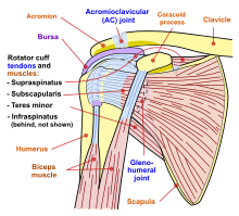
In human anatomy, the acromion is a bony process on the scapula. Together with the coracoid process, it extends laterally over the shoulder joint. The acromion is a continuation of the scapular spine, and hooks over anteriorly. It articulates with the clavicle to form the acromioclavicular joint.

<span class="mw-page-title-main">Rotator cuff</span> Group of muscles

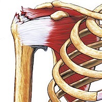
The rotator cuff is a group of muscles and their tendons that act to stabilize the human shoulder and allow for its extensive range of motion. Of the seven scapulohumeral muscles, four make up the rotator cuff. The four muscles are:

<span class="mw-page-title-main">Coracoid process</span> Small hook-like structure on the lateral edge of the superior anterior portion of the scapula

The coracoid process is a small hook-like structure on the lateral edge of the superior anterior portion of the scapula. Pointing laterally forward, it, together with the acromion, serves to stabilize the shoulder joint. It is palpable in the deltopectoral groove between the deltoid and pectoralis major muscles.

<span class="mw-page-title-main">Shoulder problem</span> Medical condition

Shoulder problems including pain, are one of the more common reasons for physician visits for musculoskeletal symptoms. The shoulder is the most movable joint in the body. However, it is an unstable joint because of the range of motion allowed. This instability increases the likelihood of joint injury, often leading to a degenerative process in which tissues break down and no longer function well.

<span class="mw-page-title-main">Shoulder</span> Part of the body

The human shoulder is made up of three bones: the clavicle (collarbone), the scapula, and the humerus as well as associated muscles, ligaments and tendons.

<span class="mw-page-title-main">Deltoid muscle</span> Shoulder muscle

The deltoid muscle is the muscle forming the rounded contour of the human shoulder. It is also known as the 'common shoulder muscle', particularly in other animals such as the domestic cat. Anatomically, the deltoid muscle appears to be made up of three distinct sets of muscle fibers, namely the

  1. anterior or clavicular part
  2. posterior or scapular part
  3. intermediate or acromial part
<span class="mw-page-title-main">Rotator cuff tear</span> Shoulder injury

Rotator cuff tendinopathy is a process of senescence. The pathophysiology is mucoid degeneration. Most people develop rotator cuff tendinopathy within their lifetime.

<span class="mw-page-title-main">Acromioclavicular joint</span> Shoulder junction between the scapula and the clavicle

The acromioclavicular joint, or AC joint, is a joint at the top of the shoulder. It is the junction between the acromion and the clavicle. It is a plane synovial joint.

<span class="mw-page-title-main">Supraspinatus muscle</span> Muscle of the upper back

The supraspinatus is a relatively small muscle of the upper back that runs from the supraspinous fossa superior portion of the scapula to the greater tubercle of the humerus. It is one of the four rotator cuff muscles and also abducts the arm at the shoulder. The spine of the scapula separates the supraspinatus muscle from the infraspinatus muscle, which originates below the spine.

<span class="mw-page-title-main">Infraspinatus muscle</span> Main external rotator of the shoulder

In human anatomy, the infraspinatus muscle is a thick triangular muscle, which occupies the chief part of the infraspinatous fossa. As one of the four muscles of the rotator cuff, the main function of the infraspinatus is to externally rotate the humerus and stabilize the shoulder joint.

<span class="mw-page-title-main">Shoulder joint</span> Synovial ball and socket joint in the shoulder

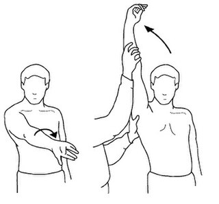
The shoulder joint is structurally classified as a synovial ball-and-socket joint and functionally as a diarthrosis and multiaxial joint. It involves an articulation between the glenoid fossa of the scapula and the head of the humerus. Due to the very loose joint capsule that gives a limited interface of the humerus and scapula, it is the most mobile joint of the human body.

<span class="mw-page-title-main">Shoulder girdle</span> Set of bones which connects the arm to the axial skeleton on each side

The shoulder girdle or pectoral girdle is the set of bones in the appendicular skeleton which connects to the arm on each side. In humans it consists of the clavicle and scapula; in those species with three bones in the shoulder, it consists of the clavicle, scapula, and coracoid. Some mammalian species have only the scapula.

<span class="mw-page-title-main">Coracoacromial ligament</span> Ligament between the coracoid process and the acromion of the scapula

The coracoacromial ligament is a strong triangular ligament between the coracoid process and the acromion. It protects the head of the humerus. Its acromial attachment may be repositioned to the clavicle during reconstructive surgery of the acromioclavicular joint.

<span class="mw-page-title-main">Separated shoulder</span> Medical condition

A separated shoulder, also known as acromioclavicular joint injury, is a common injury to the acromioclavicular joint. The AC joint is located at the outer end of the clavicle where it attaches to the acromion of the scapula. Symptoms include non-radiating pain which may make it difficult to move the shoulder. The presence of swelling or bruising and a deformity in the shoulder is also common depending on how severe the dislocation is.

<span class="mw-page-title-main">Subacromial bursitis</span> Medical condition

Subacromial bursitis is a condition caused by inflammation of the bursa that separates the superior surface of the supraspinatus tendon from the overlying coraco-acromial ligament, acromion, and coracoid and from the deep surface of the deltoid muscle. The subacromial bursa helps the motion of the supraspinatus tendon of the rotator cuff in activities such as overhead work.

Shoulder surgery is a means of treating injured shoulders. Many surgeries have been developed to repair the muscles, connective tissue, or damaged joints that can arise from traumatic or overuse injuries to the shoulder.

<span class="mw-page-title-main">Subacromial bursa</span>

The subacromial bursa is the synovial cavity located just below the acromion, which communicates with the subdeltoid bursa in most individuals, forming the so-called subacromial-subdeltoid bursa (SSB).

<span class="mw-page-title-main">Neer Impingement Test</span> Medical test

The Neer Impingement Test is a test designed to reproduce symptoms of rotator cuff impingement through flexing the shoulder and pressure application. Symptoms should be reproduced if there is a problem with the supraspinatus or biceps brachii. This test is also associated with the Hawkins-Kennedy Test and Jobe's Test.

A shoulder examination is a portion of a physical examination used to identify potential pathology involving the shoulder. It should be conducted with both shoulders exposed to assess for asymmetry and muscle wasting.

The Hawkins–Kennedy Test is a test used in the evaluation of orthopedic shoulder injury. It was first described in the 1980s by Canadians R. Hawkins and J. Kennedy, and a positive test is most likely indicative of damage to the tendon of the supraspinatus muscle.

References

  1. Thomas M DeBerardino (17 January 2023). "Supraspinatus Tendonitis". Medscape . Updated: Dec 03, 2018
  2. 1 2 3 4 5 6 7 Fongemie AE, Buss DD, Rolnick SJ (February 1998). "Management of shoulder impingement syndrome and rotator cuff tears". Am Fam Physician. 57 (4): 667–74, 680–2. PMID   9490991.
  3. SimonMoyes.com. "What is Subacromial Impingement?". Archived from the original on 2011-08-27. Retrieved 2011-03-14.{{cite journal}}: Cite journal requires |journal= (help)
  4. 1 2 3 4 5 6 7 8 9 Chen AL, Rokito AS, Zuckerman JD (April 2003). "The role of the acromioclavicular joint in impingement syndrome". Clin Sports Med . 22 (2): 343–57. doi:10.1016/S0278-5919(03)00015-2. PMID   12825535.
  5. 1 2 Habermeyer, Magosch & Lichtenberg 2006 , pp. 1–3
  6. Neer, C. S. (1983). "Impingement lesions". Clinical Orthopaedics and Related Research. 173: 70–77. doi:10.1097/00003086-198303000-00010.
  7. Schoenfeld, Brad; Kolber, Morey J; Haimes, Jonathan E (October 2011). "The Upright Row: Implications for Preventing Subacromial Impingement". Strength and Conditioning Journal. 33 (5): 25–28. doi: 10.1519/SSC.0b013e31822ec3e3 . S2CID   58333549.
  8. 1 2 Kibler, BW (1998). "The role of the scapula in athletic shoulder function". The American Journal of Sports Medicine. 26 (2): 325–337. doi:10.1177/03635465980260022801. PMID   9548131. S2CID   26608885.
  9. Marieb, E. (2005). Anatomy & Physiology (2nd ed.). San Francisco, CA: Pearson Benjamin Cummings.
  10. Cools, A.; Dewitte, V.; Lanszweert, F.; Notebaert, D.; Roets, A.; et al. (2007). "Rehabilitation of scapular muscle balance". The American Journal of Sports Medicine. 35 (10): 1744–51. doi:10.1177/0363546507303560. PMID   17606671. S2CID   4206849.
  11. Craig Hacking and Frank Gaillard (2019-03-06). "Subacromial impingement". Radiopaedia .
  12. "Shoulder Impingement Syndrome". Stanford University Medical Center . 2019-03-06.
  13. Garving, Christina; Jakob, Sascha; Bauer, Isabel; Nadjar, Rudolph; Brunner, Ulrich H. (2017). "Impingement Syndrome of the Shoulder". Deutsches Ärzteblatt Online. 114 (45): 765–776. doi:10.3238/arztebl.2017.0765. ISSN   1866-0452. PMC   5729225 . PMID   29202926.
  14. Botser, Itamar; Hershkovich, Oded; Shapira, Shachar; Sela, Yaron (2014). "Role of radiographs in shoulder pathology: a clinical review". Reports in Medical Imaging: 75. doi: 10.2147/RMI.S35587 . ISSN   1179-1586.
  15. Dhillon, KS (November 2019). "Subacromial Impingement Syndrome of the Shoulder: A Musculoskeletal Disorder or a Medical Myth?". Malaysian Orthopaedic Journal. 13 (3): 1–7. doi:10.5704/MOJ.1911.001. ISSN   1985-2533. PMC   6915323 . PMID   31890103.
  16. Koester MC, George MS, Kuhn JE (May 2005). "Shoulder impingement syndrome". The American Journal of Medicine. 118 (5): 452–5. doi: 10.1016/j.amjmed.2005.01.040 . PMID   15866244.
  17. Marreez, YM; Forman, MD; Brown, SR (May 2013). "Physical examination of the shoulder joint-Part I: Supraspinatus rotator cuff muscle clinical testing" (PDF). Osteopathic Family Physician. 5 (3): 128–134. doi:10.1016/j.osfp.2013.01.005.
  18. Oh JH, Kim SH, Kim KH, Oh CH, Gong HS (July 2010). "Modified impingement test can predict the level of pain reduction after rotator cuff repair". The American Journal of Sports Medicine. 38 (7): 1383–8. doi:10.1177/0363546509359071. PMID   20522833. S2CID   23919395.
  19. Lewis JS. (Oct 17, 2008). "Rotator cuff tendinopathy / subacromial impingement syndrome: Is it time for a new method of assessment?". British Journal of Sports Medicine. 43 (4): 259–64. doi: 10.1136/bjsm.2008.052183 . PMID   18838403.
  20. 1 2 3 Taheriazam A, Sadatsafavi M, Moayyeri A (2005). "Outcome predictors in nonoperative management of newly diagnosed subacromial impingement syndrome: a longitudinal study". MedGenMed. 7 (1): 63. PMC   1681371 . PMID   16369368.
  21. Dominguez-Romero, Juan G.; Jiménez-Rejano, José J.; Ridao-Fernández, Carmen; Chamorro-Moriana, Gema (2021-03-16). "Exercise-Based Muscle Development Programmes and Their Effectiveness in the Functional Recovery of Rotator Cuff Tendinopathy: A Systematic Review". Diagnostics. 11 (3): 529. doi: 10.3390/diagnostics11030529 . ISSN   2075-4418. PMC   8002167 . PMID   33809604.
  22. Gebremariam, Lukas; Hay, Elaine M.; van der Sande, Renske; Rinkel, Willem D.; Koes, Bart W.; Huisstede, Bionka M. A. (August 2014). "Subacromial impingement syndrome--effectiveness of physiotherapy and manual therapy". British Journal of Sports Medicine. 48 (16): 1202–1208. doi:10.1136/bjsports-2012-091802. ISSN   1473-0480. PMID   24217037. S2CID   27383041.
  23. Mohamadi, Amin; Chan, Jimmy J.; Claessen, Femke M. A. P.; Ring, David; Chen, Neal C. (January 2017). "Corticosteroid Injections Give Small and Transient Pain Relief in Rotator Cuff Tendinosis: A Meta-analysis". Clinical Orthopaedics and Related Research. 475 (1): 232–243. doi:10.1007/s11999-016-5002-1. ISSN   1528-1132. PMC   5174041 . PMID   27469590.
  24. Poolman, Rudolf W.; Agoritsas, Thomas; Guyatt, Gordon; Proulx, Laurie; Fisch, Florian; Shunjie, Sean Chua; Albin, Alexandra; Siemieniuk, Reed A. C.; Lytvyn, Lyubov; Noorduyn, Julia; Bekerom, Michel van den; Karjalainen, Teemu; Hao, Qiukui; Burgers, Jako; Brox, Jens Ivar; Moro, Jaydeep; Buchbinder, Rachelle; Ardern, Clare; Lähdeoja, Tuomas; Vandvik, Per Olav (6 February 2019). "Subacromial decompression surgery for adults with shoulder pain: a clinical practice guideline". BMJ. 364: l294. doi:10.1136/bmj.l294. hdl: 10138/313758 . ISSN   0959-8138. PMID   30728120. S2CID   73425732.
  25. 1 2 Neer CS (January 1972). "Anterior acromioplasty for the chronic impingement syndrome in the shoulder: a preliminary report". J Bone Joint Surg Am . 54 (1): 41–50. doi:10.2106/00004623-197254010-00003. PMID   5054450.
  26. Zuckerman, JD; Kummer, FJ; Cuomo, F; Greller, M (1997). "Interobserver reliability of acromial morphology classification: an anatomic study". J Shoulder Elbow Surg. 6 (3): 286–7. doi: 10.1016/s1058-2746(97)90017-3 . PMID   9219133.
  27. Peh, WC; Farmer, TH; Totty, WG (1995). "Acromial arch shape:assessment with MR imaging". Radiology. 195 (2): 501–5. doi:10.1148/radiology.195.2.7724774. PMID   7724774.
  28. Chang, EY; Moses, DA; Babb, JS; Schweitzer, ME (2006). "Shoulder impingement: objective 3D shape analysis of acromial morphologic features". Radiology. 239 (2): 497–505. doi:10.1148/radiol.2392050324. PMID   16543591.
  29. Loehr, J; Uhthoff, H (1987). "The pathogenesis of degenerative rotator cuff tears". Orthopedic Trans. 11: 237–44.
  30. Ogata, S; Uhthoff, HK (1990). "Acromial enthesopathy and rotator cuff tear. A radiologic and histologic postmortem investigation of the coracoacromial arch". Clin Orthop Relat Res. 254: 39–48. doi:10.1097/00003086-199005000-00006.
  31. Thompson, WO; Debski, RE; Boardman, ND; et al. (1996). "A biomechanical analysis of rotator cuff deficiency in a cadaveric model". Am J Sports Med. 24 (3): 286–92. doi:10.1177/036354659602400307. PMID   8734877. S2CID   27853735.
  32. Chambler, AF; Pitsillides, AA; Emery, RJ (2003). "Acromial spur formation in patients with rotator cuff tears". J Shoulder Elbow Surg. 12 (4): 314–21. doi:10.1016/s1058-2746(03)00030-2. PMID   12934022.
  33. Sarkar, K; Taine, W; Uhthoff, HK (1990). "The ultrastructure of the coracoacromial ligament in patients with chronic impingement syndrome". Clin Orthop Relat Res. 254: 49–54. doi:10.1097/00003086-199005000-00007.
  34. Ozaki J, Fujimoto S, Nakagawa Y, et al. (1988). "Tears of the rotator cuff of the shoulder associated with pathological changes in the acromion. A study in cadavera". J Bone Joint Surg Am. 70 (8): 1224–30. doi:10.2106/00004623-198870080-00015. PMID   3417708.
  35. Park, Soo Whan; Chen, Yuan Tai; Thompson, Lindsay; Kjoenoe, Andreas; Juul-Kristensen, Birgit; Cavalheri, Vinicius; McKenna, Leanda (2020-11-26). "No relationship between the acromiohumeral distance and pain in adults with subacromial pain syndrome: a systematic review and meta-analysis". Scientific Reports. 10 (1): 20611. Bibcode:2020NatSR..1020611P. doi:10.1038/s41598-020-76704-z. ISSN   2045-2322. PMC   7693267 . PMID   33244115.
  36. Pedowitz RA, Yamaguchi K, Ahmad CS, et al. (2012). "American Academy of Orthopaedic Surgeons Clinical Practice Guideline on optimizing the management of rotator cuff problems". J Bone Joint Surg Am. 94 (2): 163–7. doi:10.2106/jbjs.k.01368. PMID   22258004.
  37. Arend CF. Ultrasound of the Shoulder. Master Medical Books, 2013

Sources