
A plantar wart, or verruca vulgaris, is a wart occurring on the bottom of the foot or toes. Its color is typically similar to that of the skin. Small black dots often occur on the surface. One or more may occur in an area. They may result in pain with pressure such that walking is difficult.
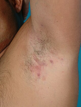
Hidradenitis suppurativa (HS), sometimes known as acne inversa or Verneuil's disease, is a long-term dermatological condition characterized by the occurrence of inflamed and swollen lumps. These are typically painful and break open, releasing fluid or pus. The areas most commonly affected are the underarms, under the breasts, and the groin. Scar tissue remains after healing. HS may significantly limit many everyday activities, for instance, walking, hugging, moving, and sitting down. Sitting disability may occur in patients with lesions in sacral, gluteal, perineal, femoral, groin or genital regions; and prolonged periods of sitting down itself can also worsen the condition of the skin of these patients.
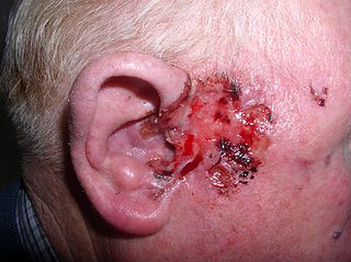
Basal-cell carcinoma (BCC), also known as basal-cell cancer, is the most common type of skin cancer. It often appears as a painless raised area of skin, which may be shiny with small blood vessels running over it. It may also present as a raised area with ulceration. Basal-cell cancer grows slowly and can damage the tissue around it, but it is unlikely to spread to distant areas or result in death.
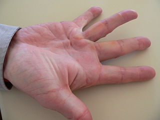
Dupuytren's contracture is a condition in which one or more fingers become progressively bent in a flexed position. It is named after Guillaume Dupuytren, who first described the underlying mechanism of action, followed by the first successful operation in 1831 and publication of the results in The Lancet in 1834. It usually begins as small, hard nodules just under the skin of the palm, then worsens over time until the fingers can no longer be fully straightened. While typically not painful, some aching or itching may be present. The ring finger followed by the little and middle fingers are most commonly affected. It can affect one or both hands. The condition can interfere with activities such as preparing food, writing, putting the hand in a tight pocket, putting on gloves, or shaking hands.

An infantile hemangioma (IH), sometimes called a strawberry mark due to appearance, is a type of benign vascular tumor or anomaly that affects babies. Other names include capillary hemangioma, strawberry hemangioma, strawberry birthmark and strawberry nevus. and formerly known as a cavernous hemangioma. They appear as a red or blue raised lesion on the skin. Typically, they begin during the first four weeks of life, growing until about five months of life, and then shrinking in size and disappearing over the next few years. Often skin changes remain after they shrink. Complications may include pain, bleeding, ulcer formation, disfigurement, or heart failure. It is the most common tumor of orbit and periorbital areas in childhood. It may occur in the skin, subcutaneous tissues and mucous membranes of oral cavities and lips as well as in extracutaneous locations including the liver and gastrointestinal tract.

Pemphigus is a rare group of blistering autoimmune diseases that affect the skin and mucous membranes. The name is derived from the Greek root pemphix, meaning "pustule".
Keratoacanthoma (KA) is a common low-grade rapidly-growing skin tumour that is believed to originate from the hair follicle and can resemble squamous cell carcinoma.

Cutis marmorata telangiectatica congenita is a rare congenital vascular disorder that usually manifests in affecting the blood vessels of the skin. The condition was first recognised and described in 1922 by Cato van Lohuizen, a Dutch pediatrician whose name was later adopted in the other common name used to describe the condition – Van Lohuizen Syndrome. CMTC is also used synonymously with congenital generalized phlebectasia, nevus vascularis reticularis, congenital phlebectasia, livedo telangiectatica, congenital livedo reticularis and Van Lohuizen syndrome.

White sponge nevus (WSN) is an autosomal dominant condition of the oral mucosa. It is caused by a mutations in certain genes coding for keratin, which causes a defect in the normal process of keratinization of the mucosa. This results in lesions which are thick, white and velvety on the inside of the cheeks within the mouth. Usually, these lesions are present from birth or develop during childhood. The condition is entirely harmless, and no treatment is required.
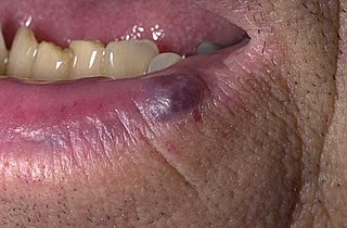
A venous lake is a generally solitary, soft, compressible, dark blue to violaceous, 0.2- to 1-cm papule commonly found on sun-exposed surfaces of the vermilion border of the lip, face and ears. Lesions generally occur among the elderly.

Cutis verticis gyrata is a medical condition usually associated with thickening of the scalp. The condition is identified by excessive thickening of the soft tissues of the scalp and characterized by ridges and furrows, which give the scalp a cerebriform appearance. Clinically, the ridges are hard and cannot be flattened on applying pressure. Patients show visible folds, ridges or creases on the surface of the top of the scalp. The number of folds can vary from two to roughly ten and are typically soft and spongy. The condition typically affects the central and rear regions of the scalp, but sometimes can involve the entire scalp.

Urbach–Wiethe disease is a very rare recessive genetic disorder, with approximately 400 reported cases since its discovery. It was first officially reported in 1929 by Erich Urbach and Camillo Wiethe, although cases may be recognized dating back as early as 1908.
Aplasia cutis congenita is a rare disorder characterized by congenital absence of skin. Ilona J. Frieden classified ACC in 1986 into 9 groups on the basis of location of the lesions and associated congenital anomalies. The scalp is the most commonly involved area with lesser involvement of trunk and extremities. Frieden classified ACC with fetus papyraceus as type 5. This type presents as truncal ACC with symmetrical absence of skin in stellate or butterfly pattern with or without involvement of proximal limbs. It is the most common congenital cicatricial alopecia, and is a congenital focal absence of epidermis with or without evidence of other layers of the skin.
Cutaneous amoebiasis, refers to a form of amoebiasis that presents primarily in the skin. It can be caused by Acanthamoeba or Entamoeba histolytica. When associated with Acanthamoeba, it is also known as "cutaneous acanthamoebiasis". Balamuthia mandrillaris can also cause cutaneous amoebiasis, but can prove fatal if the amoeba enters the bloodstream

An arsenical keratosis is a growth of keratin on the skin caused by arsenic, which occurs naturally in the earth's crust and is widely distributed in the environment, Arsenical compounds are used in industrial, agricultural, and medicinal substances. Arsenic is also found to be an environmental contaminant in drinking water and an occupational hazard for miners and glass workers. Arsenic may also causes other conditions including: Bowen's disease, cardiovascular diseases, developmental abnormalities, neurologic and neurobehavioral disorders, diabetes, hearing loss, hematologic disorders, and various types of cancer. Arsenical keratoses may persist indefinitely, and some may develop into invasive squamous cell carcinoma. Metastatic arsenic squamous cell carcinoma and arsenic-induced malignancies in internal organs such as the bladder, kidney, skin, liver, and colon, may result in death.

Frontal fibrosing alopecia is the frontotemporal hairline recession and eyebrow loss in postmenopausal women that is associated with perifollicular erythema, especially along the hairline. It is considered to be a clinical variant of lichen planopilaris.
Idiopathic scrotal calcinosis is a cutaneous condition characterized by calcification of the skin resulting from the deposition of calcium and phosphorus occurring on the scrotum. However, the levels of calcium and phosphate in the blood are normal. Idiopathic scrotal calcinosis typically affects young males, with an onset between adolescence and early adulthood. The scrotal calcinosis appears, without any symptoms, as yellowish nodules that range in size from 1 mm to several centimeters.
Garrod's pads are a cutaneous condition characterized by calluses on the dorsal aspect of the interphalangeal joints, i.e. the back side of the finger joints. They are often seen in violin, viola, and cello players, along with fiddler's neck and other dermatologic conditions peculiar to string musicians. Although Garrod's pads are conventionally described as appearing on the proximal interphalangeal joint, distal interphalangeal joint involvement has also been described.
Primary cutaneous follicle center lymphoma is a type of lymphoma. It was recognized as a distinct disease entity in the 2008 WHO classification. PCFCL had been previously conceived as a variant of follicular lymphoma (FL).