Congenital stationary night blindness (CSNB) is a rare non-progressive retinal disorder. People with CSNB often have difficulty adapting to low light situations due to impaired photoreceptortransmission. These patients may also have reduced visual acuity, myopia, nystagmus, and strabismus. CSNB has two forms -- complete, also known as type-1 (CSNB1), and incomplete, also known as type-2 (CSNB2), which are distinguished by the involvement of different retinal pathways. In CSNB1, downstream neurons called bipolar cells are unable to detect neurotransmission from photoreceptor cells. CSNB1 can be caused by mutations in various genes involved in neurotransmitter detection, including NYX. In CSNB2, the photoreceptors themselves have impaired neurotransmission function; this is caused primarily by mutations in the gene CACNA1F, which encodes a voltage-gated calcium channel important for neurotransmitter release. CSNB has been identified in horses and dogs as the result of mutations in TRPM1 (Horse, "LP")[1], GRM6 (Horse, "CSNB2")[2], and LRIT3 (Dog, CSNB)[3].
Congenital stationary night blindness (CSNB) can be inherited in an X-linked, autosomal dominant, or autosomal recessive pattern, depending on the genes involved.
Two forms of CSNB can also affect horses, one linked to the leopard complex of equine coat colors and the other found in certain horse breeds. Both are autosomal recessives.[4][5]
Symptoms and signs
The X-linked varieties of congenital stationary night blindness (CSNB) can be differentiated from the autosomal forms by the presence of myopia, which is typically absent in the autosomal forms. Patients with CSNB often have impaired night vision, myopia, reduced visual acuity, strabismus and nystagmus. Individuals with the complete form of CSNB (CSNB1) have highly impaired rod sensitivity (reduced ~300x) as well as cone dysfunction. Patients with the incomplete form can present with either myopia or hyperopia.[6]
Cause
CSNB is caused by malfunctions in neurotransmission from rod and cone photoreceptors to bipolar cells in the retina.[7] At this first synapse, information from photoreceptors is divided into two channels: ON and OFF. The ON pathway detects light onset, while the OFF pathway detects light offset.[8] The malfunctions in CSNB1 specifically affect the ON pathway, by hindering the ability of ON-type bipolar cells to detect neurotransmitter released from photoreceptors.[7] Rods, which are responsible for low-light vision, make contacts with ON-type bipolar cells only, while, cones, which are responsible for bright-light vision, make contacts with bipolar cells of both ON an OFF subtypes.[9] Because the low-light sensing rods feed only into the ON pathway, individuals with CSNB1 typically have problems with night vision, while vision in well-lit conditions is spared.[7] In CSNB2, release of neurotransmitter from photoreceptors is impaired, leading to involvement of both ON and OFF pathways.
The electroretinogram (ERG) is an important tool for diagnosing CSNB. The ERG a-wave, which reflects the function of the phototransduction cascade in response to a light flashes, is typically normal in CSNB patients, although in some cases phototransduction is also affected, leading to a reduced a-wave. The ERG b-wave, which primarily reflects the function of ON-bipolar cells, is greatly reduced in CSNB2 cases, and completely absent in CSNB1 cases.[7][10]
Genetics
Only three rhodopsin mutations have been found associated with congenital stationary night blindness (CSNB).[11] Two of these mutations are found in the second transmembrane helix of rhodopsin at Gly-90 and Thr-94. Specifically, these mutations are the Gly90Asp [12] and the Thr94Ile, which has been the most recent one reported.[13] The third mutation is Ala292Glu, and it is located in the seventh transmembrane helix, in proximity to the site of retinal attachment at Lys-296.[14] Mutations associated with CSNB affect amino acid residues near the protonated Schiff base (PSB) linkage. They are associated with changes in conformational stability and the protonated status of the PSB nitrogen.[15]
Pathophysiology
CSNB1
The complete form of X-linked congenital stationary night blindness, also known as nyctalopia, is caused by mutations in the NYX gene (Nyctalopin on X-chromosome), which encodes a small leucine-rich repeat (LRR) family protein of unknown function.[16][17] This protein consists of an N-terminal signal peptide and 11 LRRs (LRR1-11) flanked by cysteine-rich LRRs (LRRNT and LRRCT). At the C-terminus of the protein there is a putative GPI anchor site. Although the function of NYX is yet to be fully understood, it is believed to be located extracellularly. A naturally occurring deletion of 85 bases in NYX in some mice leads to the "nob" (no b-wave) phenotype, which is highly similar to that seen in CSNB1 patients.[18] NYX is expressed primarily in the rod and cone cells of the retina. There are currently almost 40 known mutations in NYX associated with CSNB1, Table 1., located throughout the protein. As the function of the nyctalopin protein is unknown, these mutations have not been further characterized. However, many of them are predicted to lead to truncated proteins that, presumably, are non-functional.
LRR: leucine-rich repeat, LRRNT and LRRCT: N- and C-terminal cysteine-rich LRRs.
CSNB2
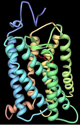
Figure 1. Schematic structure of CaV1.4 with the domains and subunits labeled.
The incomplete form of X-linked congenital stationary night blindness (CSNB2) is caused by mutations in the CACNA1F gene, which encodes the voltage-gated calcium channel CaV1.4 expressed heavily in retina.[22][23] One of the important properties of this channel is that it inactivates at an extremely low rate. This allows it to produce sustained Ca2+ entry upon depolarization. As photoreceptors depolarize in the absence of light, CaV1.4 channels operate to provide sustained neurotransmitter release upon depolarization.[24] This has been demonstrated in CACNA1F mutant mice that have markedly reduced photoreceptor calcium signals.[25] There are currently 55 mutations in CACNA1F located throughout the channel, Table 2 and Figure 1. While most of these mutations result in truncated and, likely, non-functional channels, it is expected that they prevent the ability of light to hyperpolarize photoreceptors. Of the mutations with known functional consequences, 4 produce channels that are either completely non-functional, and two that result in channels which open at far more hyperpolarized potentials than wild-type. This will result in photoreceptors that continue to release neurotransmitter even after light-induced hyperpolarization.
Table 2. Mutations in CACNA1F associated with CSNB2
1 2 3 4 Zeitz C, Robson AG, Audo I (March 2015). "Congenital stationary night blindness: an analysis and update of genotype-phenotype correlations and pathogenic mechanisms". Progress in Retinal and Eye Research. 45: 58–110. doi:10.1016/j.preteyeres.2014.09.001. PMID25307992. S2CID45696921.
↑ Euler T, Haverkamp S, Schubert T, Baden T (August 2014). "Retinal bipolar cells: elementary building blocks of vision". Nature Reviews. Neuroscience. 15 (8): 507–519. doi:10.1038/nrn3783. PMID25158357. S2CID16309488.
↑ Audo I, Robson AG, Holder GE, Moore AT (2008). "The negative ERG: clinical phenotypes and disease mechanisms of inner retinal dysfunction". Survey of Ophthalmology. 53 (1): 16–40. doi:10.1016/j.survophthal.2007.10.010. PMID18191655.
↑ N. al-Jandal, G.J. Farrar, A.S. Kiang, M.M. Humphries, N. Bannon, J.B. Findlay, P. Humphries and P.F. Kenna Hum. Mutat. 13 (1999), pp. 75–81.
↑ Dryja TP, Berson EL, Rao VR, Oprian DD (July 1993). "Heterozygous missense mutation in the rhodopsin gene as a cause of congenital stationary night blindness". Nature Genetics. 4 (3): 280–3. doi:10.1038/ng0793-280. PMID8358437. S2CID7682929.
1 2 3 4 5 Nakamura M, Ito S, Terasaki H, Miyake Y (June 2001). "Novel CACNA1F mutations in Japanese patients with incomplete congenital stationary night blindness". Investigative Ophthalmology & Visual Science. 42 (7): 1610–1616. PMID11381068.
↑ Nakamura M, Ito S, Piao CH, Terasaki H, Miyake Y (July 2003). "Retinal and optic disc atrophy associated with a CACNA1F mutation in a Japanese family". Archives of Ophthalmology. 121 (7): 1028–1033. doi:10.1001/archopht.121.7.1028. PMID12860808.
1 2 Hoda JC, Zaghetto F, Singh A, Koschak A, Striessnig J (March 2006). "Effects of congenital stationary night blindness type 2 mutations R508Q and L1364H on Cav1.4 L-type Ca2+ channel function and expression". Journal of Neurochemistry. 96 (6): 1648–1658. doi:10.1111/j.1471-4159.2006.03678.x. PMID16476079. S2CID25987619.
↑ Jacobi FK, Hamel CP, Arnaud B, Blin N, Broghammer M, Jacobi PC, etal. (May 2003). "A novel CACNA1F mutation in a french family with the incomplete type of X-linked congenital stationary night blindness". American Journal of Ophthalmology. 135 (5): 733–736. doi:10.1016/S0002-9394(02)02109-8. PMID12719097.
Rhodopsin, also known as visual purple, is a protein encoded by the RHO gene and a G-protein-coupled receptor (GPCR). It is the opsin of the rod cells in the retina and a light-sensitive receptor protein that triggers visual phototransduction in rods. Rhodopsin mediates dim light vision and thus is extremely sensitive to light. When rhodopsin is exposed to light, it immediately photobleaches. In humans, it is regenerated fully in about 30 minutes, after which the rods are more sensitive. Defects in the rhodopsin gene cause eye diseases such as retinitis pigmentosa and congenital stationary night blindness.
Achromatopsia, also known as Rod monochromacy, is a medical syndrome that exhibits symptoms relating to five conditions, most notably monochromacy. Historically, the name referred to monochromacy in general, but now typically refers only to an autosomal recessive congenital color vision condition. The term is also used to describe cerebral achromatopsia, though monochromacy is usually the only common symptom. The conditions include: monochromatic color blindness, poor visual acuity, and day-blindness. The syndrome is also present in an incomplete form that exhibits milder symptoms, including residual color vision. Achromatopsia is estimated to affect 1 in 30,000 live births worldwide.
Retinitis pigmentosa (RP) is a genetic disorder of the eyes that causes loss of vision. Symptoms include trouble seeing at night and decreasing peripheral vision. As peripheral vision worsens, people may experience "tunnel vision". Complete blindness is uncommon. Onset of symptoms is generally gradual and often begins in childhood.
A photoreceptor cell is a specialized type of neuroepithelial cell found in the retina that is capable of visual phototransduction. The great biological importance of photoreceptors is that they convert light into signals that can stimulate biological processes. To be more specific, photoreceptor proteins in the cell absorb photons, triggering a change in the cell's membrane potential.
Rod cells are photoreceptor cells in the retina of the eye that can function in lower light better than the other type of visual photoreceptor, cone cells. Rods are usually found concentrated at the outer edges of the retina and are used in peripheral vision. On average, there are approximately 92 million rod cells in the human retina. Rod cells are more sensitive than cone cells and are almost entirely responsible for night vision. However, rods have little role in color vision, which is the main reason why colors are much less apparent in dim light.
Melanopsin is a type of photopigment belonging to a larger family of light-sensitive retinal proteins called opsins and encoded by the gene Opn4. In the mammalian retina, there are two additional categories of opsins, both involved in the formation of visual images: rhodopsin and photopsin in the rod and cone photoreceptor cells, respectively.
Nyctalopia, also called night-blindness, is a condition making it difficult or impossible to see in relatively low light. It is a symptom of several eye diseases. Night blindness may exist from birth, or be caused by injury or malnutrition. It can be described as insufficient adaptation to darkness.
Visual phototransduction is the sensory transduction process of the visual system by which light is detected by photoreceptor cells in the vertebrate retina. A photon is absorbed by a retinal chromophore, which initiates a signal cascade through several intermediate cells, then through the retinal ganglion cells (RGCs) comprising the optic nerve.
Rhodopsin kinase is a serine/threonine-specific protein kinase involved in phototransduction. This enzyme catalyses the following chemical reaction:
The photoreceptor cell-specific nuclear receptor (PNR), also known as NR2E3, is a protein that in humans is encoded by the NR2E3 gene. PNR is a member of the nuclear receptor super family of intracellular transcription factors.
Transient receptor potential cation channel subfamily M member 1 is a protein that in humans is encoded by the TRPM1 gene.
Glutamate receptor, metabotropic 6, also known as GRM6 or mGluR6, is a protein which in humans is encoded by the GRM6 gene.
Cav1.4 also known as the calcium channel, voltage-dependent, L type, alpha 1F subunit (CACNA1F), is a human gene.
Rod cGMP-specific 3',5'-cyclic phosphodiesterase subunit beta is the beta subunit of the protein complex PDE6 that is encoded by the PDE6B gene. PDE6 is crucial in transmission and amplification of visual signal. The existence of this beta subunit is essential for normal PDE6 functioning. Mutations in this subunit are responsible for retinal degeneration such as retinitis pigmentosa or congenital stationary night blindness.
S-arrestin is a protein that in humans is encoded by the SAG gene.
Neural retina-specific leucine zipper protein is a protein that in humans is encoded by the NRL gene.
Nyctalopin is a protein located on the surface of photoreceptor-to-ON bipolar cell synapse in the retina. It is composed of 481 amino acids. and is encoded in human by the NYX gene. This gene is found on the chromosome X and has two exons. This protein is a leucine-rich proteoglycan which is expressed in the eye, spleen and brain in mice. Mutations in this gene cause congenital stationary night blindness in humans (CSNB). which is a stable retinal disorder. The consequence of this mutation results in an abnormal night vision. Nyctalopin is critical due to the fact that it generates a depolarizing bipolar cell response due to the mutation on the NYX gene. Most of the time, CSNB are associated to hygh myopia which is the result of a mutation on the same gene. Several mutations can occur on the NYX gene resulting on many form of night blindness in humans. Some studies show that these mutations are more present in Asian population than in Caucasian population. A mouse strain called nob carries a spontaneous mutation leading to a frameshift in this gene. These mice are used as an animal model for congenital stationary night blindness.
Nystagmus is a condition of involuntary eye movement. People can be born with it but more commonly acquire it in infancy or later in life. In many cases it may result in reduced or limited vision.
Retinal degeneration is a retinopathy which consists in the deterioration of the retina caused by the progressive death of its cells. There are several reasons for retinal degeneration, including artery or vein occlusion, diabetic retinopathy, R.L.F./R.O.P., or disease. These may present in many different ways such as impaired vision, night blindness, retinal detachment, light sensitivity, tunnel vision, and loss of peripheral vision to total loss of vision. Of the retinal degenerative diseases retinitis pigmentosa (RP) is a very important example.
Retinal gene therapy holds a promise in treating different forms of non-inherited and inherited blindness.
This page is based on this Wikipedia article Text is available under the CC BY-SA 4.0 license; additional terms may apply. Images, videos and audio are available under their respective licenses.