
In humans and other primates, the knee joins the thigh with the leg and consists of two joints: one between the femur and tibia, and one between the femur and patella. It is the largest joint in the human body. The knee is a modified hinge joint, which permits flexion and extension as well as slight internal and external rotation. The knee is vulnerable to injury and to the development of osteoarthritis.

A bunion, also known as hallux valgus, is a deformity of the joint connecting the big toe to the foot. The big toe often bends towards the other toes and the joint becomes red and painful. The onset of bunions is typically gradual. Complications may include bursitis or arthritis.
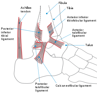
The ankle, or the talocrural region, is the region where the foot and the leg meet. The ankle includes three joints: the ankle joint proper or talocrural joint, the subtalar joint, and the inferior tibiofibular joint. The movements produced at this joint are dorsiflexion and plantarflexion of the foot. In common usage, the term ankle refers exclusively to the ankle region. In medical terminology, "ankle" can refer broadly to the region or specifically to the talocrural joint.

Kyphosis is an abnormally excessive convex curvature of the spine as it occurs in the thoracic and sacral regions. Abnormal inward concave lordotic curving of the cervical and lumbar regions of the spine is called lordosis. It can result from degenerative disc disease; developmental abnormalities, most commonly Scheuermann's disease; osteoporosis with compression fractures of the vertebra; multiple myeloma; or trauma. A normal thoracic spine extends from the 1st thoracic to the 12th thoracic vertebra and should have a slight kyphotic angle, ranging from 20° to 45°. When the "roundness" of the upper spine increases past 45° it is called kyphosis or "hyperkyphosis". Scheuermann's kyphosis is the most classic form of hyperkyphosis and is the result of wedged vertebrae that develop during adolescence. The cause is not currently known and the condition appears to be multifactorial and is seen more frequently in males than females.

Coxa vara is a deformity of the hip, whereby the angle between the head and the shaft of the femur is reduced to less than 120 degrees. This results in the leg being shortened and the development of a limp. It may be congenital and is commonly caused by injury, such as a fracture. It can also occur when the bone tissue in the neck of the femur is softer than normal, causing it to bend under the weight of the body. This may either be congenital or the result of a bone disorder. The most common cause of coxa vara is either congenital or developmental. Other common causes include metabolic bone diseases, post-Perthes deformity, osteomyelitis, and post traumatic. Shepherd's Crook deformity is a severe form of coxa vara where the proximal femur is severely deformed with a reduction in the neck shaft angle beyond 90 degrees. It is most commonly a sequela of osteogenesis imperfecta, Pagets disease, osteomyelitis, tumour and tumour-like conditions.
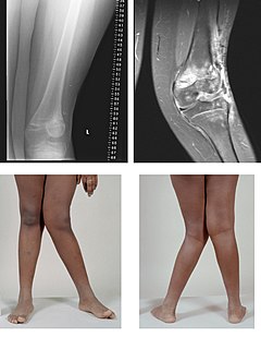
Genu valgum, commonly called "knock-knee", is a condition in which the knees angle in and touch each other when the legs are straightened. Individuals with severe valgus deformities are typically unable to touch their feet together while simultaneously straightening the legs. The term originates from the Latin genu, 'knee', and valgus which actually means 'bent outwards', but in this case, it is used to describe the distal portion of the knee joint which bends outwards and thus the proximal portion seems to be bent inwards. For citation and more information on uses of the words Valgus and Varus, see varus deformity.

Genu varum is a varus deformity marked by (outward) bowing at the knee, which means that the lower leg is angled inward (medially) in relation to the thigh's axis, giving the limb overall the appearance of an archer's bow. Usually medial angulation of both lower limb bones is involved.

A varus deformity is an excessive inward angulation of the distal segment of a bone or joint. The opposite of varus is called valgus. EX: Varus deformity results in a decreased Q angle of the knee joint.
A valgus deformity is a condition in which the bone segment distal to a joint is angled outward, that is, angled laterally, away from the body's midline. The opposite deformation, where the twist or angulation is directed medially, toward the center of the body, is called varus. Common causes of valgus knee in adults include arthritis of the knee and traumatic injuries.
Pes or the acronym PES may refer to:

A distal radius fracture, also known as wrist fracture, is a break of the part of the radius bone which is close to the wrist. Symptoms include pain, bruising, and rapid-onset swelling. The wrist may be deformed. The ulna bone may also be broken.
The knee examination, in medicine and physiotherapy, is performed as part of a physical examination, or when a patient presents with knee pain or a history that suggests a pathology of the knee joint.
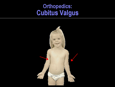
Cubitus varus is a common deformity in which the extended forearm is deviated towards midline of the body.

Hallux varus is a deformity of the great toe joint where the hallux is deviated medially away from the first metatarsal bone. The hallux usually moves in the transverse plane. Unlike hallux valgus, also known as hallux abducto valgus or bunion, hallux varus is uncommon in the West but it is common in cultures where the population remains unshod.

A supracondylar humerus fracture is a fracture of the distal humerus just above the elbow joint. The fracture is usually transverse or oblique and above the medial and lateral condyles and epicondyles. This fracture pattern is relatively rare in adults, but is the most common type of elbow fracture in children. In children, many of these fractures are non-displaced and can be treated with casting. Some are angulated or displaced and are best treated with surgery. In children, most of these fractures can be treated effectively with expectation for full recovery. Some of these injuries can be complicated by poor healing or by associated blood vessel or nerve injuries with serious complications.

A tibial plateau fracture is a break of the upper part of the tibia (shinbone) that involves the knee joint. Symptoms include pain, swelling, and a decreased ability to move the knee. People are generally unable to walk. Complication may include injury to the artery or nerve, arthritis, and compartment syndrome.
Seaver Cassidy syndrome is a very rare disorder characterized by certain facial, genital, and skeletal deformities, as well as an unusual susceptibility to bleeding. Seaver Cassidy syndrome was first described in 1991 by Laurie Seaver and Suzanne Cassidy.

Syndesmosis procedure is one of the more than 20 bunion surgeries currently being performed. While the majority of bunion surgeries involve the breaking and shifting of bones, syndesmosis procedure is one of few surgical techniques that use a soft tissue or non-osteotomy (non-bone-breaking) approach to afford the same correction. More than 130 different surgical techniques have been described for correction of one single condition of the foot: the bunion deformity.
Orthopedic surgery is the branch of surgery concerned with conditions involving the musculoskeletal system. Orthopedic surgeons use both surgical and nonsurgical means to treat musculoskeletal injuries, sports injuries, degenerative diseases, infections, bone tumours, and congenital limb deformities. Trauma surgery and traumatology is a sub-specialty dealing with the operative management of fractures, major trauma and the multiply-injured patient.

Angular limb deformity is a pathological deformity in the spatial alignment of any limb in quadrupedal animals. The term encompasses any condition in such an animal wherein a limb is not straight. It most commonly occurs in the carpal joint of the forelimbs, manifesting as the limb pointing outward or inward, deviating from normal development.
