
The tibia, also known as the shinbone or shankbone, is the larger, stronger, and anterior (frontal) of the two bones in the leg below the knee in vertebrates ; it connects the knee with the ankle. The tibia is found on the medial side of the leg next to the fibula and closer to the median plane. The tibia is connected to the fibula by the interosseous membrane of leg, forming a type of fibrous joint called a syndesmosis with very little movement. The tibia is named for the flute tibia. It is the second largest bone in the human body, after the femur. The leg bones are the strongest long bones as they support the rest of the body.

The fibula or calf bone is a leg bone on the lateral side of the tibia, to which it is connected above and below. It is the smaller of the two bones and, in proportion to its length, the most slender of all the long bones. Its upper extremity is small, placed toward the back of the head of the tibia, below the knee joint and excluded from the formation of this joint. Its lower extremity inclines a little forward, so as to be on a plane anterior to that of the upper end; it projects below the tibia and forms the lateral part of the ankle joint.

The ankle, or the talocrural region, or the jumping bone (informal) is the area where the foot and the leg meet. The ankle includes three joints: the ankle joint proper or talocrural joint, the subtalar joint, and the inferior tibiofibular joint. The movements produced at this joint are dorsiflexion and plantarflexion of the foot. In common usage, the term ankle refers exclusively to the ankle region. In medical terminology, "ankle" can refer broadly to the region or specifically to the talocrural joint.
In anatomy, a styloid process, usually serving as points of attachment for muscles, refers to the slender, pointed process (protrusion) of:
The anterior tibial artery is an artery of the leg. It carries blood to the anterior compartment of the leg and dorsal surface of the foot, from the popliteal artery.

Proximal femoral focal deficiency (PFFD), also known as Congenital Femoral Deficiency (CFD), is a rare, non-hereditary birth defect that affects the pelvis, particularly the hip bone, and the proximal femur. The disorder may affect one side or both, with the hip being deformed and the leg shortened.

Amelia is the birth defect of lacking one or more limbs. The term may be modified to indicate the number of legs or arms missing at birth, such as tetra-amelia for the absence of all four limbs. The term is from Greek ἀ- 'lack of' plus μέλος 'limb'.
Meromelia is a birth defect characterized by the lacking of a part, but not all, of one or more limbs with the presence of a hand or foot. It results in a shrunken and deformed extremity.

Dysmelia is a congenital disorder of a limb resulting from a disturbance in embryonic development.

Fibular hemimelia or longitudinal fibular deficiency is "the congenital absence of the fibula and it is the most common congenital absence of long bone of the extremities." It is the shortening of the fibula at birth, or the complete lack thereof. Fibular hemimelia often causes severe knee instability due to deficiencies of the ligaments. Severe forms of fibula hemimelia can result in a malformed ankle with limited motion and stability. Fusion or absence of two or more toes are also common. In humans, the disorder can be noted by ultrasound in utero to prepare for amputation after birth or complex bone lengthening surgery. The amputation usually takes place at six months with removal of portions of the legs to prepare them for prosthetic use. The other treatments, which include repeated corrective osteotomies and leg-lengthening surgery, are costly and associated with residual deformity.

Dror Paley is a Canadian-trained orthopedic surgeon, who specializes in limb lengthening and deformity correction procedures.
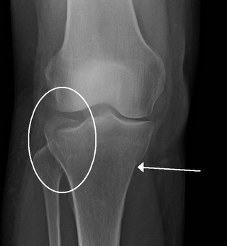
A crus fracture is a fracture of the lower legs bones meaning either or both of the tibia and fibula.

In anatomy, the fibular artery, also known as the peroneal artery, supplies blood to the lateral compartment of the leg. It arises from the tibial-fibular trunk.
Radial dysplasia, also known as radial club hand or radial longitudinal deficiency, is a congenital difference occurring in a longitudinal direction resulting in radial deviation of the wrist and shortening of the forearm. It can occur in different ways, from a minor anomaly to complete absence of the radius, radial side of the carpal bones and thumb. Hypoplasia of the distal humerus may be present as well and can lead to stiffness of the elbow. Radial deviation of the wrist is caused by lack of support to the carpus, radial deviation may be reinforced if forearm muscles are functioning poorly or have abnormal insertions. Although radial longitudinal deficiency is often bilateral, the extent of involvement is most often asymmetric.
Hecht Scott syndrome is a rare genetic disease that causes congenital limb formation. The main characterisation is the aplasia or hypoplasia of bones of the limb. It is currently presenting in less than 1 in 1,000,000 newborns. It has been known to be more commonly present in males. It was first diagnosed in 2005 by Courtens et al. who recognised the malformations with his present case and four others that were similarly described in literature.
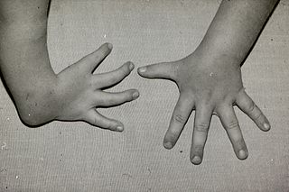
Ulnar dysplasia also known as ulnar longitudinal deficiency, ulnar club hand or ulnar aplasia/hypoplasia is a rare congenital malformation which consists of an underdeveloped or missing ulnae bone, causing an ulnar deviation of the entire wrist. The muscles and nerves in the hand may be missing or unbalanced. In severe cases, ulnar digits may be missing. Sometimes, radial dysplasia occurs alongside this malformation. This condition occurs in 1 in 100,000 live births. Sometimes, other orthopedic problems occur alongside this malformation, such as scoliosis.

Tibial hemimelia-polysyndactyly-triphalangeal thumb syndrome is a rare genetic limb malformation syndrome which is characterized by thumb triphalangy, polysyndactyly of the hand and foot, and hypoplasia/aplasia of the tibia bone. Additional features include short stature, radio-ulnar synostosis, ectrodactyly and abnormalities of the carpals and metatarsals. Only 19 affected families worldwide have been recorded in medical literature. It is associated with a heterozygous base pair substitution of A to G in position 404–406, located on intron 5 in the LMBR1 gene.
Gollop-Wolfgang complex is a very rare genetic disorder which is characterized by skeletal and digital anomalies.

