Carpenter syndrome

Last updated
Carpenter syndrome
Other namesAcrocephalopolysyndactyly type II
Carpenter Syndrome 1.jpg
Original case described by Carpenter, 1909
Specialty Medical genetics   OOjs UI icon edit-ltr-progressive.svg

Carpenter syndrome, also called acrocephalopolysyndactyly type II, [1] is an extremely rare autosomal recessive [2] congenital disorder characterized by craniofacial malformations, obesity, syndactyly, and polydactyly. [2] Acrocephalopolysyndactyly is a variation of acrocephalosyndactyly that presents with polydactyly.

Contents

It was first characterized in 1909, and is named for George Alfred Carpenter. [3] [4]

Presentation

Polydactyly in original case, 1909 Carpenter Syndrome 5.jpg
Polydactyly in original case, 1909
Symptoms of Carpenter syndrome in an 8-year-old boy, 1919 Acrocephalie 1.jpg
Symptoms of Carpenter syndrome in an 8-year-old boy, 1919

Carpenter syndrome presents several features: [5]

Intellectual disability is also common with the disorder, although some patients may have average intellectual capacity. [6]

Description

Carpenter syndrome belongs to a rare genetic disorder known as acrocephalosyndactyly, (ACPS) (RN, 2007). There were originally five types of ACPS, but this number has been decreased because they have been found to be closely related to one another or to other disorders (Paul A. Johnson, 2002). The most common physical manifestation of Carpenter syndrome is early fusing of the fibrous cranial sutures which results in an abnormally pointed head. The fusion of the skull bones is evident from birth (National Organization for Rare Disorders, Inc., 2008). Babies' mobile cranial bones form a cone shape as they pass through the birth canal and soon thereafter return to a normal shape; however, a baby affected by carpenter syndrome maintains a cone shaped head.[ citation needed ]

A baby affected by Carpenter syndrome will also display malformations of the face. An individual affected by the syndrome may have broad cheeks, a flat nasal bridge, and a wide upturned nose with abnormally large nasal openings. Their ears will most commonly be low, unevenly set, and malformed in structure. In addition to these facial abnormalities, individuals also have an underdeveloped maxilla and/or mandible with a highly arched and narrow palate which makes speech a very difficult skill to master. Teeth are usually very late to come in and will be undersized and spaced far apart.[ citation needed ]

Other physical abnormalities associated with Carpenter syndrome include extra digits. Extra toes are more commonly seen than fingers. Often both the toes and fingers are webbed, a process that occurs before the sixth week gestational period. Often their digits will be abnormally short, and the fingers are commonly missing an interphalangeal joint. Roughly half of the babies born with Carpenter syndrome have some type of heart defect, and seventy five percent of individuals with this disease will experience some degree of development delay due to mild intellectual disability (Carpenter syndrome-description).[ citation needed ]

Genetics

Carpenter syndrome has an autosomal recessive pattern of inheritance. Autorecessive.svg
Carpenter syndrome has an autosomal recessive pattern of inheritance.

Carpenter syndrome has been associated with mutations in the RAB23 gene, [7] which is located on chromosome 6 in humans. Additionally, three key SNPs in the MEGF8 gene, [8] located on chromosome 19 at 19q13.2, have been identified as primary causes of Carpenter syndrome.

Diagnosis

The diagnosis of Carpenter syndrome is made based on the presence of the bicoronal and sagittal skull malformations, which results in a pointed, cone-shaped or short, broad head. The diagnosis is also made based on the presence of extra or fused digits. X rays and/ or CT scans of the skull may be performed in order to accurately diagnose the individual; however, other genetic disorders, which have available genetic tests, are also characterized by skull malformations. A positive result on these tests can rule out a Carpenter syndrome diagnosis.[ citation needed ]

Types

The primary diagnostic factor is a malformation of the skull. The two most common types of craniosynostosis are sagittal and bicoronal. Sagittal craniosynostosis manifests itself by causing a long narrow skull, resembling a football. It is quantitatively determined by measuring the anterior to posterior (front to back) diameter of the skull. An increased A-P diameter indicates a malformed fusion of the sagittal suture. Individuals affected with sagittal craniosynostosis have narrow, prominent foreheads and the back of the head is much larger than normal. The "soft spot" is very small or missing altogether with this particular type of craniosynostosis.[ citation needed ]

The second common type of skull malformation is bicoronal craniosynostosis is characterized by a wide, short skull. In this particular type of craniosynostosis the A-P diameter is smaller than in normal individuals. These individuals have malformed eye sockets and foreheads. The eye sockets are much smaller than normal and often cause visual impairment. Complications may include damage to the optic nerve, resulting in a decrease in visual clarity, bulging eyeballs as a result of shallow eye orbits which usually causes some sort of damage to the cornea (the outer layer of the eye). Bicoronal craniosynostosis may also result in widely spaced eyes and narrowing of the sinuses and tear ducts that may result in inflammation of the mucous membranes of the exposed portion of the eye.[ citation needed ]

In addition to the previously named complications of bicoronal craniosynostosis, many babies will also be affected by hydrocephalus, more commonly known as water on the brain. Hydrocephalus results in increased pressure on the brain which can cause permanent brain damage if not treated promptly. An abnormally highly arched palate is also seen in affected individuals causing dental problems and the thrusting forward of the lower jaw. Individuals affected by Carpenter syndrome often experience cutaneous syndactyly (fusion of the digits) or polydactyly (presence of extra digits) of the toes more often than fingers. Individuals also have short fingers. Approximately one third of individuals born with Carpenter syndrome have a type of heart defect. Commonly seen heart defects may include: narrowing of the pulmonary artery, transposition of the major blood vessels, or the presence of an abnormally large vena cava, which delivers blood back to the heart from the head, neck, and upper limbs. The testes of males affected by Carpenter syndrome may also fail to descend (Paul A. Johnson, 2002).

Treatment

Operations to correct the malformations of the skull should be performed within the first year of infancy in patients affected by Carpenter syndrome. Performing surgery at a young age increases the likelihood of obtaining a greatly improved appearance of the head because modifying bone is much easier to do when the skull is still constantly growing and changing. [9] In surgery the doctor breaks the fused sutures to allow for brain growth. Doctors remove the cranial plates of the skull, reshape them and replace them back onto the skull in an attempt to reshape the head to appear more normal. Although the sutures are broken during surgery they will quickly refuse, and in some cases holes form in the plates allowing cerebral spinal fluid to escape into cyst like structures on the external surface of the head. [10]

If an individual with Carpenter syndrome has a serious heart defect they will require surgery to correct the malformation of the heart. Other elective surgeries may also be performed. Some parents opt to have their child's webbed fingers or toes separated which improves their appearance but not necessarily the functionality of the digits. In order to address the occupational challenges of the disease, many children with Carpenter Syndrome go through speech and occupational therapy in order to achieve more independence in everyday tasks and activities (RN, 2007).

In order to address the vision problems that are associated with bicoronal craniosynostosis, the individual must seek consultation from an ophthalmologist. If the palate is severely affected dental consultation may be necessary to correct the malformation. Obesity is often associated with Carpenter syndrome, so a lifelong diet plan is often utilized to maintain a healthy weight. In addition surgery must be performed if the testes fail to descend (Paul A. Johnson, 2002). If the procedure is not performed the individual will become infertile.

Occurrence

There are approximately three hundred known cases of Carpenter syndrome in the United States. Only 1 in 1 million live births will result in an infant affected by Carpenter syndrome (RN, 2007).

Carpenter syndrome is an autosomal recessive disease which means both parents must have the faulty genes in order to pass the disease onto their children. Even if both parents possess the faulty gene there is still only a twenty five percent chance that they will produce a child affected by the syndrome. Their children who do not have the disease will still be carriers and possess the ability to pass the disease onto their offspring if their spouse is also a carrier of the particular gene. [9]

See also

Related Research Articles

<span class="mw-page-title-main">Polydactyly</span> Physical anomaly involving extra fingers or toes

Polydactyly or polydactylism, also known as hyperdactyly, is an anomaly in humans and animals resulting in supernumerary fingers and/or toes. Polydactyly is the opposite of oligodactyly.

<span class="mw-page-title-main">Scaphocephaly</span> Cephalic disorder involving premature fusion of the sagittal suture

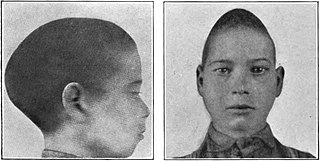
Scaphocephaly, or sagittal craniosynostosis, is a type of cephalic disorder which occurs when there is a premature fusion of the sagittal suture. Premature closure results in limited lateral expansion of the skull resulting in a characteristic long, narrow head. The skull base is typically spared.

<span class="mw-page-title-main">Crouzon syndrome</span> Genetic disorder of the skull and face

Crouzon syndrome is an autosomal dominant genetic disorder known as a branchial arch syndrome. Specifically, this syndrome affects the first branchial arch, which is the precursor of the maxilla and mandible. Since the branchial arches are important developmental features in a growing embryo, disturbances in their development create lasting and widespread effects.

<span class="mw-page-title-main">Apert syndrome</span> Congenital disorder of the skull and digits

Apert syndrome is a form of acrocephalosyndactyly, a congenital disorder characterized by malformations of the skull, face, hands and feet. It is classified as a branchial arch syndrome, affecting the first branchial arch, the precursor of the maxilla and mandible. Disturbances in the development of the branchial arches in fetal development create lasting and widespread effects.

<span class="mw-page-title-main">Craniosynostosis</span> Premature fusion of bones in the skull

Craniosynostosis is a condition in which one or more of the fibrous sutures in a young infant's skull prematurely fuses by turning into bone (ossification), thereby changing the growth pattern of the skull. Because the skull cannot expand perpendicular to the fused suture, it compensates by growing more in the direction parallel to the closed sutures. Sometimes the resulting growth pattern provides the necessary space for the growing brain, but results in an abnormal head shape and abnormal facial features. In cases in which the compensation does not effectively provide enough space for the growing brain, craniosynostosis results in increased intracranial pressure leading possibly to visual impairment, sleeping impairment, eating difficulties, or an impairment of mental development combined with a significant reduction in IQ.

<span class="mw-page-title-main">Saethre–Chotzen syndrome</span> Medical condition

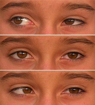
Saethre–Chotzen syndrome (SCS), also known as acrocephalosyndactyly type III, is a rare congenital disorder associated with craniosynostosis. This affects the shape of the head and face, resulting in a cone-shaped head and an asymmetrical face. Individuals with SCS also have droopy eyelids (ptosis), widely spaced eyes (hypertelorism), and minor abnormalities of the hands and feet (syndactyly). Individuals with more severe cases of SCS may have mild to moderate intellectual or learning disabilities. Depending on the level of severity, some individuals with SCS may require some form of medical or surgical intervention. Most individuals with SCS live fairly normal lives, regardless of whether medical treatment is needed or not.

<span class="mw-page-title-main">Pfeiffer syndrome</span> Genetic disorder of the skull

Pfeiffer syndrome is a rare genetic disorder, characterized by the premature fusion of certain bones of the skull (craniosynostosis), which affects the shape of the head and face. The syndrome includes abnormalities of the hands and feet, such as wide and deviated thumbs and big toes.

<span class="mw-page-title-main">Greig cephalopolysyndactyly syndrome</span> Medical condition

Greig cephalopolysyndactyly syndrome is a disorder that affects development of the limbs, head, and face. The features of this syndrome are highly variable, ranging from very mild to severe. People with this condition typically have one or more extra fingers or toes (polydactyly) or an abnormally wide thumb or big toe (hallux).

<span class="mw-page-title-main">Duane-radial ray syndrome</span> Medical condition

Duane-radial ray syndrome, also known as Okihiro Syndrome, is a rare autosomal dominant disorder that primarily affects the eyes and causes abnormalities of bones in the arms and hands. This disorder is considered to be a SALL4-related disorder due to the SALL4 gene mutations leading to these abnormalities. It is diagnosed by clinical findings on a physical exam as well as genetic testing and imaging. After being diagnosed, there are other evaluations that one may go through in order to determine the extent of the disease. There are various treatments for the symptoms of this disorder.

<span class="mw-page-title-main">Synostosis</span>

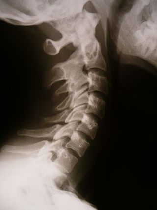
Synostosis is fusion of two or more bones. It can be normal in puberty, fusion of the epiphyseal plate to become the epiphyseal line, or abnormal. When synostosis is abnormal it is a type of dysostosis. Examples of synostoses include:

<span class="mw-page-title-main">Acrocephalosyndactyly</span> Group of diseases

Acrocephalosyndactyly is a group of autosomal dominant congenital disorders characterized by craniofacial (craniosynostosis) and hand and foot (syndactyly) abnormalities. When polydactyly is present, the classification is acrocephalopolysyndactyly. Acrocephalosyndactyly is mainly diagnosed postnatally, although prenatal diagnosis is possible if the mutation is known to be within the family genome. Treatment often involves surgery in early childhood to correct for craniosynostosis and syndactyly.

<span class="mw-page-title-main">Muenke syndrome</span> Medical condition

Muenke syndrome, also known as FGFR3-related craniosynostosis, is a human specific condition characterized by the premature closure of certain bones of the skull during development, which affects the shape of the head and face. First described by Maximilian Muenke, the syndrome occurs in about 1 in 30,000 newborns. This condition accounts for an estimated 8 percent of all cases of craniosynostosis.

Pediatric plastic surgery is plastic surgery performed on children. Its procedures are most often conducted for reconstructive or cosmetic purposes. In children, this line is often blurred, as many congenital deformities impair physical function as well as aesthetics.

<span class="mw-page-title-main">Frontonasal dysplasia</span> Medical condition

Frontonasal dysplasia (FND) is a congenital malformation of the midface. For the diagnosis of FND, a patient should present at least two of the following characteristics: hypertelorism, a wide nasal root, vertical midline cleft of the nose and/or upper lip, cleft of the wings of the nose, malformed nasal tip, encephalocele or V-shaped hair pattern on the forehead. The cause of FND remains unknown. FND seems to be sporadic (random) and multiple environmental factors are suggested as possible causes for the syndrome. However, in some families multiple cases of FND were reported, which suggests a genetic cause of FND.

<span class="mw-page-title-main">Roberts syndrome</span> Medical condition

Roberts syndrome, or sometimes called pseudothalidomide syndrome, is an extremely rare autosomal recessive genetic disorder that is characterized by mild to severe prenatal retardation or disruption of cell division, leading to malformation of the bones in the skull, face, arms, and legs.

<span class="mw-page-title-main">Ectrodactyly</span> Medical condition

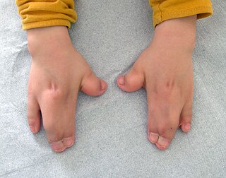
Ectrodactyly, split hand, or cleft hand involves the deficiency or absence of one or more central digits of the hand or foot and is also known as split hand/split foot malformation (SHFM). The hands and feet of people with ectrodactyly (ectrodactyls) are often described as "claw-like" and may include only the thumb and one finger with similar abnormalities of the feet.

<span class="mw-page-title-main">McGillivray syndrome</span> Medical condition

McGillivray syndrome is a rare syndrome characterized mainly by heart defects, skull and facial abnormalities and ambiguous genitalia. The symptoms of this syndrome are ventricular septal defect, patent ductus arteriosus, small jaw, undescended testes, and webbed fingers. Beside to these symptoms there are more symptoms which is related with bone structure and misshape.

<span class="mw-page-title-main">Polysyndactyly</span> Medical condition

Polysyndactyly is a congenital anomaly, combining polydactyly and syndactyly, in which affected individuals have an extra finger or toe that is connected, via fusing or webbing, to an adjacent digit.

<span class="mw-page-title-main">Kleeblattschaedel</span> Medical condition

Kleeblattschaedel is a rare malformation of the head where there is a protrusion of the skull and broadening of the face. This condition is a severe type of craniosynostosis.

Filippi syndrome, also known as Syndactyly Type I with Microcephaly and Mental Retardation, is a very rare autosomal recessive genetic disease. Only a very limited number of cases have been reported to date. Filippi Syndrome is associated with diverse symptoms of varying severity across affected individuals, for example malformation of digits, craniofacial abnormalities, intellectual disability, and growth retardation. The diagnosis of Filippi Syndrome can be done through clinical observation, radiography, and genetic testing. Filippi Syndrome cannot be cured directly as of 2022, hence the main focus of treatments is on tackling the symptoms observed on affected individuals. It was first reported in 1985.

References

  1. Online Mendelian Inheritance in Man (OMIM): 201000
  2. 1 2 Perlyn, Ca; Marsh, Jl (March 1909). "Craniofacial dysmorphology of Carpenter syndrome: lessons from three affected siblings". Plastic and Reconstructive Surgery. 121 (3): 971–81. doi:10.1097/01.prs.0000299284.92862.6c. PMID   18317146. S2CID   21493967.
  3. Carpenter G (1909). "Case of acrocephaly with other congenital malformations". Proceedings of the Royal Society of Medicine. 2 (Sect Study Dis Child): 45–53, 199–201. doi:10.1177/003591570900201418. PMC   2047261 . PMID   19974019.
  4. Beighton, Peter; Beighton, Greta (2012-12-06). The Man Behind the Syndrome. Springer Science & Business Media. p. 25. ISBN   9781447114154 . Retrieved 7 August 2018.
  5. "Carpenter syndrome". Genetic and Rare Diseases Information Center. National Center for Advancing Transnational Sciences. Retrieved 2021-02-19.
  6. Frias, Jl; Felman, Ah; Rosenbloom, Al; Finkelstein, Sn; Hoyt, Wf; Hall, Bd (1978). "Normal intelligence in two children with Carpenter syndrome". American Journal of Medical Genetics. 2 (2): 191–9. doi:10.1002/ajmg.1320020210. PMID   263437.
  7. Jenkins, D; Seelow, D; Jehee, Fs; Perlyn, Ca; Alonso, Lg; Bueno, Df; Donnai, D; Josifova, D; Mathijssen, Im; Morton, Je; Orstavik, Kh; Sweeney, E; Wall, Sa; Marsh, Jl; Nurnberg, P; Passos-Bueno, Mr; Wilkie, Ao (June 2007). "RAB23 mutations in Carpenter syndrome imply an unexpected role for hedgehog signaling in cranial-suture development and obesity". American Journal of Human Genetics. 80 (6): 1162–70. doi:10.1086/518047. PMC   1867103 . PMID   17503333.
  8. Twigg, SR; Lloyd, D; Jenkins, D; Elçioglu, NE; Cooper, CD; Al-Sannaa, N; Annagür, A; Gillessen-Kaesbach, G; Hüning, I; Knight, SJ; Goodship, JA; Keavney, BD; Beales, PL; Gileadi, O; McGowan, SJ; Wilkie, AO (Nov 2, 2012). "Mutations in multidomain protein MEGF8 identify a Carpenter syndrome subtype associated with defective lateralization". American Journal of Human Genetics. 91 (5): 897–905. doi:10.1016/j.ajhg.2012.08.027. PMC   3487118 . PMID   23063620.
  9. 1 2 Paul A. Johnson, 2002
  10. Carpenter Syndrom-What is it?, 2007