Noonan syndrome

Last updated
Noonan syndrome
Other namesMale Turner syndrome, Noonan–Ehmke syndrome, Turner-like syndrome, Ullrich–Noonan syndrome [1]
Noonan syndrome.PNG
A 12-year-old girl with Noonan syndrome, displaying typical webbed neck and double structural curve with rib deformity.
Specialty Medical genetics, pediatrics
Symptoms Mildly unusual facial features, short height, congenital heart disease, bleeding problems, skeletal malformations [1]
Complications Leukemia [1]
Usual onsetPresent at birth [2]
TypesType 1 to 6 [3]
Causes Genetic mutation (autosomal dominant) [1]
Diagnostic method Suspected based on symptoms, confirmed with genetic testing [4] [2]
Differential diagnosis Cardiofaciocutaneous syndrome, Turner syndrome, Costello syndrome, neurofibromatosis type 1 [2] [3]
TreatmentBased on the symptoms [3]
Medication Growth hormone [3]
Prognosis Depends on the severity of heart problems [3]
Frequency1 in 1000 (1 in 2,000 severe disease) [4]
Named after Jacqueline Noonan

Noonan syndrome (NS) is a genetic disorder that may present with mildly unusual facial features, short height, congenital heart disease, bleeding problems, and skeletal malformations. [1] Facial features include widely spaced eyes, light-colored eyes, low-set ears, a short neck, and a small lower jaw. [1] Heart problems may include pulmonary valve stenosis. [1] The breast bone may either protrude or be sunken, while the spine may be abnormally curved. [1] Intelligence is often normal. [1] Complications of NS can include leukemia. [1]

Contents

A number of genetic mutations can result in Noonan syndrome. [1] The condition may be inherited as an autosomal dominant condition or occur as a new mutation. [3] [1] Noonan syndrome is a type of RASopathy, the underlying mechanism for which involves attenuation of the RAS/MAPK cell signaling pathway. [1] The diagnosis may be suspected based on symptoms, medical imaging, and blood tests. [2] [4] Confirmation may be achieved with genetic testing. [2]

No cure for NS is known. [5] Treatment is based on the symptoms and underlying problems, and extra support in school may be required. [3] Growth hormone therapy during childhood can increase an affected person's final height. [3] Long-term outcomes typically depend on the severity of heart problems. [3]

An estimated 1 in 1,000 people are mildly affected by NS, while about 1 in 2,000 have a more severe form of the condition. [4] Males appear to be affected more often than females. [2] The condition was named after American pediatric cardiologist Jacqueline Noonan, who described her first case in 1963. [2]

Signs and symptoms

Abnormal features of Noonan syndrome at the age of 3 months: Note the eyebrow slant and left-side eyelid dropping. InfantWithNoonan1.jpg
Abnormal features of Noonan syndrome at the age of 3 months: Note the eyebrow slant and left-side eyelid dropping.
Abnormal features of Noonan syndrome at the age of 3 months: Note the low-set, posteriorly rotated, and abnormally formed ear. InfantWithNoonan2.jpg
Abnormal features of Noonan syndrome at the age of 3 months: Note the low-set, posteriorly rotated, and abnormally formed ear.

The most common signs leading to the diagnosis of Noonan syndrome are unique facial characteristics and musculoskeletal features. The facial characteristics are most prominent in infancy, becoming less apparent with age in many people with Noonan syndrome. [7]

Some of the characteristic features of Noonan syndrome include a large head with excess skin on the back of the neck, low hairline at the nape of the neck, high hairline at the front of the head, triangular face shape, broad forehead, and a short, webbed neck.

In the eyes, hypertelorism (widely set eyes) is a defining characteristic, present in 95% of people with Noonan syndrome. This may be accompanied by epicanthal folds (extra fold of skin at the inner corner of the eye), ptosis (drooping of the eyelids), proptosis (bulging eyes), strabismus (inward or outward turning of the eyes), nystagmus (jerking movement of the eyes) and refractive visual errors.

The nose may be small, wide, and upturned.

The development of the ears and auditory system may be affected in people with Noonan's syndrome. This can result in low-set ears (in over 90%), backward-rotated ears (over 90%), thick helix (outer rim) of ear (over 90%), incomplete folding of ears, chronic otitis media (ear infections), and hearing loss.

Development of the mouth may also be affected in Noonan syndrome. This can result in deeply grooved philtrum (top lip line) (over 90%), micrognathia (undersized lower jaw), high arched palate, articulation difficulties (teeth don't line up) which can lead to dental problems. Similar to the muscular manifestations above, in the mouth, poor tongue control may be observed.

Skin

Skin signs and symptoms in Noonan syndrome include lymphedema (lymph swelling of the extremities), keloid formation, excessive scar formation, hyperkeratosis (overdevelopment of outer skin layer), pigmented nevi (darkly pigmented skin spots), and connective tissue disease.

Musculoskeletal

Abnormalities in the limbs and extremities may occur in Noonan syndrome. This may manifest as bluntly ended fingers, extra padding on fingers and toes, edema of the back of hands and tops of feet, and cubitus valgus (wide carrying angle of the elbows).

For short stature, growth hormone is sometimes combined with IGF-1 (or as an alternative, IGF-1 as a stand-alone) can be used to achieve an increased height/final height quicker. The final adult height of individuals with Noonan syndrome is about 161–167 cm in males and 150–155 cm in females, which approaches the lower limit of normal. [8]

Spinal abnormalities may be present up to 30% of the time and this may require surgery to correct in over 60% of these cases. Other musculoskeletal manifestations in Noonan syndrome are associated with undifferentiated connective-tissue disorders which can be associated with joint contractures (tightness) or joint hypermobility (looseness). Additional factors may present in the form of winging of the scapula, scoliosis, breast bone prominence (pectus carinatum), breast bone depression (pectus excavatum). Muscle abnormalities may present as hypotonia (low muscle tone), which may lead to lordosis (increased hollow in the back) due to poor abdominal muscle tone.

Heart

Noonan syndrome is the second most common syndromic cause of congenital heart disease. 50-70% of individuals with NS are born with some form of congenital heart defect, with pulmonary valvular stenosis being the most common (50–60%). [9] Other heart defects include hypertrophic cardiomyopathy (12–35%), ventricular septal defects (5–20%), and atrial septal defects (10–25%). [10] [11] [12]

Lungs

Restrictive lung function has been reported in some people.

Gastrointestinal

A number of diverse gastrointestinal (GI) symptoms have been associated with Noonan syndrome. These include swallowing difficulties, low gut motility, gastroparesis (delayed gastric emptying), intestinal malrotation, and frequent or forceful vomiting. These digestive issues may lead to decreased appetite, failure to thrive from infancy to puberty (75%), and occasionally the need for a feeding tube.

Genitourinary system

In some males with Noonan syndrome, testicles do not descend (cryptorchidism).

Circulation

Lymphatic anomalies including Posterior cervical hygroma (webbed neck) and Lymphedema may present in people with Noonan syndrome.

A number of bleeding disorders have been associated with Noonan syndrome, these include platelet dysfunction, Blood clotting disorders, partial deficiency of factor VIII:C, partial deficiency of factor XI:C, partial deficiency of factor XII:C, and an imbalance of plasminogen activator inhibitor type-1 (PAI-1) and tissue plasminogen activator (t-PA) activity. It has been associated with Von Willebrand disease, Amegakaryocytic thrombocytopenia (low platelet count), prolonged activated partial thromboplastin time, combined coagulation defects. When present, these Noonan-syndrome accompanying disorders can be associated with a predisposition to bruise easily, or hemorrhage.

Neurological and cognitive impairment

Individuals with NS exhibit a broad range of cognitive abilities, typically ranging from mild intellectual disability to completely normal intelligence. Most patients have normal IQ levels (70-120), while around 20% may have cognitive impairment (IQ<70). [13] [14] [15] Occasionally, Chiari malformation (type 1), may occur, which can lead to hydrocephalus. [16] Seizures have also been reported. [17]

Bleeding disorders

Individuals may experience bleeding disorders of various types, often associated with thrombocytopenia, low levels of clotting factors, impaired platelet function, and more. [18] [19]

Causes

NS is typically inherited in an autosomal dominant pattern with variable expression. Autosomal dominant - en.svg
NS is typically inherited in an autosomal dominant pattern with variable expression.

Recurrence in siblings and apparent transmission from parent to child has long suggested a genetic defect with autosomal dominant inheritance and variable expression. Mutations in the Ras/mitogen activated protein kinase signaling pathways are known to be responsible for about 70% of NS cases. [20]

Individuals with NS have up to a 50% chance of transmitting it to their offspring. However, while 30-75% of cases show a noticeable inheritance from one of the parents, the rest are caused by de-novo genetic mutations occurring for the first time in the affected individual. In such cases, the risk of recurrence is less than 1%, but it still represents a higher risk than in the general population. [21] [22]

The fact that an affected parent is not always identified for children with NS suggests several possibilities:

  1. Manifestations could be so subtle as to go unrecognized (variable expressivity)
  2. NS is heterogeneous, comprising more than one similar condition of differing causes, and some of these may not be inherited.
  3. A high proportion of cases may represent new, sporadic mutations.

Correlations between phenotype and genotype

Several genes are involved in the genetic etiology of NS, with the key ones being PTPN11 accounting for 50% of genetically diagnosed cases, SOS1 responsible for 10-13% of cases, and RAF1 or RIT1 - each contributing to an additional 5% of cases. Correlations between phenotype and genotype exist, and identifying the genetic cause can shed light on expected symptoms. For example, mutations in the PTPN11 gene are associated with an increased tendency for pulmonary stenosis or leukemia, while mutations in the SOS1 gene are linked to relatively normal development and stature compared to other NS cases. About 15-20% of NS cases remain genetically undiagnosed. [23] [24] [25]

Type Online Mendelian Inheritance in Man database GeneYear foundLocus % of casesDescriptionRefs.
NS1 163950 PTPN11 200112q24.150%The PTPN11 gene encodes the protein tyrosine phosphatase SHP-2. This protein is a component of several intracellular signal transduction pathways involved in embryonic development that modulate cell division, differentiation, and migration, including one mediated by the epidermal growth factor receptor, which is important in the formation of the semilunar heart valves.
Duplication of the chromosome region containing PTPN11 can also result in NS.
[26]
[27]
NS2 605275 Unknown; autosomal recessive [28]
NS3 609942 KRAS 200612p12.1<5% [29]
NS4 610733 SOS1 20062p2210%Activating mutations in SOS1 can give rise to NS. SHP-2 and SOS1 positively regulate the Ras/MAP kinase pathway, suggesting that its dysregulation mediates NS development. [30] [31]
NS5 611553 RAF1 20073p253–17% [32]

Heterozygous mutations in NRAS , HRAS , BRAF , SHOC2 , MAP2K1 , MAP2K2 , and CBL have also been associated with a smaller percentage of NS and related phenotypes. [33]

A condition known as "neurofibromatosis–Noonan syndrome" is associated with neurofibromin. [34]

Diagnosis

Diagnosing of NS is based on the clinical symptoms presented by the individual, accompanied by attempts to confirm the diagnosis through molecular genetic tests to identify the specific genetic change leading to the condition. However, despite identification of 14 causative genes, the absence of a known mutation will not exclude the diagnosis, as more, as-yet-undiscovered genes can cause NS. Thus, the diagnosis of NS is still based on clinical features. In other words, it is made when a physician feels that a person has enough of the features to warrant the label. The principal values of making a genetic diagnosis are that it guides additional medical and developmental evaluations, it excludes other possible explanations for the features, and it allows more accurate recurrence risk estimates. With more genotype-phenotype correlation studies being performed, a positive genetic diagnosis will help the clinician to be aware of possible anomalies specific to that certain gene mutation. For example, an increase in hypertrophic cardiomyopathy is seen in people with a mutation of KRAS and an increased risk of juvenile myelomonocytic leukemia exists for a mutation of PTPN11. In the future, studies may lead to a targeted management of NS symptoms that depends on what genetic mutation a person has.

Before birth

Prenatal features that might lead physicians to consider a diagnosis of Noonan syndrome include cystic hygroma, increased nuchal translucency, pleural effusion, and edema. [35]

Differential diagnosis

While Turner syndrome has similarities with renal anomalies and developmental delay, Turner syndrome is only found in females and often expresses differently. In Turner syndrome, there is a lower incidence of developmental delays, left-sided heart defects are constant and the occurrence of renal abnormalities is much lower. [36]

Other RASopathies

Management

There is no single treatment tailored to alleviate all possible symptoms of NS. Instead, the treatment varies depending on complications but tend to be quite standard, reflecting the treatment of the general population. [36] Management guidelines, divided by systems, including general, developmental, dental, growth and feeding, cardiovascular, audiological, haematological, renal and skeletal, that account for actions to be taken at diagnosis, after diagnosis and if symptomatic, have been published by an American consortium. [35]

Specifically, treatment of cardiovascular complications resemble that of the general population and treatment of bleeding diathesis is guided by the specific factor deficiency or platelet aggregation. [36]

Anesthesia risk

Although a few people with Noonan syndrome have been reported to develop malignant hyperthermia, the gene mutation of diseases known to be associated with malignant hyperthermia is different from that of Noonan syndrome. [44]

Prognosis

The lifespan of people with Noonan's syndrome can be similar to the general population, however, Noonan syndrome can be associated with several health conditions that can contribute to mortality. The greatest contributor to mortality in individuals with Noonan syndrome is complications of cardiovascular disease. [45] [8] Prognosis is therefore largely dependent on the presence or absence of cardiac disease, as well as the type and severity of the disease (if disease is present). [8] Most notably, Noonan syndrome with hypertrophic cardiomyopathy is associated with increased mortality. [45] [8]

History

The oldest known case of NS, described in 1883 by Kobylinski Noonan1883.JPG
The oldest known case of NS, described in 1883 by Kobylinski

Jacqueline Noonan was practicing as a pediatric cardiologist at the University of Iowa when she noticed that children with a rare type of heart defect, valvular pulmonary stenosis, often had a characteristic physical appearance, with short stature, webbed neck, wide spaced eyes, and low-set ears. Both boys and girls were affected. These characteristics were sometimes seen running in families but were not associated with gross chromosomal abnormalities. She studied 833 people with Noonan syndrome at the congenital heart disease clinic, looking for other congenital abnormalities, and, in 1963, presented a paper: "Associated non-cardiac malformations in children with congenital heart disease". [46] This described nine children who in addition to congenital heart disease had characteristic facial features, chest deformities and short stature.

Dr. John Opitz, a former student of Noonan's, first began to call the condition "Noonan syndrome" when he saw children who looked like those whom Dr. Noonan had described. Noonan produced a paper titled "Hypertelorism with Turner Phenotype" in 1968 where she studied 19 patients who displayed symptoms indicative of Noonan's Syndrome. [47] In 1971, at the Symposium of Cardiovascular defects, the name "Noonan syndrome" became officially recognized.

See also

Related Research Articles

<span class="mw-page-title-main">Genetic disorder</span> Health problem caused by one or more abnormalities in the genome

A genetic disorder is a health problem caused by one or more abnormalities in the genome. It can be caused by a mutation in a single gene (monogenic) or multiple genes (polygenic) or by a chromosomal abnormality. Although polygenic disorders are the most common, the term is mostly used when discussing disorders with a single genetic cause, either in a gene or chromosome. The mutation responsible can occur spontaneously before embryonic development, or it can be inherited from two parents who are carriers of a faulty gene or from a parent with the disorder. When the genetic disorder is inherited from one or both parents, it is also classified as a hereditary disease. Some disorders are caused by a mutation on the X chromosome and have X-linked inheritance. Very few disorders are inherited on the Y chromosome or mitochondrial DNA.

<span class="mw-page-title-main">Waardenburg syndrome</span> Genetic condition involving hearing loss and depigmentation

Waardenburg syndrome is a group of rare genetic conditions characterised by at least some degree of congenital hearing loss and pigmentation deficiencies, which can include bright blue eyes, a white forelock or patches of light skin. These basic features constitute type 2 of the condition; in type 1, there is also a wider gap between the inner corners of the eyes called telecanthus, or dystopia canthorum. In type 3, which is rare, the arms and hands are also malformed, with permanent finger contractures or fused fingers, while in type 4, the person also has Hirschsprung's disease. There also exist at least two types that can result in central nervous system (CNS) symptoms such as developmental delay and muscle tone abnormalities.

<span class="mw-page-title-main">Kabuki syndrome</span> Medical condition

Kabuki syndrome is a rare congenital disorder of genetic origin. It affects multiple parts of the body, with varying symptoms and severity, although the most common is the characteristic facial appearance.

<span class="mw-page-title-main">Alagille syndrome</span> Medical condition

Alagille syndrome (ALGS) is a genetic disorder that affects primarily the liver and the heart. Problems associated with the disorder generally become evident in infancy or early childhood. The disorder is inherited in an autosomal dominant pattern, and the estimated prevalence of Alagille syndrome is 1 in every 30,000 to 1 in every 40,000 live births. It is named after the French pediatrician Daniel Alagille, who first described the condition in 1969. Children with Alagille syndrome live to the age of 18 in about 90% of the cases.

<span class="mw-page-title-main">CHARGE syndrome</span> Medical condition

CHARGE syndrome is a rare syndrome caused by a genetic disorder. First described in 1979, the acronym "CHARGE" came into use for newborn children with the congenital features of coloboma of the eye, heart defects, atresia of the nasal choanae, restricted growth and/or development, genital and/or urinary abnormalities, and ear abnormalities and deafness. These features are no longer used in making a diagnosis of CHARGE syndrome, but the name remains. About two thirds of cases are due to a CHD7 mutation. CHARGE syndrome occurs only in 0.1–1.2 per 10,000 live births; as of 2009, it was the leading cause of congenital deafblindness in the US.

<span class="mw-page-title-main">Fraser syndrome</span> Recessive genetic disorder involving eye and genital abnormalities

Fraser syndrome is an autosomal recessive congenital disorder, identified by several developmental anomalies. Fraser syndrome is named for the geneticist George R. Fraser, who first described the syndrome in 1962.

<span class="mw-page-title-main">Barakat syndrome</span> Disease characterized by hypoparathyroidism, sensorineural deafness and renal disease

Barakat syndrome is a rare disease characterized by hypoparathyroidism, sensorineural deafness and renal disease, and hence also known as HDR syndrome. It is an autosomal dominant condition with incomplete penetrance and variable expressivity that was first described by Amin J. Barakat et al. in 1977.

<span class="mw-page-title-main">Simpson–Golabi–Behmel syndrome</span> Congenital disorder

Simpson–Golabi–Behmel syndrome (SGBS) is a rare inherited congenital disorder that can cause craniofacial, skeletal, vascular, cardiac, and renal abnormalities. There is a high prevalence of cancer associated in those with SGBS which includes wilms tumors, neuroblastoma, tumors of the adrenal gland, liver, lungs and abdominal organs. The syndrome is inherited in an X-linked recessive manner. Females that possess one copy of the mutation are considered to be carriers of the syndrome but may still express varying degrees of the phenotype, suffering mild to severe malady. Males experience a higher likelihood of fetal death.

<span class="mw-page-title-main">MERRF syndrome</span> Medical condition

MERRF syndrome is a mitochondrial disease. It is extremely rare, and has varying degrees of expressivity owing to heteroplasmy. MERRF syndrome affects different parts of the body, particularly the muscles and nervous system. The signs and symptoms of this disorder appear at an early age, generally childhood or adolescence. The causes of MERRF syndrome are difficult to determine, but because it is a mitochondrial disorder, it can be caused by the mutation of nuclear DNA or mitochondrial DNA. The classification of this disease varies from patient to patient, since many individuals do not fall into one specific disease category. The primary features displayed on a person with MERRF include myoclonus, seizures, cerebellar ataxia, myopathy, and ragged red fibers (RRF) on muscle biopsy, leading to the disease's name. Secondary features include dementia, optic atrophy, bilateral deafness, peripheral neuropathy, spasticity, or multiple lipomata. Mitochondrial disorders, including MERRFS, may present at any age.

<span class="mw-page-title-main">Noonan syndrome with multiple lentigines</span> Rare autosomal dominant multi-system genetic condition

Noonan syndrome with multiple lentigines (NSML) which is part of a group called Ras/MAPK pathway syndromes, is a rare autosomal dominant, multisystem disease caused by a mutation in the protein tyrosine phosphatase, non-receptor type 11 gene (PTPN11). The disease is a complex of features, mostly involving the skin, skeletal and cardiovascular systems, which may or may not be present in all patients. The nature of how the mutation causes each of the condition's symptoms is not well known; however, research is ongoing. It is a RASopathy.

<span class="mw-page-title-main">Acrocephalosyndactyly</span> Group of diseases

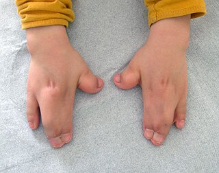
Acrocephalosyndactyly is a group of congenital conditions characterized by irregular features of the face and skull (craniosynostosis) and hands and feet (syndactyly). Craniosynostosis occurs when the cranial sutures, the fibrous tissue connecting the skull bones, fuse the cranial bones early in development. Cranial sutures allow the skull bones to continue growing until they fuse at age 24. Premature fusing of the cranial sutures can result in alterations to the skull shape and interfere with brain growth. Syndactyly occurs when digits of the hands or feet are fused together. When polydactyly is also present, the classification is acrocephalopolysyndactyly. Polydactyly occurs when the hands or feet possess additional digits. Acrocephalosyndactyly is usually diagnosed after birth, although prenatal diagnosis is sometimes possible if the genetic variation is present in family members, as the conditions are typically inherited in an autosomal dominant pattern Treatment often involves surgery in early childhood to correct for craniosynostosis and syndactyly.

<span class="mw-page-title-main">Axenfeld–Rieger syndrome</span> Medical condition

Axenfeld–Rieger syndrome is a rare autosomal dominant disorder, which affects the development of the teeth, eyes, and abdominal region.

<span class="mw-page-title-main">3C syndrome</span> Medical condition

3C syndrome is a rare condition whose symptoms include heart defects, cerebellar hypoplasia, and cranial dysmorphism. It was first described in the medical literature in 1987 by Ritscher and Schinzel, for whom the disorder is sometimes named.

<span class="mw-page-title-main">Antley–Bixler syndrome</span> Congenital disorder

Antley–Bixler syndrome is a rare, severe autosomal recessive congenital disorder characterized by malformations and deformities affecting the majority of the skeleton and other areas of the body.

The RASopathies are a group of developmental syndromes caused by germline mutations in genes belonging to the Ras/MAPK pathway. Common features include intellectual disability, congenital heart defects, skin abnormalities, and craniofacial abnormalities.

<span class="mw-page-title-main">Floating–Harbor syndrome</span> Medical condition

Floating–Harbor syndrome, also known as Pelletier–Leisti syndrome, is a rare disease with fewer than 50 cases described in the literature. It is usually diagnosed in early childhood and is characterized by the triad of proportionate short stature with delayed bone age, characteristic facial appearance, and delayed speech development. Although its cause is unknown, it is thought to result from genetic mutation, and diagnosis is established by the presence of a heterozygous SRCAP mutation in those with clinical findings of FHS.

XK aprosencephaly is an extremely rare congenital disorder characterized by the absence of the embryonic forebrain. Because the prosencephalon gives way to the cerebral cortex, survival with aprosencephaly is not possible outside utero. The external symptoms are similar to holoprosencephaly, a related disorder, including a smaller than normal head (microcephaly), small eyeballs (microphthalmia), a small mouth (microstomia), anal atresia, and abnormalities of the external genitalia, radius, nostrils, and pharynx (throat).

<span class="mw-page-title-main">Waardenburg syndrome type 1</span> Congenital disorder

Waardenburg syndrome type 1 is a congenital disorder that caused by a mutation in the PAX3 gene that results in abnormal development in the neural crest during early development. Type 1 results in early graying and white forelock and a notable distance between the eyes, noted as dystopia canthorum. Common symptoms of the disease also includes non-progressive hearing loss in majority of patients with type 1. Patients can display complete or partial heterochromia and hypoplastic blue irides and congenital leukemia.

<span class="mw-page-title-main">Pentasomy X</span> Chromosomal disorder

Pentasomy X, also known as 49,XXXXX, is a chromosomal disorder in which a female has five, rather than two, copies of the X chromosome. Pentasomy X is associated with short stature, intellectual disability, characteristic facial features, heart defects, skeletal anomalies, and pubertal and reproductive abnormalities. The condition is exceptionally rare, with an estimated prevalence between 1 in 85,000 and 1 in 250,000.

Filippi syndrome, also known as Syndactyly Type I with Microcephaly and Mental Retardation, is a very rare autosomal recessive genetic disease. Only a very limited number of cases have been reported to date. Filippi Syndrome is associated with diverse symptoms of varying severity across affected individuals, for example malformation of digits, craniofacial abnormalities, intellectual disability, and growth retardation. The diagnosis of Filippi Syndrome can be done through clinical observation, radiography, and genetic testing. Filippi Syndrome cannot be cured directly as of 2022, hence the main focus of treatments is on tackling the symptoms observed on affected individuals. It was first reported in 1985.

References

  1. 1 2 3 4 5 6 7 8 9 10 11 12 13 "Noonan syndrome". Genetics Home Reference. Retrieved 24 December 2018.
  2. 1 2 3 4 5 6 7 "Noonan Syndrome". NORD (National Organization for Rare Disorders). 2016. Retrieved 24 December 2018.
  3. 1 2 3 4 5 6 7 8 9 "Noonan syndrome". Genetic and Rare Diseases Information Center (GARD) – an NCATS Program. Retrieved 25 December 2018.
  4. 1 2 3 4 Bhambhani V, Muenke M (January 2014). "Noonan syndrome". American Family Physician. 89 (1): 37–43. PMC   4099190 . PMID   24444506.
  5. "Noonan Syndrome - Children's Health Issues". Merck Manuals Consumer Version. Retrieved 25 December 2018.
  6. 1 2 Nosan G, Bertok S, Vesel S, Yntema HG, Paro-Panjan D (December 2013). "A lethal course of hypertrophic cardiomyopathy in Noonan syndrome due to a novel germline mutation in the KRAS gene: case study". Croatian Medical Journal. 54 (6): 574–8. doi:10.3325/cmj.2013.54.574. PMC   3893993 . PMID   24382853.
  7. Romano, Alicia A.; Allanson, Judith E.; Dahlgren, Jovanna; Gelb, Bruce D.; Hall, Bryan; Pierpont, Mary Ella; Roberts, Amy E.; Robinson, Wanda; Takemoto, Clifford M.; Noonan, Jacqueline A. (October 2010). "Noonan syndrome: clinical features, diagnosis, and management guidelines". Pediatrics. 126 (4): 746–759. doi:10.1542/peds.2009-3207. ISSN   1098-4275. PMID   20876176. S2CID   11297756.
  8. 1 2 3 4 5 Allanson, Judith E.; Roberts, Amy E. (1993), Adam, Margaret P.; Ardinger, Holly H.; Pagon, Roberta A.; Wallace, Stephanie E. (eds.), "Noonan Syndrome", GeneReviews®, University of Washington, Seattle, PMID   20301303 , retrieved 2019-11-18
  9. Anderson, Kailyn; Cnota, James; James, Jeanne; Miller, Erin M.; Parrott, Ashley; Pilipenko, Valentina; Weaver, Kathryn Nicole; Shikany, Amy (2018-12-16). "Prevalence of Noonan spectrum disorders in a pediatric population with valvar pulmonary stenosis". Congenital Heart Disease. 14 (2): 264–273. doi:10.1111/chd.12721. ISSN   1747-079X. PMID   30556322. S2CID   58767341.
  10. Rivero-García, Pamela; Campuzano-Estrada, Isabel del Carmen; Hernandez-Felix, Jorge Humberto (2023). "Hypertrophic cardiomyopathy in an adult patient with Noonan syndrome with multiple lentigines". Clinical Case Reports. 11 (6): e7607. doi: 10.1002/ccr3.7607 . ISSN   2050-0904. PMC   10290193 . PMID   37361648.
  11. Marino, Bruno; Digilio, Maria Cristina; Toscano, Alessandra; Giannotti, Aldo; Dallapiccola, Bruno (December 1999). "Congenital heart diseases in children with Noonan syndrome: An expanded cardiac spectrum with high prevalence of atrioventricular canal". The Journal of Pediatrics. 135 (6): 703–706. doi:10.1016/s0022-3476(99)70088-0. ISSN   0022-3476. PMID   10586172.
  12. Linglart, Léa; Gelb, Bruce D. (2020-02-05). "Congenital heart defects in Noonan syndrome: Diagnosis, management, and treatment". American Journal of Medical Genetics Part C: Seminars in Medical Genetics. 184 (1): 73–80. doi:10.1002/ajmg.c.31765. ISSN   1552-4868. PMC   7682536 . PMID   32022400.
  13. Roelofs, Renée L.; Janssen, Nikki; Wingbermühle, Ellen; Kessels, Roy P.C.; Egger, Jos I.M. (2016-05-03). "Intellectual development in Noonan syndrome: a longitudinal study". Brain and Behavior. 6 (7): e00479. doi: 10.1002/brb3.479 . ISSN   2162-3279. PMC   4864201 . PMID   27247851.
  14. van der Burgt, Ineke; Thoonen, Geert; Roosenboom, Natasja; Assman-Hulsmans, Claire; Gabreels, Fons; Otten, Barto; Brunner, Han G. (1999). "Patterns of cognitive functioning in school-aged children with Noonan syndrome associated with variability in phenotypic expression". The Journal of Pediatrics. 135 (6): 707–713. doi:10.1016/s0022-3476(99)70089-2. ISSN   0022-3476. PMID   10586173.
  15. Wingbermühle, E.; Roelofs, R. L.; van der Burgt, I.; Souren, P. M.; Verhoeven, W. M. A.; Kessels, R. P. C.; Egger, J. I. M. (2012-08-10). "Cognitive functioning of adults with Noonan syndrome: a case-control study". Genes, Brain and Behavior. 11 (7): 785–793. doi:10.1111/j.1601-183x.2012.00821.x. ISSN   1601-1848. PMID   22783933. S2CID   31704142.
  16. Galarza, Marcelo; Martínez-Lage, Juan F.; Ham, Steven; Sood, Sandeep (2010). "Cerebral Anomalies and Chiari Type 1 Malformation". Pediatric Neurosurgery. 46 (6): 442–449. doi:10.1159/000327220. ISSN   1016-2291. PMID   21540621. S2CID   11867857.
  17. Davico, Chiara; D'Alessandro, Rossella; Borgogno, Marta; Campagna, Filippa; Torta, Francesca; Ricci, Federica; Amianto, Federico; Vittorini, Roberta; Carli, Diana; Mussa, Alessandro; Vitiello, Benedetto; Ferrero, Giovanni Battista (2022-05-16). "Epilepsy in a cohort of children with Noonan syndrome and related disorders". European Journal of Pediatrics. 181 (8): 2919–2926. doi:10.1007/s00431-022-04497-6. ISSN   1432-1076. PMID   35575813. S2CID   248800780.
  18. Unuma, Kana; Tomomasa, Dan; Noma, Kosuke; Yamamoto, Kouhei; Matsuyama, Taka-aki; Makino, Yohsuke; Hijikata, Atsushi; Wen, Shuheng; Ogata, Tsutomu; Okamoto, Nobuhiko; Okada, Satoshi; Ohashi, Kenichi; Uemura, Koichi; Kanegane, Hirokazu (2023-04-18). "Case Report: Molecular autopsy underlie COVID-19-associated sudden, unexplained child mortality". Frontiers in Immunology. 14. doi: 10.3389/fimmu.2023.1121059 . ISSN   1664-3224. PMC   10151512 . PMID   37143668.
  19. Fernández, Delia I.; Diender, Marije; Hermida-Nogueira, Lidia; Huang, Jingnan; Veiras, Sonia; Henskens, Yvonne M.C.; te Loo, Maroeska W.M.; Heemskerk, Johan W.M.; Kuijpers, Marijke J.E.; García, Ángel (2023). "Role of SHP2 (PTPN11) in glycoprotein VI-dependent thrombus formation: Improved platelet responsiveness by the allosteric drug SHP099 in Noonan syndrome patients". Thrombosis Research. 228: 105–116. doi: 10.1016/j.thromres.2023.06.001 . ISSN   0049-3848. PMID   37302266.
  20. Razzaque MA, Komoike Y, Nishizawa T, Inai K, Furutani M, Higashinakagawa T, Matsuoka R (March 2012). "Characterization of a novel KRAS mutation identified in Noonan syndrome". American Journal of Medical Genetics. Part A. 158A (3): 524–32. doi:10.1002/ajmg.a.34419. PMID   22302539. S2CID   34135931.
  21. Sharland, Michael; Morgan, Maureen; Smith, Gill; Burch, Michael; Patton, Michael A. (1993-02-15). "Genetic counselling in Noonan syndrome". American Journal of Medical Genetics. 45 (4): 437–440. doi:10.1002/ajmg.1320450407. ISSN   0148-7299. PMID   8465845.
  22. El Bouchikhi, Ihssane; Belhassan, Khadija; Moufid, Fatima Zohra; Iraqui Houssaini, Mohammed; Bouguenouch, Laila; Samri, Imane; Atmani, Samir; Ouldim, Karim (2016). "Noonan syndrome-causing genes: Molecular update and an assessment of the mutation rate". International Journal of Pediatrics and Adolescent Medicine. 3 (4): 133–142. doi: 10.1016/j.ijpam.2016.06.003 . ISSN   2352-6467. PMC   6372459 . PMID   30805484.
  23. Zenker, Martin; Buheitel, Gernot; Rauch, Ralf; Koenig, Rainer; Bosse, Kirstin; Kress, Wolfram; Tietze, Hans-Ulrich; Doerr, Helmuth-Guenther; Hofbeck, Michael; Singer, Helmut; Reis, André; Rauch, Anita (2004). "Genotype-phenotype correlations in Noonan syndrome". The Journal of Pediatrics. 144 (3): 368–374. doi:10.1016/j.jpeds.2003.11.032. ISSN   0022-3476. PMID   15001945.
  24. Ko, Jung Min; Kim, Jae-Min; Kim, Gu-Hwan; Yoo, Han-Wook (2008-11-20). "PTPN11, SOS1, KRAS, and RAF1 gene analysis, and genotype–phenotype correlation in Korean patients with Noonan syndrome". Journal of Human Genetics. 53 (11–12): 999–1006. doi: 10.1007/s10038-008-0343-6 . ISSN   1434-5161. PMID   19020799.
  25. Lee, Beom Hee; Kim, Jae-Min; Jin, Hye Young; Kim, Gu-Hwan; Choi, Jin-Ho; Yoo, Han-Wook (2011). "Spectrum of Mutations in Noonan Syndrome and Their Correlation with Phenotypes". The Journal of Pediatrics. 159 (6): 1029–1035. doi:10.1016/j.jpeds.2011.05.024. ISSN   0022-3476. PMID   21784453.
  26. Tartaglia M, Mehler EL, Goldberg R, Zampino G, Brunner HG, Kremer H, et al. (December 2001). "Mutations in PTPN11, encoding the protein tyrosine phosphatase SHP-2, cause Noonan syndrome". Nature Genetics. 29 (4): 465–8. doi:10.1038/ng772. PMID   11704759. S2CID   14627986.
  27. Shchelochkov OA, Patel A, Weissenberger GM, Chinault AC, Wiszniewska J, Fernandes PH, et al. (April 2008). "Duplication of chromosome band 12q24.11q24.23 results in apparent Noonan syndrome". American Journal of Medical Genetics. Part A. 146A (8): 1042–8. doi:10.1002/ajmg.a.32215. PMID   18348260. S2CID   205309115.
  28. van Der Burgt I, Brunner H (September 2000). "Genetic heterogeneity in Noonan syndrome: evidence for an autosomal recessive form". American Journal of Medical Genetics. 94 (1): 46–51. doi:10.1002/1096-8628(20000904)94:1<46::AID-AJMG10>3.0.CO;2-I. PMID   10982482.
  29. Schubbert S, Zenker M, Rowe SL, Böll S, Klein C, Bollag G, et al. (March 2006). "Germline KRAS mutations cause Noonan syndrome". Nature Genetics. 38 (3): 331–6. doi:10.1038/ng1748. PMID   16474405. S2CID   8193354.
  30. Bentires-Alj M, Kontaridis MI, Neel BG (March 2006). "Stops along the RAS pathway in human genetic disease". Nature Medicine. 12 (3): 283–5. doi:10.1038/nm0306-283. PMID   16520774. S2CID   6989331.
  31. Roberts AE, Araki T, Swanson KD, Montgomery KT, Schiripo TA, Joshi VA, et al. (January 2007). "Germline gain-of-function mutations in SOS1 cause Noonan syndrome". Nature Genetics. 39 (1): 70–4. doi:10.1038/ng1926. PMID   17143285. S2CID   10222262.
  32. Razzaque MA, Nishizawa T, Komoike Y, Yagi H, Furutani M, Amo R, et al. (August 2007). "Germline gain-of-function mutations in RAF1 cause Noonan syndrome". Nature Genetics. 39 (8): 1013–7. doi:10.1038/ng2078. PMID   17603482. S2CID   29753972.
  33. "Download Catalog - Mayo Medical Laboratories". www.mayomedicallaboratories.com. Archived from the original on 2017-05-17. Retrieved 2014-07-08.
  34. De Luca A, Bottillo I, Sarkozy A, Carta C, Neri C, Bellacchio E, et al. (December 2005). "NF1 gene mutations represent the major molecular event underlying neurofibromatosis–Noonan syndrome". American Journal of Human Genetics. 77 (6): 1092–101. doi:10.1086/498454. PMC   1285166 . PMID   16380919.
  35. 1 2 Roberts, Amy E.; Allanson, Judith E.; Tartaglia, Marco; Gelb, Bruce D. (2013-01-26). "Noonan syndrome". Lancet. 381 (9863): 333–342. doi:10.1016/S0140-6736(12)61023-X. ISSN   1474-547X. PMC   4267483 . PMID   23312968.
  36. 1 2 3 4 5 6 7 Allanson, Judith E.; Roberts, Amy E. (1993), Adam, Margaret P.; Ardinger, Holly H.; Pagon, Roberta A.; Wallace, Stephanie E. (eds.), "Noonan Syndrome", GeneReviews®, University of Washington, Seattle, PMID   20301303 , retrieved 2019-11-25
  37. Allanson, J. E.; Upadhyaya, M.; Watson, G. H.; Partington, M.; MacKenzie, A.; Lahey, D.; MacLeod, H.; Sarfarazi, M.; Broadhead, W.; Harper, P. S. (November 1991). "Watson syndrome: is it a subtype of type 1 neurofibromatosis?". Journal of Medical Genetics. 28 (11): 752–756. doi:10.1136/jmg.28.11.752. ISSN   0022-2593. PMC   1017110 . PMID   1770531.
  38. Armour, C. M.; Allanson, J. E. (April 2008). "Further delineation of cardio-facio-cutaneous syndrome: clinical features of 38 individuals with proven mutations". Journal of Medical Genetics. 45 (4): 249–254. doi:10.1136/jmg.2007.054460. ISSN   1468-6244. PMID   18039946. S2CID   9742395.
  39. Kerr, B.; Delrue, M.-A.; Sigaudy, S.; Perveen, R.; Marche, M.; Burgelin, I.; Stef, M.; Tang, B.; Eden, O. B.; O'Sullivan, J.; De Sandre-Giovannoli, A. (May 2006). "Genotype-phenotype correlation in Costello syndrome: HRAS mutation analysis in 43 cases". Journal of Medical Genetics. 43 (5): 401–405. doi:10.1136/jmg.2005.040352. ISSN   1468-6244. PMC   2564514 . PMID   16443854.
  40. Gripp, Karen W.; Lin, Angela E.; Stabley, Deborah L.; Nicholson, Linda; Scott, Charles I.; Doyle, Daniel; Aoki, Yoko; Matsubara, Yoichi; Zackai, Elaine H.; Lapunzina, Pablo; Gonzalez-Meneses, Antonio (2006-01-01). "HRAS mutation analysis in Costello syndrome: Genotype and phenotype correlation". American Journal of Medical Genetics Part A. 140A (1): 1–7. doi:10.1002/ajmg.a.31047. ISSN   1552-4825. PMID   16329078. S2CID   27334655.
  41. Bertola, Debora R.; Pereira, Alexandre C.; Passetti, Fábio; de Oliveira, Paulo S.L.; Messiaen, Ludwine; Gelb, Bruce D.; Kim, Chong A.; Krieger, José Eduardo (2005-07-30). "Neurofibromatosis–Noonan syndrome: Molecular evidence of the concurrence of both disorders in a patient". American Journal of Medical Genetics Part A. 136A (3): 242–245. doi:10.1002/ajmg.a.30813. ISSN   1552-4825. PMID   15948193. S2CID   40235559.
  42. Morris, Colleen A. (1993), Adam, Margaret P.; Ardinger, Holly H.; Pagon, Roberta A.; Wallace, Stephanie E. (eds.), "Williams Syndrome", GeneReviews®, University of Washington, Seattle, PMID   20301427 , retrieved 2019-11-25
  43. "Noonan syndrome - Symptoms, diagnosis and treatment | BMJ Best Practice". bestpractice.bmj.com. Retrieved 2019-11-18.
  44. "Does Noonan Syndrome Increase Malignant Hyperthermia Susceptibility? - MHAUS". www.mhaus.org. Retrieved 2019-11-25.
  45. 1 2 "DynaMed". www.dynamed.com. Retrieved 2019-11-11.
  46. Noonan JA, Ehmke DA (1963). "Associated noncardiac malformations in children with congenital heart disease". Midwest Soc Pediatr Res. 63: 468–70.
  47. Noonan, Jacqueline A. (1968-10-01). "Hypertelorism With Turner Phenotype: A New Syndrome With Associated Congenital Heart Disease". American Journal of Diseases of Children. 116 (4): 373–80. doi:10.1001/archpedi.1968.02100020377005. ISSN   0002-922X. PMID   4386970.