CHARGE syndrome

Last updated
CHARGE syndrome
Charge.jpg
"Lop ear" phenotype characteristic of a person with CHARGE syndrome, along with her cochlear implant.
Specialty Medical genetics   OOjs UI icon edit-ltr-progressive.svg

CHARGE syndrome (formerly known as CHARGE association) is a rare syndrome caused by a genetic disorder. First described in 1979, the acronym "CHARGE" came into use for newborn children with the congenital features of coloboma of the eye, heart defects, atresia of the nasal choanae, restricted growth and/or development, genital and/or urinary abnormalities, and ear abnormalities and deafness. [1] These features are no longer used in making a diagnosis of CHARGE syndrome, but the name remains. About two thirds of cases are due to a CHD7 mutation. CHARGE syndrome occurs only in 0.1–1.2 per 10,000 live births; as of 2009, it was the leading cause of congenital deafblindness in the US. [2]

Contents

Genetics

CHARGE syndrome was formerly referred to as CHARGE association, which indicates a non-random pattern of congenital anomalies that occurs together more frequently than one would expect on the basis of chance, but for which a common cause has not been identified. Very few people with CHARGE will have 100% of its known features. In 2004, mutations on the CHD7 gene (located on Chromosome 8) were found in 10 of 17 patients in the Netherlands, making CHARGE an official syndrome. [3] A 2006 US study of 110 individuals with CHARGE syndrome showed that 60% of those tested had a mutation of the CHD7 gene. [4]

In 2010, a review of 379 clinically diagnosed cases of CHARGE syndrome, in which CHD7 mutation testing was undertaken found that 67% of cases were due to a CHD7 mutation. [5]

Diagnosis

The diagnosis of CHARGE syndrome is often difficult, because it is rare. The syndrome spans many disciplines, and as such, the symptoms may be recognized by a pediatrician, family medicine physician, oral and maxillofacial surgeon, ENT specialist, ophthalmologist, endocrinologist, cardiologist, urologist, developmental specialist, radiologist, geneticist, physiotherapist, occupational therapist, speech therapist, or orthopedic specialist. [6]

Signs

Although genetic testing positively identifies nearly two thirds of children with CHARGE syndrome, diagnosis is still largely clinical. [6] The following signs were originally identified in children with this syndrome, but are no longer used to make the diagnosis alone.

Genetic testing

Genetic testing for CHARGE syndrome involves specific genetic testing for the CHD7 gene. The test is available[ when? ] at most major genetic testing laboratories. Insurance companies sometimes do not pay for such genetic tests, though this is changing rapidly as genetic testing is becoming standard across all aspects of medicine. CHARGE syndrome is a clinical diagnosis, which means genetic testing is not required in order to make the diagnosis. Rather, the diagnosis can be made based on clinical features alone.

Screening other organ systems

Once the diagnosis is made based on clinical signs, it is important to investigate other body systems that may be involved. For example, if the diagnosis is made based on the abnormal appearance of the ears and developmental delay, it is important to check the child's hearing, vision, heart, nose, and urogenital system. Ideally, every child newly diagnosed with CHARGE syndrome should have a complete evaluation by an ENT specialist, audiologist, ophthalmologist, pediatric cardiologist, developmental therapist, and pediatric urologist.

Recent research led by Yu Liang from Shijiazhuang Fourth Hospital and Sijie He from BGI Genomics, highlighted the use of prenatal ultrasound to detect CHARGE syndrome. Key findings indicated the difficulty in diagnosing CHARGE syndrome without genetic testing, [7] as mutations in the CHD7 gene are not always listed in public databases. [8] and predicted to be deleterious by CADD [9] and MutationTaster. [10]

Phenotypic variability further complicates diagnosis, with no clear correlation between mutation severity and clinical presentation. Trio-WES analysis confirmed a de novo mutation in the newborn, emphasizing the importance of early diagnosis for effective management. The study underscores the significance of early prenatal diagnosis through ultrasound examinations and specialized genetic testing. [7] This approach facilitates timely interventions and enhances understanding of rare genetic conditions, improving diagnostic protocols for CHARGE syndrome and similar disorders.

Treatment

Children with CHARGE syndrome may have a number of life-threatening medical conditions; with advances in medical care, these children can survive and can thrive with the support of a multidisciplinary team of medical professionals. Therapies and education must take into consideration hearing impairment, vision problems, and any others. Early intervention, such as occupational, speech-language, and physical therapy, to improve static posture, ambulation, and self-care skills is important. The intelligence of children with multiple health impairments, such as combined deafblindness, can be underestimated in the absence of early intervention. [11]

Education

Children with CHARGE syndrome will vary greatly in their abilities in the classroom: some may need little support, while some may require full-time support and individualized programs. Taking each of the various affected body systems into account is vital to the success of the child in the educational setting.

An important step in dealing with abnormal behavior is understanding why it is occurring and helping the child learn more appropriate methods of communicating. [12]

Epidemiology

The incidence is estimated to range from 0.1 to 1.2 per 10,000 live births, though the true incidence is unknown. [13] As of 2005, the highest prevalence was found in Canada and estimated at 1 in 8,500 live births. [14]

History

B.D. Hall first described the CHARGE association in a 1979 journal paper of about 17 children who had been born with choanal atresia. [15] During the same year, H.M. Hittner described 10 children who had choanal atresia as well as coloboma, congenital heart defect, and hearing loss. [16] Using both coloboma or choanal atresia and some of the other related characteristic malformations, R. A. Pagon first coined the acronym CHARGE in 1981 to emphasize that this cluster of associated malformations occurred together. [1] It came to be recognised[ when? ] as a syndrome within the umbrella of the CHARGE association, a set of apparently random signs occurring together. Since the signs seen in CHARGE are caused by a genetic anomaly, its name was eventually[ when? ] changed to 'CHARGE syndrome'.[ citation needed ]

The CHARGE Syndrome Foundation, formally incorporated in 1993, [17] is a US-based organization for individuals with CHARGE syndrome, families, researchers, and clinicians to further research and scientific knowledge about CHARGE Syndrome. The Foundation holds a biennial international conference that was first held in 1993. [18]

See also

Related Research Articles

<span class="mw-page-title-main">Choanal atresia</span> Congenital disorder where the nasal passage is blocked

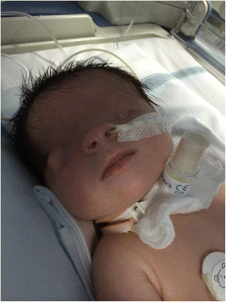
Choanal atresia is a congenital disorder where the back of the nasal passage (choana) is blocked, usually by abnormal bony or soft tissue (membranous) due to failed hole development of the nasal fossae during prenatal development. It causes persistent rhinorrhea, and with bilateral choanal atresia and obstructed airway that can cause cyanosis and hypoxia.

<span class="mw-page-title-main">Kabuki syndrome</span> Medical condition

Kabuki syndrome is a rare congenital disorder of genetic origin. It affects multiple parts of the body, with varying symptoms and severity, although the most common is the characteristic facial appearance.

<span class="mw-page-title-main">Noonan syndrome</span> Genetic condition involving facial, heart, blood and skeletal features

Noonan syndrome (NS) is a genetic disorder that may present with mildly unusual facial features, short height, congenital heart disease, bleeding problems, and skeletal malformations. Facial features include widely spaced eyes, light-colored eyes, low-set ears, a short neck, and a small lower jaw. Heart problems may include pulmonary valve stenosis. The breast bone may either protrude or be sunken, while the spine may be abnormally curved. Intelligence is often normal. Complications of NS can include leukemia.

<span class="mw-page-title-main">Treacher Collins syndrome</span> Human genetic disorder

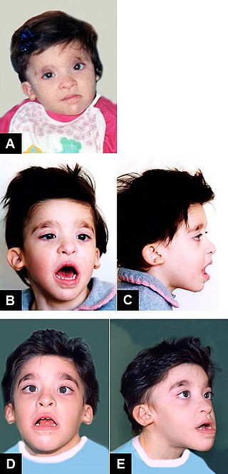
Treacher Collins syndrome (TCS) is a genetic disorder characterized by deformities of the ears, eyes, cheekbones, and chin. The degree to which a person is affected, however, may vary from mild to severe. Complications may include breathing problems, problems seeing, cleft palate, and hearing loss. Those affected generally have normal intelligence.

<span class="mw-page-title-main">Coloboma</span> Hole in one of the structures of the eye

A coloboma is a hole in one of the structures of the eye, such as the iris, retina, choroid, or optic disc. The hole is present from birth and can be caused when a gap called the choroid fissure, which is present during early stages of prenatal development, fails to close up completely before a child is born. Ocular coloboma is relatively uncommon, affecting less than one in every 10,000 births.

Microphthalmia, also referred as microphthalmos, is a developmental disorder of the eye in which one or both eyes are abnormally small and have anatomic malformations. Microphthalmia is a distinct condition from anophthalmia and nanophthalmia. Although sometimes referred to as 'simple microphthalmia', nanophthalmia is a condition in which the size of the eye is small but no anatomical alterations are present.

Atresia is a condition in which an orifice or passage in the body is closed or absent.

<span class="mw-page-title-main">Microtia</span> Medical condition

Microtia is a congenital deformity where the auricle is underdeveloped. A completely undeveloped auricle is referred to as anotia. Because microtia and anotia have the same origin, it can be referred to as microtia-anotia. Microtia can be unilateral or bilateral. Microtia occurs in 1 out of about 8,000–10,000 births. In unilateral microtia, the right ear is most commonly affected. It may occur as a complication of taking Accutane (isotretinoin) during pregnancy.

<span class="mw-page-title-main">Fraser syndrome</span> Recessive genetic disorder involving eye and genital abnormalities

Fraser syndrome is an autosomal recessive congenital disorder, identified by several developmental anomalies. Fraser syndrome is named for the geneticist George R. Fraser, who first described the syndrome in 1962.

<span class="mw-page-title-main">Robinow syndrome</span> Rare genetic disorder characterized by a fetal face

Robinow syndrome is an extremely rare genetic disorder characterized by short-limbed dwarfism, abnormalities in the head, face, and external genitalia, as well as vertebral segmentation. The disorder was first described in 1969 by human geneticist Meinhard Robinow, along with physicians Frederic N. Silverman and Hugo D. Smith, in the American Journal of Diseases of Children. By 2002, over 100 cases had been documented and introduced into medical literature.

Abruzzo–Erickson syndrome is an extremely rare disorder characterized by deafness, protruding ears, coloboma, a cleft palate or palatal rugosity, radial synostosis, and short stature. It was first characterized by Abruzzo and Erickson in 1977 as a CHARGE like syndrome as variably expressed among a family of two brothers, their mother, and their maternal uncle. Members of this family exhibited many of the CHARGE symptoms, but notably did not have choanal atresia and the brothers experienced typical genital development. Due to the recent discovery of this disorder, its etiology is not fully known but it is understood that it arises from mutations on the TBX22 gene on the X-chromosome. The disorder is inherited in an X-linked recessive manner. There is currently no known cure but its symptoms can be treated.

<span class="mw-page-title-main">Duane-radial ray syndrome</span> Medical condition

Duane-radial ray syndrome, also known as Okihiro syndrome, is a rare autosomal dominant disorder that primarily affects the eyes and causes abnormalities of bones in the arms and hands. This disorder is considered to be a SALL4-related disorder due to the SALL4 gene mutations leading to these abnormalities. It is diagnosed by clinical findings on a physical exam as well as genetic testing and imaging. After being diagnosed, there are other evaluations that one may go through in order to determine the extent of the disease. There are various treatments for the symptoms of this disorder.

<span class="mw-page-title-main">Mowat–Wilson syndrome</span> Medical condition

Mowat–Wilson syndrome is a rare genetic disorder that was clinically delineated by David R. Mowat and Meredith J. Wilson in 1998. The condition affects both males and females, has been described in various countries and ethnic groups around the world, and occurs in approximately 1 in 50,000–100,000 births.

<span class="mw-page-title-main">Papillorenal syndrome</span> Medical condition

Papillorenal syndrome is an autosomal dominant genetic disorder marked by underdevelopment (hypoplasia) of the kidney and colobomas of the optic nerve.

<span class="mw-page-title-main">3C syndrome</span> Medical condition

3C syndrome is a rare condition whose symptoms include heart defects, cerebellar hypoplasia, and cranial dysmorphism. It was first described in the medical literature in 1987 by Ritscher and Schinzel, for whom the disorder is sometimes named.

<span class="mw-page-title-main">Cat eye syndrome</span> Genetic partial duplication of chromosome 22

Cat-eye syndrome (CES) or Schmid–Fraccaro syndrome is a rare condition caused by an abnormal extra chromosome, i.e. a small supernumerary marker chromosome. This chromosome consists of the entire short arm and a small section of the long arm of chromosome 22. In consequence, individuals with the cat-eye syndrome have three (trisomic) or four (tetrasomic) copies of the genetic material contained in the abnormal chromosome instead of the normal two copies. The prognosis for patients with CES varies depending on the severity of the condition and their associated signs and symptoms, especially when heart or kidney abnormalities are seen.

<span class="mw-page-title-main">CHD7</span> Protein-coding gene in the species Homo sapiens

Chromodomain-helicase-DNA-binding protein 7 is an ATP-dependent 'chromatin' or 'nucleosome' remodeling factor that in humans is encoded by the CHD7 gene.

<span class="mw-page-title-main">13q deletion syndrome</span> Medical condition

13q deletion syndrome is a rare genetic disease caused by the deletion of some or all of the large arm of human chromosome 13. Depending upon the size and location of the deletion on chromosome 13, the physical and mental manifestations will vary. It has the potential to cause intellectual disability and congenital malformations that affect a variety of organ systems. Because of the rarity of the disease in addition to the variations in the disease, the specific genes that cause this disease are unknown. This disease is also known as:

<span class="mw-page-title-main">Strømme syndrome</span> Rare genetic condition involving intestinal atresia, eye abnormalities and microcephaly

Strømme syndrome is a very rare autosomal recessive genetic condition characterised by intestinal atresia, eye abnormalities and microcephaly. The intestinal atresia is of the "apple-peel" type, in which the remaining intestine is twisted around its main artery. The front third of the eye is typically underdeveloped, and there is usually moderate developmental delay. Less common features include an atrial septal defect, increased muscle tone or skeletal abnormalities. Physical features may include short stature, large, low-set ears, a small jaw, a large mouth, epicanthic folds, or fine, sparse hair.

<span class="mw-page-title-main">Mandibulofacial dysostosis-microcephaly syndrome</span> Medical condition

Mandibulofacial dysostosis with microcephaly syndrome, also known as growth delay-intellectual disability-mandibulofacial dysostosis-microcephaly-cleft palate syndrome, mandibulofacial dysostosis, guion-almeida type, or simply as MFDM syndrome is a rare genetic disorder which is characterized by developmental delays, intellectual disabilities, and craniofacial dysmorphisms.

References

  1. 1 2 Pagon RA, Graham JM, Zonana J, Yong SL (August 1981). "Coloboma, congenital heart disease, and choanal atresia with multiple anomalies: CHARGE association". The Journal of Pediatrics. 99 (2): 223–227. doi:10.1016/S0022-3476(81)80454-4. PMID   6166737.
  2. "The 2008 National Child Count of Children and Youth who are Deaf-Blind" (PDF). The National Consortium on Deaf-Blindness. 2009. p. 30. Archived from the original (PDF) on 2012-03-15.
  3. Vissers LE, van Ravenswaaij CM, Admiraal R, Hurst JA, de Vries BB, Janssen IM, et al. (September 2004). "Mutations in a new member of the chromodomain gene family cause CHARGE syndrome". Nature Genetics. 36 (9): 955–957. doi: 10.1038/ng1407 . PMID   15300250.
  4. Lalani SR, Safiullah AM, Fernbach SD, Harutyunyan KG, Thaller C, Peterson LE, et al. (February 2006). "Spectrum of CHD7 mutations in 110 individuals with CHARGE syndrome and genotype-phenotype correlation". American Journal of Human Genetics. 78 (2): 303–314. doi:10.1086/500273. PMC   1380237 . PMID   16400610.
  5. Zentner GE, Layman WS, Martin DM, Scacheri PC (March 2010). "Molecular and phenotypic aspects of CHD7 mutation in CHARGE syndrome". American Journal of Medical Genetics. Part A. 152A (3): 674–686. doi:10.1002/ajmg.a.33323. PMC   2918278 . PMID   20186815.
  6. 1 2 "Genetics: Diagnosis, Testing & Recurrence | Charge Syndrome Foundation". Charge Syndrome Foundation | A better world for people with CHARGE Syndrome. 2016-06-20. Retrieved 2022-02-20.
  7. 1 2 Liang Y, He S, Yang L, Li T, Zhao L, Sun CX (March 2024). "CHARGE syndrome with early fetal ear abnormalities: A case report". Clinical Case Reports. 12 (3): e8670. doi:10.1002/ccr3.8670. PMC   10948373 . PMID   38505478.Lay summary in: "Study Reveals How Fetal Ear Shape Could Signal Rare Genetic Condition". BGI Insight. Shenzhen, China. 2 May 2024.
  8. "Genome Aggregation Database".
  9. "CADD".
  10. "MutationTaster".
  11. "Therapies | Charge Syndrome Foundation". Charge Syndrome Foundation | A better world for people with CHARGE Syndrome. 2016-05-31. Retrieved 2022-02-20.
  12. "Education | Charge Syndrome Foundation". Charge Syndrome Foundation | A better world for people with CHARGE Syndrome. Retrieved 2022-02-20.
  13. Blake KD, Prasad C (September 2006). "CHARGE syndrome". Orphanet Journal of Rare Diseases. 1: 34. doi: 10.1186/1750-1172-1-34 . PMC   1586184 . PMID   16959034.
  14. Issekutz KA, Graham JM, Prasad C, Smith IM, Blake KD (March 2005). "An epidemiological analysis of CHARGE syndrome: preliminary results from a Canadian study". American Journal of Medical Genetics. Part A. 133A (3): 309–317. doi: 10.1002/ajmg.a.30560 . PMID   15637722. S2CID   23647735.
  15. Hall BD (September 1979). "Choanal atresia and associated multiple anomalies". The Journal of Pediatrics. 95 (3): 395–398. doi:10.1016/S0022-3476(79)80513-2. PMID   469662.
  16. Hittner HM, Hirsch NJ, Kreh GM, Rudolph AJ (1979). "Colobomatous microphthalmia, heart disease, hearing loss, and mental retardation--a syndrome". Journal of Pediatric Ophthalmology and Strabismus. 16 (2): 122–128. doi:10.3928/0191-3913-19790301-10. PMID   458518.
  17. "Foundation History | Charge Syndrome Foundation". Charge Syndrome Foundation | A better world for people with CHARGE Syndrome. Retrieved 2021-12-09.
  18. "Conferences | Charge Syndrome Foundation". Charge Syndrome Foundation | A better world for people with CHARGE Syndrome. Retrieved 2021-12-09.