Dihydrolipoamide dehydrogenase

Last updated
DLD
PDB 1zy8 EBI.png
Available structures
PDB Ortholog search: PDBe RCSB
Identifiers
Aliases DLD , DLDD, DLDH, E3, GCSL, LAD, PHE3, dihydrolipoamide dehydrogenase, Dihydrolipoamide dehydrogenase, OGDC-E3
External IDs OMIM: 238331 MGI: 107450 HomoloGene: 84 GeneCards: DLD
Orthologs
SpeciesHumanMouse
Entrez
Ensembl
UniProt
RefSeq (mRNA)

NM_001289752
NM_000108
NM_001289750
NM_001289751

NM_007861

RefSeq (protein)

NP_000099
NP_001276679
NP_001276680
NP_001276681

NP_031887

Location (UCSC) Chr 7: 107.89 – 107.93 Mb Chr 12: 31.38 – 31.4 Mb
PubMed search [3] [4]
Wikidata
View/Edit Human View/Edit Mouse

Dihydrolipoamide dehydrogenase (DLD), also known as dihydrolipoyl dehydrogenase, mitochondrial, is an enzyme that in humans is encoded by the DLD gene. [5] [6] [7] [8] DLD is a flavoprotein enzyme that oxidizes dihydrolipoamide to lipoamide.

Contents

Dihydrolipoamide dehydrogenase (DLD) is a mitochondrial enzyme that plays a vital role in energy metabolism in eukaryotes. This enzyme is required for the complete reaction of at least five different multi-enzyme complexes. [9] Additionally, DLD is a flavoenzyme oxidoreductase that contains a reactive disulfide bridge and a FAD cofactor that are directly involved in catalysis. The enzyme associates into tightly bound homodimers required for its enzymatic activity. [10]

Structure

The protein encoded by the DLD gene comes together with another protein to form a dimer in the central metabolic pathway. Several amino acids within the catalytic pocket have been identified as important to DLD function, including R281 and N473. [11] [12] Although the overall fold of the human enzyme is similar to that of yeast, the human structure is different in that it has two loops that extend from the general protein structure and into the FAD binding sites when bound the NAD+ molecule, required for catalysis, is not close to the FAD moiety. However, when NADH is bound instead, it is stacked directly op top of the FAD central structure. The current hE3 structures show directly that the disease-causing mutations occur at three locations in the human enzyme: the dimer interface, the active site, and the FAD and NAD(+)-binding sites. [13]

Function

The DLD homodimer functions as the E3 component of the pyruvate, α-ketoglutarate, α-adipate and branched-chain amino acid-dehydrogenase complexes and the glycine cleavage system, all in the mitochondrial matrix. In these complexes, DLD converts dihydrolipoic acid and NAD+ into lipoic acid and NADH. [14] DLD also has diaphorase activity, being able to catalyze the oxidation of NADH to NAD+ by using different electron acceptors such as O2, labile ferric iron, nitric oxide, and ubiquinone. [9] DLD is thought to have a pro-oxidant role by reducing oxygen to a superoxide or ferric to ferrous iron, which then catalyzes production of hydroxyl radicals. [15] [16] Diaphorase activity of DLD may have an antioxidant role through its ability to scavenge nitric oxide and to reduce ubiquinone to ubiquinol. [17] [18] [19] The dihyrolipamide dehydrogenase gene is known to have multiple splice variants.

Moonlighting function

Certain DLD mutations can simultaneously induce the loss of a primary metabolic activity and the gain of a moonlighting proteolytic activity. The moonlighting proteolytic activity of DLD is revealed by conditions that destabilize the DLD homodimer and decrease its DLD activity. [9] Acidification of the mitochondrial matrix, as a result of ischemia-reperfusion injury, can disrupt the quaternary structure of DLD leading to decreased dehydrogenase activity and increased diaphorase activity. [20] The moonlighting proteolytic activity of DLD could also arise under pathological conditions. Proteolytic activity can further complicate the reduction in energy metabolism and an increase in oxidative damage as a result of decreased DLD activity and an increase in diaphorase activity respectively. [19] With its proteolytic function, DLD removes a functionally vital domain from the N-terminus of frataxin, a mitochondrial protein involved in iron metabolism and antioxidant protection. [21] [22]

Clinical significance

In humans, mutations in DLD are linked to a severe disorder of infancy with failure to thrive, hypotonia, and metabolic acidosis. [23] DLD deficiency manifests itself in a great degree of variability, which has been attributed to varying effects of different DLD mutations on the stability of the protein and its ability to dimerize or interact with other components of the three α-ketoacid dehydrogenase complexes. [23] With its proteolytic function, DLD causes a deficiency in frataxin, which leads to the neurodegenerative and cardiac disease, Friedreich's ataxia. [24]

Interactive pathway map

Click on genes, proteins and metabolites below to link to respective articles. [§ 1]
[[File:
TCACycle WP78.png Go to articleGo to articleGo to articleGo to articleGo to articleGo to articleGo to articleGo to articleGo to articleGo to articleGo to articleGo to articleGo to articleGo to articleGo to articleGo to articleGo to articleGo to articleGo to articleGo to articleGo to articleGo to articleGo to articleGo to articleGo to articleGo to articleGo to articleGo to articleGo to articleGo to articleGo to articleGo to articleGo to articleGo to articleGo to articleGo to articleGo to articleGo to articleGo to articleGo to articleGo to articleGo to HMDBGo to articleGo to articleGo to articleGo to articleGo to articleGo to articleGo to articleGo to articleGo to articleGo to article
[[
]]
[[
]]
[[
]]
[[
]]
[[
]]
[[
]]
[[
]]
[[
]]
[[
]]
[[
]]
[[
]]
[[
]]
[[
]]
[[
]]
[[
]]
[[
]]
[[
]]
[[
]]
[[
]]
[[
]]
[[
]]
[[
]]
[[
]]
[[
]]
[[
]]
[[
]]
[[
]]
[[
]]
[[
]]
[[
]]
[[
]]
[[
]]
[[
]]
[[
]]
[[
]]
[[
]]
[[
]]
[[
]]
[[
]]
[[
]]
[[
]]
[[
]]
[[
]]
[[
]]
[[
]]
[[
]]
[[
]]
[[
]]
[[
]]
[[
]]
[[
]]
[[
]]
[[
]]
[[
]]
[[
]]
[[
]]
[[
]]
[[
]]
[[
]]
[[
]]
[[
]]
[[
]]
[[
]]
[[
]]
TCACycle WP78.png Go to articleGo to articleGo to articleGo to articleGo to articleGo to articleGo to articleGo to articleGo to articleGo to articleGo to articleGo to articleGo to articleGo to articleGo to articleGo to articleGo to articleGo to articleGo to articleGo to articleGo to articleGo to articleGo to articleGo to articleGo to articleGo to articleGo to articleGo to articleGo to articleGo to articleGo to articleGo to articleGo to articleGo to articleGo to articleGo to articleGo to articleGo to articleGo to articleGo to articleGo to articleGo to HMDBGo to articleGo to articleGo to articleGo to articleGo to articleGo to articleGo to articleGo to articleGo to articleGo to article
|alt=TCACycle_WP78 edit]]
TCACycle_WP78 edit
  1. The interactive pathway map can be edited at WikiPathways: "TCACycle_WP78".
Click on genes, proteins and metabolites below to link to respective articles. [§ 1]
[[File:
WP534.png go to articlego to articlego to articlego to articlego to articlego to articlego to articlego to articlego to articlego to articlego to articlego to articlego to articlego to articlego to articlego to articlego to articlego to articlego to articlego to articlego to articlego to articlego to articlego to articlego to articlego to articlego to articlego to articlego to articlego to articlego to articlego to articlego to articlego to articlego to articlego to articlego to articlego to articlego to articlego to articlego to articlego to articlego to articlego to articlego to articlego to articlego to articlego to articlego to articlego to articlego to articlego to articlego to articlego to articlego to articlego to articlego to articlego to articlego to articlego to articlego to articlego to articlego to articlego to articlego to articlego to articlego to articlego to articlego to articlego to articlego to articlego to article
[[
]]
[[
]]
[[
]]
[[
]]
[[
]]
[[
]]
[[
]]
[[
]]
[[
]]
[[
]]
[[
]]
[[
]]
[[
]]
[[
]]
[[
]]
[[
]]
[[
]]
[[
]]
[[
]]
[[
]]
[[
]]
[[
]]
[[
]]
[[
]]
[[
]]
[[
]]
[[
]]
[[
]]
[[
]]
[[
]]
[[
]]
[[
]]
[[
]]
[[
]]
[[
]]
[[
]]
[[
]]
[[
]]
[[
]]
[[
]]
[[
]]
[[
]]
[[
]]
[[
]]
[[
]]
[[
]]
[[
]]
[[
]]
[[
]]
[[
]]
[[
]]
[[
]]
[[
]]
[[
]]
[[
]]
[[
]]
[[
]]
[[
]]
[[
]]
[[
]]
[[
]]
[[
]]
[[
]]
[[
]]
[[
]]
[[
]]
[[
]]
[[
]]
[[
]]
[[
]]
[[
]]
[[
]]
[[
]]
[[
]]
[[
]]
WP534.png go to articlego to articlego to articlego to articlego to articlego to articlego to articlego to articlego to articlego to articlego to articlego to articlego to articlego to articlego to articlego to articlego to articlego to articlego to articlego to articlego to articlego to articlego to articlego to articlego to articlego to articlego to articlego to articlego to articlego to articlego to articlego to articlego to articlego to articlego to articlego to articlego to articlego to articlego to articlego to articlego to articlego to articlego to articlego to articlego to articlego to articlego to articlego to articlego to articlego to articlego to articlego to articlego to articlego to articlego to articlego to articlego to articlego to articlego to articlego to articlego to articlego to articlego to articlego to articlego to articlego to articlego to articlego to articlego to articlego to articlego to articlego to article
|alt=Glycolysis and Gluconeogenesis edit]]
Glycolysis and Gluconeogenesis edit
  1. The interactive pathway map can be edited at WikiPathways: "GlycolysisGluconeogenesis_WP534".

Enzyme regulation

This protein may use the morpheein model of allosteric regulation. [25]

See also

Related Research Articles

<span class="mw-page-title-main">Citric acid cycle</span> Series of interconnected biochemical reactions

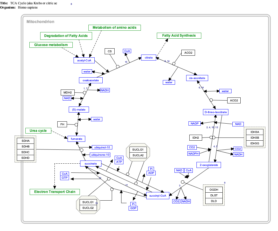
The citric acid cycle —also known as the Krebs cycle, Szent-Györgyi-Krebs cycle or the TCA cycle (tricarboxylic acid cycle)—is a series of chemical reactions to release stored energy through the oxidation of acetyl-CoA derived from carbohydrates, fats, and proteins. The Krebs cycle is used by organisms that respire (as opposed to organisms that ferment) to generate energy, either by anaerobic respiration or aerobic respiration. In addition, the cycle provides precursors of certain amino acids, as well as the reducing agent NADH, that are used in numerous other reactions. Its central importance to many biochemical pathways suggests that it was one of the earliest components of metabolism. Even though it is branded as a 'cycle', it is not necessary for metabolites to follow only one specific route; at least three alternative segments of the citric acid cycle have been recognized.

<span class="mw-page-title-main">Pyruvate dehydrogenase complex</span> Three-enzyme complex responsible for pyruvate decarboxylation

Pyruvate dehydrogenase complex (PDC) is a complex of three enzymes that converts pyruvate into acetyl-CoA by a process called pyruvate decarboxylation. Acetyl-CoA may then be used in the citric acid cycle to carry out cellular respiration, and this complex links the glycolysis metabolic pathway to the citric acid cycle. Pyruvate decarboxylation is also known as the "pyruvate dehydrogenase reaction" because it also involves the oxidation of pyruvate.

<span class="mw-page-title-main">Mitochondrial matrix</span> Space within the inner membrane of the mitochondrion

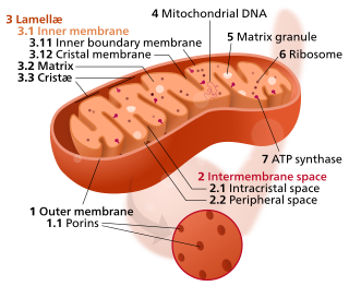
In the mitochondrion, the matrix is the space within the inner membrane. The word "matrix" stems from the fact that this space is viscous, compared to the relatively aqueous cytoplasm. The mitochondrial matrix contains the mitochondrial DNA, ribosomes, soluble enzymes, small organic molecules, nucleotide cofactors, and inorganic ions.[1] The enzymes in the matrix facilitate reactions responsible for the production of ATP, such as the citric acid cycle, oxidative phosphorylation, oxidation of pyruvate, and the beta oxidation of fatty acids.

<span class="mw-page-title-main">Pyruvate dehydrogenase lipoamide kinase isozyme 1</span> Protein-coding gene in the species Homo sapiens

Pyruvate dehydrogenase lipoamide kinase isozyme 1, mitochondrial is an enzyme that in humans is encoded by the PDK1 gene. It codes for an isozyme of pyruvate dehydrogenase kinase (PDK).

The branched-chain α-ketoacid dehydrogenase complex is a multi-subunit complex of enzymes that is found on the mitochondrial inner membrane. This enzyme complex catalyzes the oxidative decarboxylation of branched, short-chain alpha-ketoacids. BCKDC is a member of the mitochondrial α-ketoacid dehydrogenase complex family comprising pyruvate dehydrogenase and alpha-ketoglutarate dehydrogenase, key enzymes that function in the Krebs cycle.

Pyruvate dehydrogenase deficiency is a rare neurodegenerative disorder associated with abnormal mitochondrial metabolism. PDCD is a genetic disease resulting from mutations in one of the components of the pyruvate dehydrogenase complex (PDC). The PDC is a multi-enzyme complex that plays a vital role as a key regulatory step in the central pathways of energy metabolism in the mitochondria. The disorder shows heterogeneous characteristics in both clinical presentation and biochemical abnormality.

<span class="mw-page-title-main">Dihydrolipoyl transacetylase</span>

Dihydrolipoyl transacetylase is an enzyme component of the multienzyme pyruvate dehydrogenase complex. The pyruvate dehydrogenase complex is responsible for the pyruvate decarboxylation step that links glycolysis to the citric acid cycle. This involves the transformation of pyruvate from glycolysis into acetyl-CoA which is then used in the citric acid cycle to carry out cellular respiration.

Oxidative decarboxylation is a decarboxylation reaction caused by oxidation. Most are accompanied by α- Ketoglutarate α- Decarboxylation caused by dehydrogenation of hydroxyl carboxylic acids such as carbonyl carboxylic acid, malic acid, isocitric acid, etc.

<span class="mw-page-title-main">Pyruvate dehydrogenase</span> Class of enzymes

Pyruvate dehydrogenase is an enzyme that catalyzes the reaction of pyruvate and a lipoamide to give the acetylated dihydrolipoamide and carbon dioxide. The conversion requires the coenzyme thiamine pyrophosphate.

<span class="mw-page-title-main">Pyruvate dehydrogenase kinase</span> Class of enzymes

Pyruvate dehydrogenase kinase is a kinase enzyme which acts to inactivate the enzyme pyruvate dehydrogenase by phosphorylating it using ATP.

<span class="mw-page-title-main">E3 binding protein</span> Protein-coding gene in the species Homo sapiens

E3 binding protein also known as pyruvate dehydrogenase protein X component, mitochondrial is a protein that in humans is encoded by the PDHX gene. The E3 binding protein is a component of the pyruvate dehydrogenase complex found only in eukaryotes. Defects in this gene are a cause of pyruvate dehydrogenase deficiency which results in neurological dysfunction and lactic acidosis in infancy and early childhood. This protein is also a minor antigen for antimitochondrial antibodies. These autoantibodies are present in nearly 95% of patients with primary biliary cholangitis, an autoimmune disease of the liver. In primary biliary cholangitis, activated T lymphocytes attack and destroy epithelial cells in the bile duct where this protein is abnormally distributed and overexpressed. Primary biliary cholangitis eventually leads to liver failure.

<span class="mw-page-title-main">Glycerol-3-phosphate dehydrogenase</span> Class of enzymes

Glycerol-3-phosphate dehydrogenase (GPDH) is an enzyme that catalyzes the reversible redox conversion of dihydroxyacetone phosphate to sn-glycerol 3-phosphate.

<span class="mw-page-title-main">Glycerate dehydrogenase</span>

In enzymology, a glycerate dehydrogenase (EC 1.1.1.29) is an enzyme that catalyzes the chemical reaction

<span class="mw-page-title-main">Pyruvate dehydrogenase (lipoamide) alpha 1</span> Protein-coding gene in the species Homo sapiens

Pyruvate dehydrogenase E1 component subunit alpha, somatic form, mitochondrial is an enzyme that in humans is encoded by the PDHA1 gene.The pyruvate dehydrogenase complex is a nuclear-encoded mitochondrial matrix multienzyme complex that provides the primary link between glycolysis and the tricarboxylic acid (TCA) cycle by catalyzing the irreversible conversion of pyruvate into acetyl-CoA. The PDH complex is composed of multiple copies of 3 enzymes: E1 (PDHA1); dihydrolipoyl transacetylase (DLAT) ; and dihydrolipoyl dehydrogenase (DLD). The E1 enzyme is a heterotetramer of 2 alpha and 2 beta subunits. The E1-alpha subunit contains the E1 active site and plays a key role in the function of the PDH complex.

<span class="mw-page-title-main">DBT (gene)</span> Mammalian protein found in Homo sapiens

Lipoamide acyltransferase component of branched-chain alpha-keto acid dehydrogenase complex, mitochondrial is an enzyme that in humans is encoded by the DBT gene.

<span class="mw-page-title-main">PDK2</span> Protein-coding gene in the species Homo sapiens

Pyruvate dehydrogenase kinase isoform 2 (PDK2) also known as pyruvate dehydrogenase lipoamide kinase isozyme 2, mitochondrial is an enzyme that in humans is encoded by the PDK2 gene. PDK2 is an isozyme of pyruvate dehydrogenase kinase.

<span class="mw-page-title-main">PDK3</span> Protein-coding gene in the species Homo sapiens

Pyruvate dehydrogenase lipoamide kinase isozyme 3, mitochondrial is an enzyme that in humans is encoded by the PDK3 gene. It codes for an isozyme of pyruvate dehydrogenase kinase.The pyruvate dehydrogenase (PDH) complex is a nuclear-encoded mitochondrial multienzyme complex that catalyzes the overall conversion of pyruvate to acetyl-CoA and CO2. It provides the primary link between glycolysis and the tricarboxylic acid (TCA) cycle, and thus is one of the major enzymes responsible for the regulation of glucose metabolism. The enzymatic activity of PDH is regulated by a phosphorylation/dephosphorylation cycle, and phosphorylation results in inactivation of PDH. The protein encoded by this gene is one of the four pyruvate dehydrogenase kinases that inhibits the PDH complex by phosphorylation of the E1 alpha subunit. This gene is predominantly expressed in the heart and skeletal muscles. Alternatively spliced transcript variants encoding different isoforms have been found for this gene.

<span class="mw-page-title-main">NDUFB7</span> Protein-coding gene in the species Homo sapiens

NADH dehydrogenase [ubiquinone] 1 beta subcomplex subunit 7, also known as complex I-B18, is an enzyme that in humans is encoded by the NDUFB7 gene. NADH dehydrogenase (ubiquinone) 1 beta subcomplex subunit 7 is an accessory subunit of the NADH dehydrogenase (ubiquinone) complex, located in the mitochondrial inner membrane. It is also known as Complex I and is the largest of the five complexes of the electron transport chain.

<span class="mw-page-title-main">Pyruvate dehydrogenase (lipoamide) alpha 2</span> Protein-coding gene in the species Homo sapiens

Pyruvate dehydrogenase (lipoamide) alpha 2, also known as pyruvate dehydrogenase E1 component subunit alpha, testis-specific form, mitochondrial or PDHE1-A type II, is an enzyme that in humans is encoded by the PDHA2 gene.

<span class="mw-page-title-main">Pyruvate dehydrogenase (lipoamide) beta</span> Protein-coding gene in the species Homo sapiens

Pyruvate dehydrogenase (lipoamide) beta, also known as pyruvate dehydrogenase E1 component subunit beta, mitochondrial or PDHE1-B is an enzyme that in humans is encoded by the PDHB gene. The pyruvate dehydrogenase (PDH) complex is a nuclear-encoded mitochondrial multienzyme complex that catalyzes the overall conversion of pyruvate to acetyl-CoA and CO2, and provides the primary link between glycolysis and the tricarboxylic acid (TCA) cycle. The PDH complex is composed of multiple copies of three enzymatic components: pyruvate dehydrogenase (E1), dihydrolipoamide acetyltransferase (E2) and lipoamide dehydrogenase (E3). The E1 enzyme is a heterotetramer of two alpha and two beta subunits. This gene encodes the E1 beta subunit. Mutations in this gene are associated with pyruvate dehydrogenase E1-beta deficiency.

References

  1. 1 2 3 GRCh38: Ensembl release 89: ENSG00000091140 - Ensembl, May 2017
  2. 1 2 3 GRCm38: Ensembl release 89: ENSMUSG00000020664 - Ensembl, May 2017
  3. "Human PubMed Reference:". National Center for Biotechnology Information, U.S. National Library of Medicine.
  4. "Mouse PubMed Reference:". National Center for Biotechnology Information, U.S. National Library of Medicine.
  5. "Entrez Gene: dihydrolipoamide dehydrogenase".
  6. Otulakowski G, Robinson BH (December 1987). "Isolation and sequence determination of cDNA clones for porcine and human lipoamide dehydrogenase. Homology to other disulfide oxidoreductases". The Journal of Biological Chemistry. 262 (36): 17313–8. doi: 10.1016/S0021-9258(18)45379-3 . PMID   3693355.
  7. Pons G, Raefsky-Estrin C, Carothers DJ, Pepin RA, Javed AA, Jesse BW, et al. (March 1988). "Cloning and cDNA sequence of the dihydrolipoamide dehydrogenase component human alpha-ketoacid dehydrogenase complexes". Proceedings of the National Academy of Sciences of the United States of America. 85 (5): 1422–6. Bibcode:1988PNAS...85.1422P. doi: 10.1073/pnas.85.5.1422 . PMC   279783 . PMID   3278312.
  8. Scherer SW, Otulakowski G, Robinson BH, Tsui LC (1991). "Localization of the human dihydrolipoamide dehydrogenase gene (DLD) to 7q31----q32". Cytogenetics and Cell Genetics. 56 (3–4): 176–7. doi:10.1159/000133081. hdl: 10722/42531 . PMID   2055113.
  9. 1 2 3 Babady NE, Pang YP, Elpeleg O, Isaya G (April 2007). "Cryptic proteolytic activity of dihydrolipoamide dehydrogenase". Proceedings of the National Academy of Sciences of the United States of America. 104 (15): 6158–63. Bibcode:2007PNAS..104.6158B. doi: 10.1073/pnas.0610618104 . PMC   1851069 . PMID   17404228.
  10. Ciszak EM, Makal A, Hong YS, Vettaikkorumakankauv AK, Korotchkina LG, Patel MS (January 2006). "How dihydrolipoamide dehydrogenase-binding protein binds dihydrolipoamide dehydrogenase in the human pyruvate dehydrogenase complex". The Journal of Biological Chemistry. 281 (1): 648–55. doi: 10.1074/jbc.M507850200 . PMID   16263718.
  11. Kim H (March 2005). "Asparagine-473 residue is important to the efficient function of human dihydrolipoamide dehydrogenase". Journal of Biochemistry and Molecular Biology. 38 (2): 248–52. doi: 10.5483/bmbrep.2005.38.2.248 . PMID   15826505.
  12. Wang YC, Wang ST, Li C, Chen LY, Liu WH, Chen PR, et al. (January 2008). "The role of amino acids T148 and R281 in human dihydrolipoamide dehydrogenase". Journal of Biomedical Science. 15 (1): 37–46. doi:10.1007/s11373-007-9208-9. PMID   17960497.
  13. Brautigam CA, Chuang JL, Tomchick DR, Machius M, Chuang DT (July 2005). "Crystal structure of human dihydrolipoamide dehydrogenase: NAD+/NADH binding and the structural basis of disease-causing mutations". Journal of Molecular Biology. 350 (3): 543–52. doi:10.1016/j.jmb.2005.05.014. PMID   15946682.
  14. Carothers DJ, Pons G, Patel MS (February 1989). "Dihydrolipoamide dehydrogenase: functional similarities and divergent evolution of the pyridine nucleotide-disulfide oxidoreductases". Archives of Biochemistry and Biophysics. 268 (2): 409–25. doi:10.1016/0003-9861(89)90309-3. PMID   2643922.
  15. Petrat F, Paluch S, Dogruöz E, Dörfler P, Kirsch M, Korth HG, et al. (November 2003). "Reduction of Fe(III) ions complexed to physiological ligands by lipoyl dehydrogenase and other flavoenzymes in vitro: implications for an enzymatic reduction of Fe(III) ions of the labile iron pool". The Journal of Biological Chemistry. 278 (47): 46403–13. doi: 10.1074/jbc.M305291200 . PMID   12963736.
  16. Yoneyama K, Shibata R, Igarashi A, Kojima S, Kodani Y, Nagata K, et al. (September 2014). "Proteomic identification of dihydrolipoamide dehydrogenase as a target of autoantibodies in patients with endometrial cancer". Anticancer Research. 34 (9): 5021–7. PMID   25202086.
  17. Igamberdiev AU, Bykova NV, Ens W, Hill RD (June 2004). "Dihydrolipoamide dehydrogenase from porcine heart catalyzes NADH-dependent scavenging of nitric oxide". FEBS Letters. 568 (1–3): 146–50. doi: 10.1016/j.febslet.2004.05.024 . PMID   15196936. S2CID   20180110.
  18. Olsson JM, Xia L, Eriksson LC, Björnstedt M (April 1999). "Ubiquinone is reduced by lipoamide dehydrogenase and this reaction is potently stimulated by zinc". FEBS Letters. 448 (1): 190–2. doi: 10.1016/s0014-5793(99)00363-4 . PMID   10217438. S2CID   34370150.
  19. 1 2 Xia L, Björnstedt M, Nordman T, Eriksson LC, Olsson JM (March 2001). "Reduction of ubiquinone by lipoamide dehydrogenase. An antioxidant regenerating pathway". European Journal of Biochemistry. 268 (5): 1486–90. doi: 10.1046/j.1432-1327.2001.02013.x . PMID   11231302.
  20. Klyachko NL, Shchedrina VA, Efimov AV, Kazakov SV, Gazaryan IG, Kristal BS, Brown AM (April 2005). "pH-dependent substrate preference of pig heart lipoamide dehydrogenase varies with oligomeric state: response to mitochondrial matrix acidification". The Journal of Biological Chemistry. 280 (16): 16106–14. doi: 10.1074/jbc.M414285200 . PMID   15710613.
  21. Al-Karadaghi S, Franco R, Hansson M, Shelnutt JA, Isaya G, Ferreira GC (March 2006). "Chelatases: distort to select?". Trends in Biochemical Sciences. 31 (3): 135–42. doi:10.1016/j.tibs.2006.01.001. PMC   2997100 . PMID   16469498.
  22. O'Neill HA, Gakh O, Park S, Cui J, Mooney SM, Sampson M, et al. (January 2005). "Assembly of human frataxin is a mechanism for detoxifying redox-active iron". Biochemistry. 44 (2): 537–45. doi:10.1021/bi048459j. PMID   15641778.
  23. 1 2 Quinonez SC, Thoene JG (9 July 2020). "Dihydrolipoamide Dehydrogenase Deficiency". In Adam MP, Ardinger HH, Pagon RA, Wallace SE, Bean LJ, Mirzaa G, Amemiya A (eds.). GeneReviews. University of Washington, Seattle. PMID   25032271.
  24. Ambrus A, Adam-Vizi V (July 2018). "Human dihydrolipoamide dehydrogenase (E3) deficiency: Novel insights into the structural basis and molecular pathomechanism" (PDF). Neurochemistry International. 117: 5–14. doi:10.1016/j.neuint.2017.05.018. PMID   28579060. S2CID   38777180.
  25. Selwood T, Jaffe EK (March 2012). "Dynamic dissociating homo-oligomers and the control of protein function". Archives of Biochemistry and Biophysics. 519 (2): 131–43. doi:10.1016/j.abb.2011.11.020. PMC   3298769 . PMID   22182754.

Further reading

This article incorporates text from the United States National Library of Medicine, which is in the public domain.