Fibularis brevis

Last updated
Fibularis brevis muscle
Gray441.png
The mucous sheaths of the tendons around the ankle. Lateral aspect. (Fibularis [peroneus] brevis labeled at bottom left.)
Fibularis brevis muscle - animation.gif
Animation
Details
Origin Lower two-thirds of lateral fibula
Insertion Fifth metatarsal
Artery Fibular (peroneal) artery
Nerve Superficial fibular nerve
Actions Plantarflexion, eversion
Identifiers
Latin musculus fibularis brevis
TA98 A04.7.02.042
TA2 2653
FMA 22540
Anatomical terms of muscle

In human anatomy, the fibularis brevis (or peroneus brevis) is a muscle that lies underneath the fibularis longus within the lateral compartment of the leg. It acts to tilt the sole of the foot away from the midline of the body (eversion) and to extend the foot downward away from the body at the ankle (plantar flexion).

Contents

Structure

The fibularis brevis (bottom-most label) is a muscle of the lower leg and aids in plantar flexion and eversion of the foot. 1123 Muscles of the Leg that Move the Foot and Toes c.png
The fibularis brevis (bottom-most label) is a muscle of the lower leg and aids in plantar flexion and eversion of the foot.

The fibularis brevis arises from the lower two-thirds of the lateral, or outward, surface of the fibula (inward in relation to the fibularis longus) and from the connective tissue between it and the muscles on the front and back of the leg.

The muscle passes downward and ends in a tendon that runs behind the lateral malleolus of the ankle in a groove that it shares with the tendon of the fibularis longus; the groove is converted into a canal by the superior fibular retinaculum, and the tendons in it are contained in a common mucous sheath.

The tendon then runs forward along the lateral side of the calcaneus, above the calcaneal tubercle and the tendon of the fibularis longus. It inserts into the tuberosity at the base of the fifth metatarsal on its lateral side.

The fibularis brevis is supplied by the superficial fibular (peroneal) nerve. [1]

Function

The fibularis brevis is the strongest abductor of the foot. [2] Together with the fibularis longus and the tibialis posterior, it extends the foot downward away from the body at the ankle (plantar flexion). It opposes the tibialis anterior and the fibularis tertius, which pull the foot upward toward the body (dorsiflexion). [3] The fibularis longus also tilts the sole of the foot away from the midline of the body (eversion). [3]

Together, the fibularis muscles help to steady the leg upon the foot, especially in standing on one leg. [3]

Clinical significance

When the base of the fifth metatarsal is fractured, the fibularis brevis may pull on and displace the upper fragment (known as a Jones fracture). An inversion sprain of the foot may pull the tendon such that it avulses the tuberosity at the base of the fifth metatarsal.

Fibularis brevis split tears are not uncommon a source of lateral ankle pain. These tears are easily diagnosed with MRI imaging and sometimes with ultrasound. The tendon itself can develop tendinopathy or the common peroneal sheath develop tenosynovitis.

Nomenclature and etymology

Terminologia Anatomica designates "fibularis" as the preferred word over "peroneus.". [4]

The word "peroneus" comes from the Greek word "perone," meaning pin of a brooch or a buckle. In medical terminology, the word refers to being of or relating to the fibula or to the outer portion of the leg.

Additional images

See also

Related Research Articles

<span class="mw-page-title-main">Foot</span> Anatomical structure found in vertebrates

The foot is an anatomical structure found in many vertebrates. It is the terminal portion of a limb which bears weight and allows locomotion. In many animals with feet, the foot is a separate organ at the terminal part of the leg made up of one or more segments or bones, generally including claws and/or nails.

<span class="mw-page-title-main">Human leg</span> Lower extremity or limb of the human body (foot, lower leg, thigh and hip)

The leg is the entire lower limb of the human body, including the foot, thigh or sometimes even the hip or buttock region. The major bones of the leg are the femur, tibia, and adjacent fibula. The thigh is between the hip and knee, while the calf (rear) and shin (front) are between the knee and foot.

<span class="mw-page-title-main">Fibularis longus</span> Superficial muscle in the lateral compartment of the leg

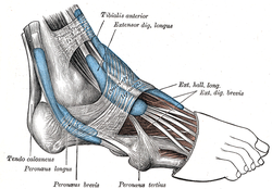
In human anatomy, the fibularis longus is a superficial muscle in the lateral compartment of the leg. It acts to tilt the sole of the foot away from the midline of the body (eversion) and to extend the foot downward away from the body at the ankle.

<span class="mw-page-title-main">Cuboid bone</span> Bone of the ankle

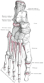
In the human body, the cuboid bone is one of the seven tarsal bones of the foot.

<span class="mw-page-title-main">Fibula</span> Leg bone in vertebrates

The fibula or calf bone is a leg bone on the lateral side of the tibia, to which it is connected above and below. It is the smaller of the two bones and, in proportion to its length, the most slender of all the long bones. Its upper extremity is small, placed toward the back of the head of the tibia, below the knee joint and excluded from the formation of this joint. Its lower extremity inclines a little forward, so as to be on a plane anterior to that of the upper end; it projects below the tibia and forms the lateral part of the ankle joint.

<span class="mw-page-title-main">Ankle</span> Region where the foot and the leg meet

The ankle, the talocrural region or the jumping bone (informal) is the area where the foot and the leg meet. The ankle includes three joints: the ankle joint proper or talocrural joint, the subtalar joint, and the inferior tibiofibular joint. The movements produced at this joint are dorsiflexion and plantarflexion of the foot. In common usage, the term ankle refers exclusively to the ankle region. In medical terminology, "ankle" can refer broadly to the region or specifically to the talocrural joint.

<span class="mw-page-title-main">Calcaneus</span> Bone of the tarsus of the foot

In humans and many other primates, the calcaneus or heel bone is a bone of the tarsus of the foot which constitutes the heel. In some other animals, it is the point of the hock.

<span class="mw-page-title-main">Flexor hallucis longus muscle</span> One of the three deep muscles in the lower leg

The flexor hallucis longus muscle (FHL) attaches to the plantar surface of phalanx of the great toe and is responsible for flexing that toe. The FHL is one of the three deep muscles of the posterior compartment of the leg, the others being the flexor digitorum longus and the tibialis posterior. The tibialis posterior is the most powerful of these deep muscles. All three muscles are innervated by the tibial nerve which comprises half of the sciatic nerve.

<span class="mw-page-title-main">Extensor digitorum longus muscle</span> Pennate muscle, situated at the lateral part of the front of the leg

The extensor digitorum longus is a pennate muscle, situated at the lateral part of the front of the leg.

<span class="mw-page-title-main">Fibularis tertius</span> Muscle of the human body located in the lower limb

In human anatomy, the fibularis tertius is a muscle in the anterior compartment of the leg. It acts to tilt the sole of the foot away from the midline of the body (eversion) and to pull the foot upward toward the body (dorsiflexion).

<span class="mw-page-title-main">Superficial fibular nerve</span> Nerve in the human leg and foot

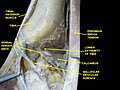
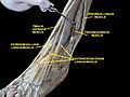
The superficial fibular nerve is a mixed nerve that provides motor innervation to the fibularis longus and fibularis brevis muscles, and sensory innervation to skin over the antero-lateral aspect of the leg along with the greater part of the dorsum of the foot.

<span class="mw-page-title-main">Deep fibular nerve</span> Type of nerve

The deep fibular nerve begins at the bifurcation of the common fibular nerve between the fibula and upper part of the fibularis longus, passes infero-medially, deep to the extensor digitorum longus, to the anterior surface of the interosseous membrane, and comes into relation with the anterior tibial artery above the middle of the leg; it then descends with the artery to the front of the ankle-joint, where it divides into a lateral and a medial terminal branch.

<span class="mw-page-title-main">Fibular retinacula</span> Anatomical structure of the ankle

The fibular retinacula are fibrous retaining bands that bind down the tendons of the fibularis longus and fibularis brevis muscles as they run across the side of the ankle.

<span class="mw-page-title-main">Inferior extensor retinaculum of foot</span> Y-shaped band placed in front of the ankle-joint

The inferior extensor retinaculum of the foot is a Y-shaped band placed in front of the ankle-joint, the stem of the Y being attached laterally to the upper surface of the calcaneus, in front of the depression for the interosseous talocalcaneal ligament; it is directed medialward as a double layer, one lamina passing in front of, and the other behind, the tendons of the peroneus tertius and extensor digitorum longus.

<span class="mw-page-title-main">Fibularis muscles</span> Group of muscles in the leg

The fibularis muscles are a group of muscles in the lower leg.

<span class="mw-page-title-main">First metatarsal bone</span>

The first metatarsal bone is the bone in the foot just behind the big toe. The first metatarsal bone is the shortest of the metatarsal bones and by far the thickest and strongest of them.

<span class="mw-page-title-main">Anterior compartment of leg</span> Part of the fascial compartments of leg

The anterior compartment of the leg is a fascial compartment of the lower leg. It contains muscles that produce dorsiflexion and participate in inversion and eversion of the foot, as well as vascular and nervous elements, including the anterior tibial artery and veins and the deep fibular nerve.

<span class="mw-page-title-main">Lateral compartment of leg</span>

The lateral compartment of the leg is a fascial compartment of the lower leg. It contains muscles which make eversion and plantarflexion of the foot.

<span class="mw-page-title-main">Deep fascia of leg</span> Forms a complete investment to the muscles

The deep fascia of leg or crural fascia forms a complete investment to the muscles, and is fused with the periosteum over the subcutaneous surfaces of the bones.

References

PD-icon.svgThis article incorporates text in the public domain from page 487 of the 20th edition of Gray's Anatomy (1918)

  1. Apaydin, Nihal (2015-01-01), Tubbs, R. Shane; Rizk, Elias; Shoja, Mohammadali M.; Loukas, Marios (eds.), "Chapter 47 - Variations of the Lumbar and Sacral Plexuses and Their Branches", Nerves and Nerve Injuries, San Diego: Academic Press, pp. 627–645, doi:10.1016/b978-0-12-410390-0.00049-4, ISBN   978-0-12-410390-0 , retrieved 2021-02-20
  2. Sammarco, Vincent James; James Sammarco, G. (2008-01-01), Porter, David A.; Schon, Lew C. (eds.), "Chapter 6 - Injuries to the Tibialis Anterior, Peroneal Tendons, and Long Flexors of the Toes", Baxter's the Foot and Ankle in Sport (Second Edition), Philadelphia: Mosby, pp. 121–146, doi:10.1016/b978-032302358-0.10006-5, ISBN   978-0-323-02358-0 , retrieved 2021-02-20
  3. 1 2 3 Gray's Anatomy (1918), see infobox
  4. FIPAT (2019). Terminologia Anatomica. Federative International Programme for Anatomical Terminology.