Gluteus maximus

Last updated
Gluteus maximus
Sobo 1909 294.png
The gluteus maximus, with surrounding fascia. Right buttock, viewed from behind, skin covering removed.
Posterior Hip Muscles 3.PNG
The gluteus medius and nearby muscles.
Details
Origin Gluteal surface of ilium, lumbar fascia, sacrum, sacrotuberous ligament
Insertion Gluteal tuberosity of the femur and iliotibial tract
Artery Superior and inferior gluteal arteries
Nerve Inferior gluteal nerve (L5, S1 and S2 nerve roots)
Actions External rotation and extension of the hip joint, supports the extended knee through the iliotibial tract, chief antigravity muscle in sitting and abduction of the hip
Antagonist Iliacus, psoas major and psoas minor
Identifiers
Latin musculus glutaeus maximus
TA98 A04.7.02.006
TA2 2598
FMA 22314
Anatomical terms of muscle

The gluteus maximus is the main extensor muscle of the hip in humans. It is the largest and outermost of the three gluteal muscles and makes up a large part of the shape and appearance of each side of the hips. It is the single largest muscle in the human body. [1] Its thick fleshy mass, in a quadrilateral shape, forms the prominence of the buttocks. The other gluteal muscles are the medius and minimus, and sometimes informally these are collectively referred to as the glutes.

Contents

Its large size is one of the most characteristic features of the muscular system in humans, [2] connected as it is with the power of maintaining the trunk in the erect posture. Other primates have much flatter hips and cannot sustain standing erectly.

The muscle is made up of muscle fascicles lying parallel with one another, and are collected together into larger bundles separated by fibrous septa.

Structure

Muscles of the gluteal and posterior femoral regions, showing origin and insertion of gluteus maximus muscle. Gluteus maximus muscle.PNG
Muscles of the gluteal and posterior femoral regions, showing origin and insertion of gluteus maximus muscle.

The gluteus maximus (or buttock) is the outermost muscle of the buttocks. It arises from connections to nearby structures in this area. It arises from the posterior gluteal line of the inner upper ilium, a bone of the pelvis, as well as above it to the iliac crest and slightly below it; from the lower part of the sacrum and the side of the coccyx, the tailbone; from the aponeurosis of the erector spinae (lumbodorsal fascia), the sacrotuberous ligament, and the fascia covering the gluteus medius (gluteal aponeurosis). [3]

The fibers are directed obliquely inferiorly and laterally;

The gluteus maximus ends in two main areas:

Bursae

Three bursae are usually found in relation with the deep surface of this muscle:

Function

The gluteus maximus straightens the leg at the hip; when the leg is flexed at the hip, the gluteus maximus extends it to bring the leg into a straight line with the body. [3] The anus also aligns when the leg is flexed at the hip, making the muscle tighten and the pelvis tilt forward.

Taking its fixed point from below, it acts upon the pelvis, supporting it and the trunk upon the head of the femur; this is particularly obvious in standing on one leg. Its most powerful action is to cause the body to regain the erect position after stooping, by drawing the pelvis backward, being assisted in this action by the biceps femoris (long head), semitendinosus, semimembranosus, and adductor magnus.

The gluteus maximus is a tensor of the fascia lata, and by its connection with the iliotibial band steadies the femur on the articular surfaces of the tibia during standing, when the extensor muscles are relaxed.

The lower part of the muscle also acts as an adductor and external rotator of the limb. The upper fibers act as abductors of the hip joints.

Society and culture

Training

Unilateral barbell back squat squatting.jpg
Unilateral barbell back squat standing.jpg
A unilateral (single leg) barbell back squat with the raised foot positioned behind. The bar is held in the low bar style. Squats precipitate high levels of gluteal muscle activation. [4]

The gluteus maximus is involved in several sports, from running to weight-lifting. A number of exercises focus on the gluteus maximus and other muscles of the upper leg:

In art

In cultural terms, the glutes are often considered a symbol of health and strength, and aesthetically appealing. They frequently feature in artwork which seeks to emphasise and celebrate physicality, and the ability to move dynamically and powerfully. They are usually shown to be efficiently proportioned and prominent.

Evidence of such depictions of the gluteal muscles extends from at least Ancient Greece to the modern day. [5] [6]

Clinical significance

Functional assessment can be useful in assessing injuries to the gluteus maximus and surrounding muscles.

The 30-second chair-to-stand test measures a participant's ability to stand up from a seated position as many times as possible in a thirty-second period of time. [7] Testing the number of times a person can stand up in a thirty-second period helps assess strength, flexibility, pain, and endurance, [7] which can help determine how far along a person is in rehabilitation, or how much work is still to be done.

The piriformis test measures flexibility of the gluteus maximus. This requires a trained professional and is based on the angle of external and internal rotation in relation to normal range of motion without injury or impingement. [8]

Other animals

The gluteus maximus is larger in size and thicker in humans than in other primates. [3] Its large size is one of the most characteristic features of the muscular system in humans, [2] connected as it is with the power of maintaining the trunk in the erect posture. Other primates have much flatter hips and cannot sustain standing erectly. [9] [10]

In other primates, the correlate to the human gluteus maximus consists of the ischiofemoralis, a small muscle that corresponds to the human gluteus maximus and originates from the ilium and the ligaments of the sacroiliac, and the gluteus maximus proprius, a large muscle that extends from the ischial tuberosity to a relatively more distant insertion on the femur. In adapting to bipedal gait, reorganization of the attachment of the muscle as well as the moment arm was required. [9]

Additional images

See also

Related Research Articles

<span class="mw-page-title-main">Human leg</span> Lower extremity or limb of the human body (foot, lower leg, thigh and hip)

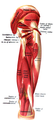
The human leg is the entire lower limb of the human body, including the foot, thigh or sometimes even the hip or buttock region. The major bones of the leg are the femur, tibia, and adjacent fibula. The thigh is between the hip and knee, while the calf (rear) and shin (front) are between the knee and foot.

<span class="mw-page-title-main">Gluteus medius</span> One of the three gluteal muscles

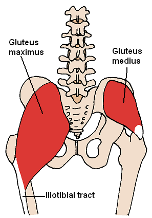
The gluteus medius, one of the three gluteal muscles, is a broad, thick, radiating muscle. It is situated on the outer surface of the pelvis.

<span class="mw-page-title-main">Gluteus minimus</span> Smallest of the three gluteal muscles

The gluteus minimus, or glutæus minimus, the smallest of the three gluteal muscles, is situated immediately beneath the gluteus medius.

<span class="mw-page-title-main">Greater trochanter</span> Quadrilateral part of the thigh bone

The greater trochanter of the femur is a large, irregular, quadrilateral eminence and a part of the skeletal system.

<span class="mw-page-title-main">Hip</span> Anatomical region between the torso and the legs, holding the buttocks and genital region

In vertebrate anatomy, hip refers to either an anatomical region or a joint.

<span class="mw-page-title-main">Buttock augmentation</span> Cosmetic and corrective surgery

Gluteoplasty denotes the plastic surgery and the liposuction procedures for the correction of the congenital, traumatic, and acquired defects and deformities of the buttocks and the anatomy of the gluteal region; and for the aesthetic enhancement of the contour of the buttocks.

The biceps femoris is a muscle of the thigh located to the posterior, or back. As its name implies, it consists of two heads; the long head is considered part of the hamstring muscle group, while the short head is sometimes excluded from this characterization, as it only causes knee flexion and is activated by a separate nerve.

<span class="mw-page-title-main">Adductor magnus muscle</span> Muscle in the thigh

The adductor magnus is a large triangular muscle, situated on the medial side of the thigh.

<span class="mw-page-title-main">Vastus lateralis muscle</span> Human muscle in the thigh

The vastus lateralis, also called the vastus externus, is the largest and most powerful part of the quadriceps femoris, a muscle in the thigh. Together with other muscles of the quadriceps group, it serves to extend the knee joint, moving the lower leg forward. It arises from a series of flat, broad tendons attached to the femur, and attaches to the outer border of the patella. It ultimately joins with the other muscles that make up the quadriceps in the quadriceps tendon, which travels over the knee to connect to the tibia. The vastus lateralis is the recommended site for intramuscular injection in infants less than 7 months old and those unable to walk, with loss of muscular tone.

<span class="mw-page-title-main">Tensor fasciae latae muscle</span> Muscle of the thigh

The tensor fasciae latae is a muscle of the thigh. Together with the gluteus maximus, it acts on the iliotibial band and is continuous with the iliotibial tract, which attaches to the tibia. The muscle assists in keeping the balance of the pelvis while standing, walking, or running.

<span class="mw-page-title-main">Gluteal muscles</span> Group of three muscles which make up the buttocks

The gluteal muscles, often called glutes, are a group of three muscles which make up the gluteal region commonly known as the buttocks: the gluteus maximus, gluteus medius and gluteus minimus. The three muscles originate from the ilium and sacrum and insert on the femur. The functions of the muscles include extension, abduction, external rotation, and internal rotation of the hip joint.

<span class="mw-page-title-main">Muscles of the hip</span> Causes movement in the hip

In human anatomy, the muscles of the hip joint are those muscles that cause movement in the hip. Most modern anatomists define 17 of these muscles, although some additional muscles may sometimes be considered. These are often divided into four groups according to their orientation around the hip joint: the gluteal group; the lateral rotator group; the adductor group; and the iliopsoas group.

The gluteal tuberosity is the lateral one of the three upward prolongations of the linea aspera of the femur, extending to the base of the greater trochanter. It serves as the principal insertion site for the gluteus maximus muscle.

<span class="mw-page-title-main">Ischium</span> Lower and back region of the hip bone

The ischium forms the lower and back region of the hip bone.

<span class="mw-page-title-main">Fascia lata</span> Deep fascia of the thigh

The fascia lata is the deep fascia of the thigh. It encloses the thigh muscles and forms the outer limit of the fascial compartments of thigh, which are internally separated by the medial intermuscular septum and the lateral intermuscular septum. The fascia lata is thickened at its lateral side where it forms the iliotibial tract, a structure that runs to the tibia and serves as a site of muscle attachment.

<span class="mw-page-title-main">Superior gluteal artery</span>

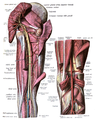
The superior gluteal artery is the terminal branch of the posterior division of the internal iliac artery. It exits the pelvis through the greater sciatic foramen before splitting into a superficial branch and a deep branch.

<span class="mw-page-title-main">Inferior gluteal artery</span>

The inferior gluteal artery is a terminal branch of the anterior trunk of the internal iliac artery. It exits the pelvis through the greater sciatic foramen. It is distributed chiefly to the buttock and the back of the thigh.

<span class="mw-page-title-main">Third trochanter</span>

In human anatomy, the third trochanter is a bony projection occasionally present on the proximal femur near the superior border of the gluteal tuberosity. When present, it is oblong, rounded, or conical in shape and sometimes continuous with the gluteal ridge. It generally occurs bilaterally without significant side to side dimorphism. A structure of minor importance in humans, the incidence of the third trochanter varies from 17 to 72% between ethnic groups and it is frequently reported as more common in females than in males. Structures analogous to the third trochanter are present in other mammals, including some primates. It is called the third trochanter in reference to the greater and lesser trochanters that are always present on the femur.

<span class="mw-page-title-main">Hip bone</span> Bone of the pelvis

The hip bone is a large flat bone, constricted in the center and expanded above and below. In some vertebrates it is composed of three parts: the ilium, ischium, and the pubis.

<span class="mw-page-title-main">Pelvis</span> Lower torso of the human body

The pelvis is the lower part of the trunk, between the abdomen and the thighs, together with its embedded skeleton.

References

PD-icon.svgThis article incorporates text in the public domain from page 474 of the 20th edition of Gray's Anatomy (1918)

  1. "What is the strongest muscle in the human body?". Library of Congress. 19 November 2019. Retrieved 2023-05-28.
  2. 1 2 Norman Eizenberg et al., General Anatomy: Principles and Applications (2008), p. 17.
  3. 1 2 3 Standring, Susan, ed. (2016). "Pelvic girdle, gluteal region and thigh: Gluteus maximus". Gray's anatomy: The anatomical basis of clinical practice (41st ed.). Philadelphia: Elsevier Limited. pp. 1357–1358. ISBN   9780702052309. OCLC   920806541.
  4. Contreras B, Cordoza G (2019). The Glute Lab. Victory Belt Publishing. p. 426. ISBN   978-1628603-46-0.
  5. Bret Contreras, Glen Cordoza (2019). The Glute Lab. Victory Belt Publishing. p. 5. ISBN   9781628603460.
  6. Metraux, Guy P. R. (1995). Sculptures and Physicians in Fifth-Century Greece. Montreal & Kingston: McGill-Queen's University Press. pp. 40–41. ISBN   0773512314.
  7. 1 2 Dobson, F.; Bennell, K.; Hinman, R.; Abbott, H.; Roos, E. (2013). "OARSI recommended performance-based tests to assess physical function in people diagnosed with hip or knee osteoarthritis" (PDF). Osteoarthritis and Cartilage. 21 (8): 1042–52. doi: 10.1016/j.joca.2013.05.002 . PMID   23680877.
  8. "Passive Piriformis ROM". Exrx.net. Retrieved February 19, 2015.
  9. 1 2 Hogervorst, T.; Vereecke, E. E. (January 2015). "Evolution of the human hip. Part 2: muscling the double extension". Journal of Hip Preservation Surgery. 2 (1): 3–14. doi:10.1093/jhps/hnu014. PMC   4718477 . PMID   27011809.
  10. Stern, Jack T.; Susman, Randall L. (June 1981). "Electromyography of the gluteal muscles in Hylobates, Pongo , and Pan: Implications for the evolution of hominid bipedality". American Journal of Physical Anthropology. 55 (2): 153–166. doi:10.1002/ajpa.1330550203.