Flexor hallucis longus muscle

Last updated
Flexor hallucis longus muscle
Posterior compartment of leg - flexor hallucis longus.png
Flexor hallucis longus. Seen from behind.
Sobo 1909 316.png
The mucous sheaths of the tendons around the ankle. Medial aspect. (Tendon of flexor hallucis longus labeled at bottom left.)
Details
Origin Fibula, posterior aspect of middle 1/3
Insertion Plantar surface; base of distal phalanx of hallux
Artery (Muscular branch of peroneal artery (peroneal branch of the posterior tibial artery)
Nerve Tibial nerve, S2 and S3 [1]
Actions Flexes all joints of the big toe, plantar flexion of the ankle joint
Antagonist Extensor hallucis longus muscle
Identifiers
Latin musculus flexor hallucis longus
Acronym(s)FHL
TA98 A04.7.02.053
TA2 2668
FMA 22593
Anatomical terms of muscle

The flexor hallucis longus muscle (FHL) attaches to the plantar surface of phalanx of the great toe and is responsible for flexing that toe. The FHL is one of the three deep muscles of the posterior compartment of the leg, the others being the flexor digitorum longus and the tibialis posterior. The tibialis posterior is the most powerful of these deep muscles. All three muscles are innervated by the tibial nerve which comprises half of the sciatic nerve. [2]

Contents

Structure

The flexor hallucis longus is situated on the fibular side of the leg. It arises from the inferior two-thirds of the posterior surface of the body of the fibula, with the exception of 2.5 cm. at its lowest part; from the lower part of the interosseous membrane; from an intermuscular septum between it and the peroneus muscles, laterally, and from the fascia covering the tibialis posterior, medially.

The fibers pass obliquely downward and backward, where it passes through the tarsal tunnel on the medial side of the foot and end in a tendon which occupies nearly the whole length of the posterior surface of the muscle.

This tendon lies in a groove which crosses the posterior surface of the lower end of the tibia, between the medial and lateral tubercles of the posterior surface of the talus, and the under surface of the sustentaculum tali of the calcaneus; in the sole of the foot it runs forward between the two heads of the flexor hallucis brevis, and is inserted into the base of the last phalanx of the great toe. The grooves on the talus and calcaneus, which contain the tendon of the muscle, are converted by tendinous fibers into distinct canals, lined by a mucous sheath.

As the tendon passes forward in the sole of the foot, it is situated above, and crosses from the lateral to the medial side of the tendon of the flexor digitorum longus, to which it is connected by a fibrous slip.

Variation

Usually a slip runs to the flexor digitorum and frequently an additional slip runs from the flexor digitorum to the flexor hallucis. Peroneocalcaneus internus, rare,[ clarification needed ] arises below or outside the flexor hallucis from the back of the fibula, passes over the sustentaculum tali with the flexor hallucis and inserts into the calcaneum.

Function

Similar to the flexor digitorum longus and tibialis posterior muscles, the flexor hallucis longus muscle functions to plantar flex and invert the foot. However, it is unique in that it also functions to flex the great toe and helps supinate the ankle. [3]

Injury and treatment

Common injuries associated with the FHL tendon are tenosynovitis, tendinopathies, and muscle strains. Because the FHL muscle is small, injuries associated with this muscle and its tendon are often overlooked. An MRI can be used to evaluate the cause and condition of the FHL tendon. Tears and areas of impingement can be found using this method. A diagnostic ultrasound can also be used to diagnose FHL injuries, as it shows the muscle in movement and potential areas of impingement. Conservatively, an FHL injury can be evaluated by determining if movements caused by the FHL muscle cause pain along the inner ankle or under the big toe.[ citation needed ]

After passing through the tarsal tunnel, the flexor hallucis longus tendon must curve around a bony landmark called the sustentaculum tali. Friction at this site is likely to cause pain on the posteromedial aspect of the ankle. While commonly referred to as "dancer's tendinitis," FHL tendinitis occurs commonly in ballet dancers, gymnasts, and runners. Due to their excessive use of toe flexion, which results in ten times their body weight being applied to this small muscle and tendon, inflammation and irritation is common at the site of the sustentaculum tali.

Hallux saltans is a condition that develops as a result of overusing the FHL muscle. With this condition, a nodule develops along the FHL tendon which may produce a popping effect during contraction because it drags along surrounding tissues. If left untreated and continually irritated, stenosis of the tendon may occur, resulting in the big toe becoming stiff and relatively immobile. This condition is known as Hallux Rigidus.

Most FHL injuries can be managed through conservative treatment. Rest is usually the first indicated intervention for minor FHL injuries. Ice and ultrasound therapy can also help with the inflammation and pain. Physical therapy exercises and stretches can help rehabilitate the muscle and tendon and potentially address biomechanical errors that cause the inflammation and microtears in the tendon. Some FHL injuries can be treated through rest, physical therapy, splints, and anti-inflammatory medication. However, more serious or chronic injuries may require surgery. If surgery is indicated, tears in the FHL will be repaired, and debris will be removed from the area. It is worth noting that an os trigonum may cause similar symptoms to the ones caused by FHL tendinitis or tenosynovitis. A radiograph should be taken to rule out this condition. [4]

Additional images

Related Research Articles

<span class="mw-page-title-main">Foot</span> Anatomical structure found in vertebrates

The foot is an anatomical structure found in many vertebrates. It is the terminal portion of a limb which bears weight and allows locomotion. In many animals with feet, the foot is a separate organ at the terminal part of the leg made up of one or more segments or bones, generally including claws and/or nails.

<span class="mw-page-title-main">Human leg</span> Lower extremity or limb of the human body (foot, lower leg, thigh and hip)

The leg is the entire lower limb of the human body, including the foot, thigh or sometimes even the hip or buttock region. The major bones of the leg are the femur, tibia, and adjacent fibula. The thigh is between the hip and knee, while the calf (rear) and shin (front) are between the knee and foot.

<span class="mw-page-title-main">Toe</span> Digit of a foot

Toes are the digits of the foot of a tetrapod. Animal species such as cats that walk on their toes are described as being digitigrade. Humans, and other animals that walk on the soles of their feet, are described as being plantigrade; unguligrade animals are those that walk on hooves at the tips of their toes.

<span class="mw-page-title-main">Cuboid bone</span> Bone of the ankle

In the human body, the cuboid bone is one of the seven tarsal bones of the foot.

<span class="mw-page-title-main">Ankle</span> Region where the foot and the leg meet

The ankle, the talocrural region or the jumping bone (informal) is the area where the foot and the leg meet. The ankle includes three joints: the ankle joint proper or talocrural joint, the subtalar joint, and the inferior tibiofibular joint. The movements produced at this joint are dorsiflexion and plantarflexion of the foot. In common usage, the term ankle refers exclusively to the ankle region. In medical terminology, "ankle" can refer broadly to the region or specifically to the talocrural joint.

<span class="mw-page-title-main">Extensor hallucis longus muscle</span> Thin muscle, situated between the tibialis anterior and the extensor digitorum longus

The extensor hallucis longus muscle is a thin skeletal muscle, situated between the tibialis anterior and the extensor digitorum longus. It extends the big toe and dorsiflects the foot. It also assists with foot eversion and inversion.

<span class="mw-page-title-main">Calcaneus</span> Bone of the tarsus of the foot

In humans and many other primates, the calcaneus or heel bone is a bone of the tarsus of the foot which constitutes the heel. In some other animals, it is the point of the hock.

<span class="mw-page-title-main">Tibialis posterior muscle</span> Muscle in the most central of all the leg muscles

The tibialis posterior muscle is the most central of all the leg muscles, and is located in the deep posterior compartment of the leg. It is the key stabilizing muscle of the lower leg.

<span class="mw-page-title-main">Tibial nerve</span> Branch of the sciatic nerve

The tibial nerve is a branch of the sciatic nerve. The tibial nerve passes through the popliteal fossa to pass below the arch of soleus.

<span class="mw-page-title-main">Flexor digitorum longus muscle</span> Muscle located on the tibial side of the leg

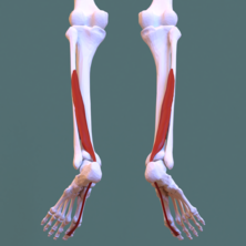
The flexor digitorum longus muscle is situated on the tibial side of the leg. At its origin it is thin and pointed, but it gradually increases in size as it descends. It serves to flex the second, third, fourth, and fifth toes.

<span class="mw-page-title-main">Extensor digitorum brevis muscle</span> Muscle on the upper surface of the foot that helps extend digits 2 through 4

The extensor digitorum brevis muscle is a muscle on the upper surface of the foot that helps extend digits 2 through 4.

<span class="mw-page-title-main">Fibularis brevis</span> Shorter and smaller of the fibularis (peroneus) muscles

In human anatomy, the fibularis brevis is a muscle that lies underneath the fibularis longus within the lateral compartment of the leg. It acts to tilt the sole of the foot away from the midline of the body (eversion) and to extend the foot downward away from the body at the ankle.

<span class="mw-page-title-main">Flexor hallucis brevis muscle</span> Muscle in sole of the foot that leads to the big toe

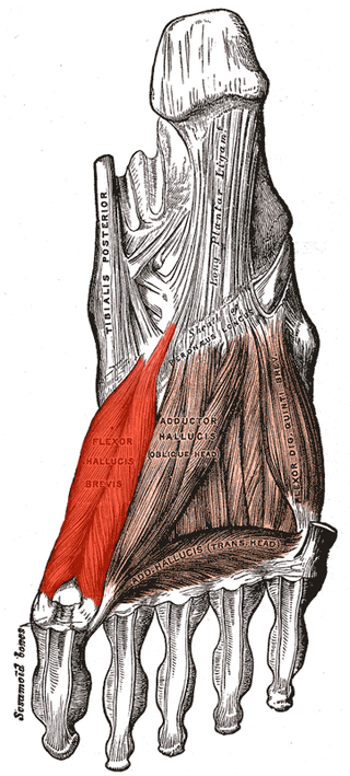
Flexor hallucis brevis muscle is a muscle of the foot that flexes the big toe.

<span class="mw-page-title-main">Quadratus plantae muscle</span> Muscles of the sole of the foot

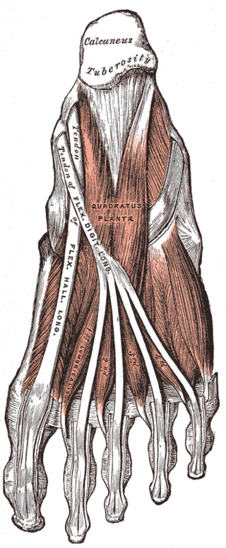
The quadratus plantae is separated from the muscles of the first layer by the lateral plantar vessels and nerve. It acts to aid in flexing the 2nd to 5th toes and is one of the few muscles in the foot with no homolog in the hand.

<span class="mw-page-title-main">Deep fibular nerve</span> Type of nerve

The deep fibular nerve begins at the bifurcation of the common fibular nerve between the fibula and upper part of the fibularis longus, passes infero-medially, deep to the extensor digitorum longus, to the anterior surface of the interosseous membrane, and comes into relation with the anterior tibial artery above the middle of the leg; it then descends with the artery to the front of the ankle-joint, where it divides into a lateral and a medial terminal branch.

<span class="mw-page-title-main">Sole (foot)</span> Bottom part of foot

In humans, the sole of the foot is anatomically referred to as the plantar aspect.

The flexor retinaculum of foot is a strong fibrous band in the foot.

<span class="mw-page-title-main">Deltoid ligament</span> Anatomical detail in the ankle

The deltoid ligament is a strong, flat, triangular band, attached, above, to the apex and anterior and posterior borders of the medial malleolus. The deltoid ligament supports the ankle joint and also resists excessive eversion of the foot. The deltoid ligament is composed of 4 fibers:

  1. Anterior tibiotalar ligament
  2. Tibiocalcaneal ligament
  3. Posterior tibiotalar ligament
  4. Tibionavicular ligament.
<span class="mw-page-title-main">Malleolus</span> Ankle bone protrusion

A malleolus is the bony prominence on each side of the human ankle.

<span class="mw-page-title-main">Tarsal tunnel</span> Canal in the ankle area

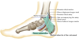
The tarsal tunnel is a passage found along the inner leg underneath the medial malleolus of the ankle.

References

PD-icon.svgThis article incorporates text in the public domain from page 485 of the 20th edition of Gray's Anatomy (1918)

  1. Aids to the Examination of the Peripheral Nervous System, 5th edition
  2. "Peripheral Nerve Injury". www.hopkinsmedicine.org. Retrieved 20 October 2020.
  3. "Flexor Hallucis Longus" . Retrieved 20 October 2020.
  4. "Flexor Hallucis Longus Dysfunction". Massage Today. Retrieved 20 October 2020.