Gluteal muscles

Last updated
Gluteal muscles
Gluteus muscles.PNG
Right leg, Calves rear view: Muscles of the gluteal and posterior femoral regions. Gluteus minimus and the origins and insertions of medius and maximus shown in red & also lower legs
Gluteus maximus.png
Gluteus maximus
Details
Parts Gluteus maximus, gluteus medius and gluteus minimus
Artery Superior and inferior gluteal arteries
Vein Superior and inferior gluteal veins
Nerve Superior and inferior gluteal nerves (L4, L5, S1 and S2 nerve roots)
Anatomical terminology

The gluteal muscles, often called glutes, are a group of three muscles which make up the gluteal region commonly known as the buttocks: the gluteus maximus, gluteus medius and gluteus minimus. The three muscles originate from the ilium and sacrum and insert on the femur. The functions of the muscles include extension, abduction, external rotation, and internal rotation of the hip joint. [1]

Contents

Structure

The gluteus maximus is the largest and most superficial of the three gluteal muscles. It makes up a large part of the shape and appearance of the hips. It is a narrow and thick fleshy mass of a quadrilateral shape, and forms the prominence of the buttocks. The gluteus medius is a broad, thick, radiating muscle, situated on the outer surface of the pelvis. It lies profound to the gluteus maximus and its posterior third is covered by the gluteus maximus, its anterior two-thirds by the gluteal aponeurosis, which separates it from the superficial fascia and skin. The gluteus minimus is the smallest of the three gluteal muscles and is situated immediately beneath the gluteus medius.

The bulk of the gluteal muscle mass contributes only partially to shape of the buttocks. The other major contributing factor is that of the panniculus adiposus (fatty layer) of the buttocks, which is very well developed in this area, and gives the buttock its characteristic rounded shape. Exercise in general (not only of the gluteal muscles but of the body in general) which can contribute to fat loss can lead to reduction of mass in subcutaneal fat storage locations on the body which includes the panniculus, so for leaner and more active individuals, the glutes will more predominantly contribute to the shape than someone less active with a fattier composition.[ citation needed ] The degree of body fat stored in various locations such as the panniculus is dictated by genetic and hormonal profiles.[ citation needed ]

Gluteus maximus

The gluteus maximus arises from the posterior gluteal line of the inner upper ilium, and the rough portion of bone including the crest, immediately above and behind it; from the posterior surface of the lower part of the sacrum and the side of the coccyx; from the aponeurosis of the erector spinae (lumbodorsal fascia), the sacrotuberous ligament, and the fascia covering the gluteus medius. The fibers are directed obliquely downward and lateralward; the muscle has two insertions: Those forming the upper and larger portion of the muscle, together with the superficial fibers of the lower portion, end in a thick tendinous lamina, which passes across the greater trochanter, and inserts into the iliotibial band of the fascia lata; and the deeper fibers of the lower portion of the muscle are inserted into the gluteal tuberosity between the vastus lateralis and adductor magnus. Its action is to extend and to laterally rotate the hip, and also to extend the trunk.[ citation needed ]

Gluteus medius

The gluteus medius muscle originates on the outer surface of the ilium between the iliac crest and the posterior gluteal line above, and the anterior gluteal line below; the gluteus medius also originates from the gluteal aponeurosis that covers its outer surface. The fibers of the muscle converge into a strong flattened tendon that inserts on the lateral surface of the greater trochanter. More specifically, the muscle's tendon inserts into an oblique ridge that runs downward and forward on the lateral surface of the greater trochanter.

Gluteus minimus

The gluteus minimus is fan-shaped, arising from the outer surface of the ilium, between the anterior and inferior gluteal lines, and behind, from the margin of the greater sciatic notch. The fibers converge to the deep surface of a radiated aponeurosis, and this ends in a tendon which is inserted into an impression on the anterior border of the greater trochanter, and gives an expansion to the capsule of the hip joint.

Function

The functions of muscles includes extension, abduction and internal as well as external rotation of the hip joint. The gluteus maximus also supports the extended knee through the iliotibial tract.

Clinical significance

Sitting for long periods can lead to the gluteal muscles atrophying through constant pressure and disuse. This may be associated with (although not necessarily the cause of) lower back pain, difficulty with some movements that naturally require the gluteal muscles such as rising from the seated position, and climbing stairs.

Exercise and stretching

Any exercise that works and/or stretches the buttocks is suitable, for example lunges, hip thrusts, climbing stairs, fencing, bicycling, rowing, squats, arabesque, aerobics, and various specific exercises for the bottom. Weight training exercises which are known to significantly strengthen the gluteal muscles include the squat, deadlift, leg press, any other movements involving external hip rotation and hip extension.

Society and culture

Cultural significance

Well formed gluteal muscles have long been associated with health, strength and sexual attractiveness. In terms of health, they act as a sign of 'being in shape'. This usually means a person is also eating, sleeping and exercising properly, all of which are beneficial to health. In terms of strength, the glutes are among the largest and most powerful muscles in the body. If they are well developed then a person is more likely to be strong. They are also key contributors to movement ranges of fundamental importance, such as bending and straightening the legs, and bending, straightening and twisting at the waist. These movement ranges are key in a person's ability to move in a powerful, dynamic fashion and they are powered to a significant extent by the glutes. If they are well formed then a person is much more likely to be able to move efficiently. In terms of sexual attractiveness, the glute specialist Bret Contreras considers in physio-anthropological terms that this is based upon a sub-conscious assessment of the relationship between a physical capability to survive and prosper, and the ability to raise a family. [2]

'It stands to reason that both males and females were attracted to nice glutes, instinctively making the connection to big, strong glutes and survival, reproduction, hunting and protection.' [3]

Artistic representation

Prominent gluteal muscles are often used in art in order to imply an ability to move in a powerful, dynamic fashion; virility and fertility; and to meet aesthetic considerations in these regards.

See also

Additional images

Related Research Articles

<span class="mw-page-title-main">Human leg</span> Lower extremity or limb of the human body (foot, lower leg, thigh and hip)

The leg is the entire lower limb of the human body, including the foot, thigh or sometimes even the hip or buttock region. The major bones of the leg are the femur, tibia, and adjacent fibula. The thigh is between the hip and knee, while the calf (rear) and shin (front) are between the knee and foot.

<span class="mw-page-title-main">Gluteus maximus</span> Largest and most superficial of the three gluteal muscles

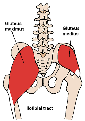
The gluteus maximus is the main extensor muscle of the hip in humans. It is the largest and outermost of the three gluteal muscles and makes up a large part of the shape and appearance of each side of the hips. It is the single largest muscle in the human body. Its thick fleshy mass, in a quadrilateral shape, forms the prominence of the buttocks. The other gluteal muscles are the medius and minimus, and sometimes informally these are collectively referred to as the glutes.

<span class="mw-page-title-main">Gluteus medius</span> One of the three gluteal muscles

The gluteus medius, one of the three gluteal muscles, is a broad, thick, radiating muscle. It is situated on the outer surface of the pelvis.

<span class="mw-page-title-main">Gluteus minimus</span> Smallest of the three gluteal muscles

The gluteus minimus, or glutæus minimus, the smallest of the three gluteal muscles, is situated immediately beneath the gluteus medius.

<span class="mw-page-title-main">Piriformis muscle</span> Hip muscle in the lateral rotator group

The piriformis muscle is a flat, pyramidally-shaped muscle in the gluteal region of the lower limbs. It is one of the six muscles in the lateral rotator group.

<span class="mw-page-title-main">Hip</span> Anatomical region between the torso and the legs, holding the buttocks and genital region

In vertebrate anatomy, the hip, or coxa in medical terminology, refers to either an anatomical region or a joint on the outer (lateral) side of the pelvis.

<span class="mw-page-title-main">Buttock augmentation</span> Cosmetic and corrective surgery

Gluteoplasty denotes the plastic surgery and the liposuction procedures for the correction of congenital, traumatic, and acquired defects/deformities of the buttocks and the anatomy of the gluteal region; and for the aesthetic enhancement of the contour of the buttocks.

<span class="mw-page-title-main">Gluteal sulcus</span> Fold separating the thigh from the buttock

The gluteal sulcus is an area of the body of humans and anthropoid apes, described by a horizontal crease formed by the inferior aspect of the buttocks and the posterior upper thigh. The gluteal sulcus is formed by the posterior horizontal skin crease of the hip joint and overlying fat and is not formed by the lower border of the gluteus maximus muscle, which crosses the fold obliquely. It is one of the major defining features of the buttocks. Children with developmental dysplasia of the hips are born with uneven gluteal folds and can be diagnosed with a physical examination and sonogram.

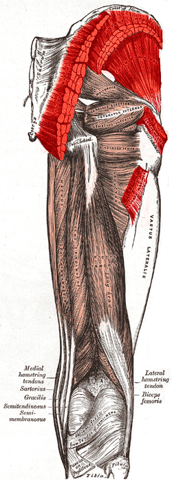
The biceps femoris is a muscle of the thigh located to the posterior, or back. As its name implies, it consists of two heads; the long head is considered part of the hamstring muscle group, while the short head is sometimes excluded from this characterization, as it only causes knee flexion and is activated by a separate nerve.

<span class="mw-page-title-main">Quadratus lumborum muscle</span> Muscle in the lower back

The quadratus lumborum muscle, informally called the QL, is a paired muscle of the left and right posterior abdominal wall. It is the deepest abdominal muscle, and commonly referred to as a back muscle. Each is irregular and quadrilateral in shape.

<span class="mw-page-title-main">Adductor magnus muscle</span> Muscle in the thigh

The adductor magnus is a large triangular muscle, situated on the medial side of the thigh.

<span class="mw-page-title-main">Tensor fasciae latae muscle</span> Muscle of the thigh

The tensor fasciae latae is a muscle of the thigh. Together with the gluteus maximus, it acts on the iliotibial band and is continuous with the iliotibial tract, which attaches to the tibia. The muscle assists in keeping the balance of the pelvis while standing, walking, or running.

<span class="mw-page-title-main">Muscles of the hip</span> Causes movement in the hip

In human anatomy, the muscles of the hip joint are those muscles that cause movement in the hip. Most modern anatomists define 17 of these muscles, although some additional muscles may sometimes be considered. These are often divided into four groups according to their orientation around the hip joint: the gluteal group; the lateral rotator group; the adductor group; and the iliopsoas group.

<span class="mw-page-title-main">Erector spinae muscles</span> Human muscle group

The erector spinae or spinal erectors is a set of muscles that straighten and rotate the back. The spinal erectors work together with the glutes to maintain stable posture standing or sitting.

<span class="mw-page-title-main">Superior gluteal artery</span>

The superior gluteal artery is the terminal branch of the posterior division of the internal iliac artery. It exits the pelvis through the greater sciatic foramen before splitting into a superficial branch and a deep branch.

<span class="mw-page-title-main">Wing of ilium</span> Flat portion of the hip bone

The wing(ala)of ilium is the large expanded portion of the ilium, the bone which bounds the greater pelvis laterally. It presents for examination two surfaces—an external and an internal—a crest, and two borders—an anterior and a posterior.

<span class="mw-page-title-main">Buttocks</span> Anatomical feature on the posterior of some primates

The buttocks are two rounded portions of the exterior anatomy of most mammals, located on the posterior of the pelvic region. In humans, the buttocks are located between the lower back and the perineum. They are composed of a layer of exterior skin and underlying subcutaneous fat superimposed on a left and right gluteus maximus and gluteus medius muscles. The two gluteus maximus muscles are the largest muscles in the human body. They are responsible for movements such as straightening the body into the upright (standing) posture when it is bent at the waist; maintaining the body in the upright posture by keeping the hip joints extended; and propelling the body forward via further leg (hip) extension when walking or running.

<span class="mw-page-title-main">Hip bone</span> Bone of the pelvis

The hip bone is a large flat bone, constricted in the center and expanded above and below. In some vertebrates it is composed of three parts: the ilium, ischium, and the pubis.

<span class="mw-page-title-main">Gluteal lines</span> Lines of the human ilium

The gluteal lines are three curved lines outlined from three bony ridges on the exterior surface of the ilium in the gluteal region. They are the anterior gluteal line; the inferior gluteal line, and the posterior gluteal line.

References

  1. Moore, Keith L.; Dalley II, Arthur F.; Agur, Anne M. R. (2018). Clinically Oriented Anatomy (Eighth ed.). Philadelphia: Wolters Kluwer. pp. 722–723. ISBN   9781496347213.
  2. Bret Contreras, Glen Cordoza (2019). The Glute Lab. Victory Belt Publishing. pp. 5, 25. ISBN   9781628603460.
  3. Bret Contreras, Glen Cordoza (2019). The Glute Lab. Victory Belt Publishing. p. 25. ISBN   9781628603460.