Glass ionomer cement

Last updated
Glass ionomer Glass ionomer.png
Glass ionomer

A glass ionomer cement (GIC) is a dental restorative material used in dentistry as a filling material and luting cement, [1] including for orthodontic bracket attachment. [2] Glass-ionomer cements are based on the reaction of silicate glass-powder (calciumaluminofluorosilicate glass [3] ) and polyacrylic acid, an ionomer. Occasionally water is used instead of an acid, [2] altering the properties of the material and its uses. [4] This reaction produces a powdered cement of glass particles surrounded by matrix of fluoride elements and is known chemically as glass polyalkenoate. [5] There are other forms of similar reactions which can take place, for example, when using an aqueous solution of acrylic/itaconic copolymer with tartaric acid, this results in a glass-ionomer in liquid form. An aqueous solution of maleic acid polymer or maleic/acrylic copolymer with tartaric acid can also be used to form a glass-ionomer in liquid form. Tartaric acid plays a significant part in controlling the setting characteristics of the material. [5] Glass-ionomer based hybrids incorporate another dental material, for example resin-modified glass ionomer cements (RMGIC) and compomers (or modified composites). [5]

Contents

Non-destructive neutron scattering has evidenced GIC setting reactions to be non-monotonic, with eventual fracture toughness dictated by changing atomic cohesion, fluctuating interfacial configurations and interfacial terahertz (THz) dynamics. [6]

It is on the World Health Organization's List of Essential Medicines. [7]

Background

Glass ionomer cement is primarily used in the prevention of dental caries. This dental material has good adhesive bond properties to tooth structure, [8] allowing it to form a tight seal between the internal structures of the tooth and the surrounding environment. Dental caries are caused by bacterial production of acid during their metabolic actions. The acid produced from this metabolism results in the breakdown of tooth enamel and subsequent inner structures of the tooth, if the disease is not intervened by a dental professional, or if the carious lesion does not arrest and/or the enamel re-mineralises by itself. Glass ionomer cements act as sealants when pits and fissures in the tooth occur and release fluoride to prevent further enamel demineralisation and promote remineralisation. Fluoride can also hinder bacterial growth, by inhibiting their metabolism of ingested sugars in the diet. It does this by inhibiting various metabolic enzymes within the bacteria. This leads to a reduction in the acid produced during the bacteria's digestion of food, preventing a further drop in pH and therefore preventing caries.[ citation needed ]

There is evidence that when using sealants, only 6% of people develop tooth decay over a 2-year period, in comparison to 40% of people when not using a sealant. [9] However, it is recommended that the use of fluoride varnish alongside glass ionomer sealants should be applied in practice to further reduce the risk of secondary dental caries. [10]

Resin-modified glass ionomers

The addition of resin to glass ionomers improves them significantly, allowing them to be more easily mixed and placed. [3] Resin-modified glass ionomers allow equal or higher fluoride release and there is evidence of higher retention, higher strength and lower solubility. [3] Resin-based glass ionomers have two setting reactions: an acid-base setting and a free-radical polymerisation. The free-radical polymerisation is the predominant mode of setting, as it occurs more rapidly than the acid-base mode. Only the material properly activated by light will be optimally cured. The presence of resin protects the cement from water contamination. Due to the shortened working time, it is recommended that placement and shaping of the material occurs as soon as possible after mixing. [5]

History

Dental sealants were first introduced as part of the preventative programme, in the late 1960s, in response to increasing cases of pits and fissures on occlusal surfaces due to caries. [9] This led to glass ionomer cements to be introduced in 1972 by Wilson and Kent as derivative of the silicate cements and the polycarboxylate cements. [5] The glass ionomer cements incorporated the fluoride releasing properties of the silicate cements with the adhesive qualities of polycarboxylate cements. [4] This incorporation allowed the material to be stronger, less soluble and more translucent (and therefore more aesthetic) than its predecessors. [5]

Glass ionomer cements were initially intended to be used for the aesthetic restoration of anterior teeth and were recommended for restoring Class III and Class V cavity preparations. [8] There have now been further developments in the material's composition to improve properties. For example, the addition of metal or resin particles into the sealant is favoured due to the longer working time and the material being less sensitive to moisture during setting. [8]

When glass ionomer cements were first used, they were mainly used for the restoration of abrasion/erosion lesions and as a luting agent for crown and bridge reconstructions. However, this has now been extended to occlusal restorations in deciduous dentition, restoration of proximal lesions and cavity bases and liners. [4] This is made possible by the ever-increasing new formulations of glass ionomer cements.

One of the early commercially successful GICs, employing G338 glass and developed by Wilson and Kent, served purpose as non-load bearing restorative materials. However, this glass resulted in a cement too brittle for use in load-bearing applications such as in molar teeth. The properties of G338 being shown to be related to its phase-composition, specifically the interplay between its three amorphous phases Ca/Na-Al-Si-O, Ca-Al-F and Ca-P-O-F, as characterised by mechanical testing, differential scanning calorimetry (DSC) and X-ray diffraction (XRD), [11] as well as quantum chemical modelling and ab initio molecular dynamics simulations. [12]

Glass ionomer versus resin-based sealants

When the two dental sealants are compared, there has always been a contradiction as to which materials is more effective in caries reduction. Therefore, there are claims against replacing resin-based sealants, the current gold standard, with glass ionomer. [13] [14] [15]

Advantages

Glass ionomer sealants are thought to prevent caries through a steady fluoride release over a prolonged period and the fissures are more resistant to demineralization, even after the visible loss of sealant material, [9] however, a systemic review found no difference in caries development when GICs was used as a fissure sealing material compared to the conventional resin based sealants, in addition, it has less retention to the tooth structure than the resin based sealants. [16]

These sealants have hydrophilic properties, allowing them to be an alternative of the hydrophobic resin in the generally wet oral cavity. Resin-based sealants are easily destroyed by saliva contamination.

They chemically bond with both enamel and dentin and do not necessarily require preparation/mechanical retention and can therefore be applied without harming existing tooth structure. This makes them ideal in many situations when tooth preservation is foremost and with minimally invasive techniques, particularly Class V fillings where there is a larger area of exposed dentin with only a thin ring of enamel. This often results in longer retention and service life than resin Class V fillings.

They chemically bond to enamel and dentin leaving a smaller gap for bacteria to enter. Particularly when paired with silver diamine fluoride this can arrest caries and harden active caries and prevent further damage.

They can be placed and cured outside of clinical settings and do not require a curing light.

Chemically curable glass ionomer cements are considered safe from allergic reactions but a few have been reported with resin-based materials. Nevertheless, allergic reactions are very rarely associated with both sealants. [9]

Disadvantages

The main disadvantage of glass ionomer sealants or cements has been inadequate retention or simply lack of strength, toughness, and limited wear resistance. [17] [18] For instance, due to its poor retention rate, periodic recalls are necessary, even after 6 months, to eventually replace the lost sealant. [9] [19] Different methods have been used to address the physical shortcomings of the glass ionomer cements such as thermo-light curing (polymerization), [20] [21] or addition of the zirconia, hydroxyapatite, N-vinyl pyrrolidone, N-vinyl caprolactam, and fluoroapatite to reinforce the glass ionomer cements. [22]

Clinical applications

Glass ionomers are widely used due to their versatile properties and ease of use. Prior to procedures, starter materials for glass ionomers are supplied as a powder and liquid or as a powder mixed with water. These materials can be mixed and encapsulated. [23]

Preparation of the material should involve following manufacture instructions. A paper pad or cool dry glass slab may be used for mixing the raw materials though it is important to note that the use of the glass slab will retard the reaction and hence increase the working time. [23] The raw materials in liquid and powder form should not be dispensed onto the chosen surface until the mixture is required in the clinical procedure the glass ionomer is being used for, as a prolonged exposure to the atmosphere could interfere with the ratio of chemicals in the liquid. At the stage of mixing, a spatula should be used to rapidly incorporate the powder into the liquid for a duration of 45–60 seconds depending on manufacture instructions and the individual products. [24]

Once mixed together to form a paste, an acid-base reaction occurs which allows the glass ionomer complex to set over a certain period of time and this reaction involves four overlapping stages:

It is important to note that glass ionomers have a long setting time and need protection from the oral environment in order to minimize interference with dissolution and prevent contamination. [25]

The type of application for glass ionomers depends on the cement consistency as varying levels of viscosity from very high viscosity to low viscosity, can determine whether the cement is used as luting agents, orthodontic bracket adhesives, pit and fissure sealants, liners and bases, core build-ups, or intermediate restorations. [23]

Clinical uses

The different clinical uses of glass ionomer compounds as restorative materials include;

Chemistry and setting reaction

All GICs contain a basic glass and an acidic polymer liquid, which set by an acid-base reaction. The polymer is an ionomer, containing a small proportion – some 5 to 10% – of substituted ionic groups. These allow it to be acid decomposable and clinically set readily.[ citation needed ]

The glass filler is generally a calcium alumino fluorosilicate powder, which upon reaction with a polyalkenoic acid gives a glass polyalkenoate-glass residue set in an ionised, polycarboxylate matrix.[ citation needed ]

The acid base setting reaction begins with the mixing of the components. The first phase of the reaction involves dissolution. The acid begins to attack the surface of the glass particles, as well as the adjacent tooth substrate, thus precipitating their outer layers but also neutralising itself. As the pH of the aqueous solution rises, the polyacrylic acid begins to ionise, and becoming negatively charged it sets up a diffusion gradient and helps draw cations out of the glass and dentine. The alkalinity also induces the polymers to dissociate, increasing the viscosity of the aqueous solution.[ citation needed ]

The second phase is gelation, where as the pH continues to rise and the concentration of the ions in solution to increase, a critical point is reached and insoluble polyacrylates begin to precipitate. These polyanions have carboxylate groups whereby cations bind them, especially Ca2+ in this early phase, as it is the most readily available ion, crosslinking into calcium polyacrylate chains that begin to form a gel matrix, resulting in the initial hard set, within five minutes. Crosslinking, H bonds and physical entanglement of the chains are responsible for gelation. During this phase, the GIC is still vulnerable and must be protected from moisture. If contamination occurs, the chains will degrade and the GIC lose its strength and optical properties. Conversely, dehydration early on will crack the cement and make the surface porous.[ citation needed ]

Over the next twenty four hours maturation occurs. The less stable calcium polyacrylate chains are progressively replaced by aluminium polyacrylate, allowing the calcium to join the fluoride and phosphate and diffuse into the tooth substrate, forming polysalts, which progressively hydrate to yield a physically stronger matrix. [31]

The incorporation of fluoride delays the reaction, increasing the working time. Other factors are the temperature of the cement, and the powder to liquid ratio – more powder or heat speeding up the reaction.[ citation needed ]

GICs have good adhesive relations with tooth substrates, uniquely chemically bonding to dentine and, to a lesser extend, to enamel. During initial dissolution, both the glass particles and the hydroxyapatite structure are affected, and thus as the acid is buffered the matrix reforms, chemically welded together at the interface into a calcium phosphate polyalkenoate bond. In addition, the polymer chains are incorporated into both, weaving cross links, and in dentine the collagen fibres also contribute, both linking physically and H-bonding to the GIC salt precipitates. There is also microretention from porosities occurring in the hydroxyapatite. [32]

Works employing non-destructive neutron scattering and terahertz (THz) spectroscopy have evidenced that GIC's developing fracture toughness during setting is related to interfacial THz dynamics, changing atomic cohesion and fluctuating interfacial configurations. Setting of GICs is non-monotonic, characterised by abrupt features, including a glass–polymer coupling point, an early setting point, where decreasing toughness unexpectedly recovers, followed by stress-induced weakening of interfaces. Subsequently, toughness declines asymptotically to long-term fracture test values. [6]

Glass ionomer cement as a permanent material

Fluoride release and remineralisation

The pattern of fluoride release from glass ionomer cement is characterised by an initial rapid release of appreciable amounts of fluoride, followed by a taper in the release rate over time. [33]   An initial fluoride “burst” effect is desirable to reduce the viability of remaining bacteria in the inner carious dentin, hence, inducing enamel or dentin remineralization. [33]  The constant fluoride release during the following days are attributed to the fluoride ability to diffuse through cement pores and fractures. Thus, continuous small amounts of fluoride surrounding the teeth reduces demineralization of the tooth tissues. [33] A study by Chau et al. shows a negative correlation between acidogenicity of the biofilm and the fluoride release by GIC, [34] suggestive that enough fluoride release may decrease the virulence of cariogenic biofilms. [35]  In addition, Ngo et al. (2006) studied the interaction between demineralised dentine and Fuji IX GP which includes a strontium – containing glass as opposed to the more conventional calcium-based glass in other GICs. A substantial amount of both strontium and fluoride ions was found to cross the interface into the partially demineralised dentine affected by caries. [35] This promoted mineral depositions in these areas where calcium ion levels were low. Hence, this study supports the idea of glass ionomers contributing directly to remineralisation of carious dentine, provided that good seal is achieved with intimate contact between the GIC and partly demineralised dentine. This, then raises a question, “Is glass ionomer cement a suitable material for permanent restorations?” due to the desirable effects of fluoride release by glass ionomer cement.

Glass Ionomer Cement in Primary Teeth

Numerous studies and reviews have been published with respect to GIC used in primary teeth restorations. Findings of a systematic review and meta-analysis suggested that conventional glass ionomers were not recommended for Class II restorations in primary molars. [36]  This material showed poor anatomical form and marginal integrity, and composite restorations were shown to be more successful than GIC when good moisture control could be achieved. [36]  Resin modified glass ionomer cements (RMGIC) were developed to overcome the limitations of the conventional glass ionomer as a restorative material. A systematic review supports the use of RMGIC in small to moderate sized class II cavities, as they are able to withstand the occlusal forces on primary molars for at least one year. [36]  With their desirable fluoride releasing effect, RMGIC may be considered for Class I and Class II restorations of primary molars in high caries risk population.

Glass Ionomer Cement in Permanent Teeth

With regard to permanent teeth, there is insufficient evidence to support the use of RMGIC as long term restorations in permanent teeth. Despite the low number of randomised control trials, a meta- analysis review by Bezerra et al. [2009] reported significantly fewer carious lesions on the margins of glass ionomer restorations in permanent teeth after six years as compared to amalgam restorations. [37]  In addition, adhesive ability and longevity of GIC from a clinical standpoint can be best studied with restoration of non- carious cervical lesions. A systematic review shows GIC has higher retention rates than resin composite in follow up periods of up to 5 years. [38] Unfortunately, reviews for Class II restorations in permanent teeth with glass ionomer cement are scarce with high bias or short study periods. However, a study [39]  [2003] of the compressive strength and the fluoride release was done on 15 commercial fluoride- releasing restorative materials. A negative linear correlation was found between the compressive strength and fluoride release (r2=0.7741), i.e., restorative materials with high fluoride release have lower mechanical properties. [39]

Related Research Articles

<span class="mw-page-title-main">Tooth decay</span> Deformation of teeth due to acids produced by bacteria

Tooth decay, also known as cavities or caries, is the breakdown of teeth due to acids produced by bacteria. The cavities may be a number of different colors from yellow to black. Symptoms may include pain and difficulty with eating. Complications may include inflammation of the tissue around the tooth, tooth loss and infection or abscess formation.

Dental products are specially fabricated materials, designed for use in dentistry. There are many different types of dental products, and their characteristics vary according to their intended purpose.

Dental restoration, dental fillings, or simply fillings are treatments used to restore the function, integrity, and morphology of missing tooth structure resulting from caries or external trauma as well as to the replacement of such structure supported by dental implants. They are of two broad types—direct and indirect—and are further classified by location and size. A root canal filling, for example, is a restorative technique used to fill the space where the dental pulp normally resides.

Dental sealants are a dental treatment intended to prevent tooth decay. Teeth have recesses on their biting surfaces; the back teeth have fissures (grooves) and some front teeth have cingulum pits. It is these pits and fissures that are most vulnerable to tooth decay because food and bacteria stick in them and because they are hard-to-clean areas. Dental sealants are materials placed in these pits and fissures to fill them in, creating a smooth surface which is easy to clean. Dental sealants are mainly used in children who are at higher risk of tooth decay, and are usually placed as soon as the adult molar teeth come through.

<span class="mw-page-title-main">Dental composite</span> Substance used to fill cavities in teeth

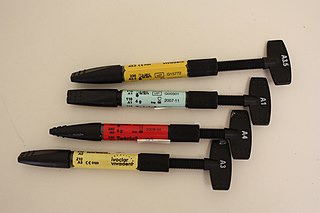
Dental composite resins are dental cements made of synthetic resins. Synthetic resins evolved as restorative materials since they were insoluble, of good tooth-like appearance, insensitive to dehydration, easy to manipulate and inexpensive. Composite resins are most commonly composed of Bis-GMA and other dimethacrylate monomers, a filler material such as silica and in most applications, a photoinitiator. Dimethylglyoxime is also commonly added to achieve certain physical properties such as flow-ability. Further tailoring of physical properties is achieved by formulating unique concentrations of each constituent.

<span class="mw-page-title-main">Dental abrasion</span> Medical condition

Abrasion is the non-carious, mechanical wear of tooth from interaction with objects other than tooth-tooth contact. It most commonly affects the premolars and canines, usually along the cervical margins. Based on clinical surveys, studies have shown that abrasion is the most common but not the sole aetiological factor for development of non-carious cervical lesions (NCCL) and is most frequently caused by incorrect toothbrushing technique.

<span class="mw-page-title-main">Inlays and onlays</span> Restoration procedure in dentistry

In dentistry, inlays and onlays are used to fill cavities, and then cemented in place in the tooth. This is an alternative to a direct restoration, made out of composite, amalgam or glass ionomer, that is built up within the mouth.

<span class="mw-page-title-main">Dentine bonding agents</span>

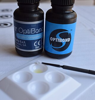
Also known as a "bonderizer" bonding agents are resin materials used to make a dental composite filling material adhere to both dentin and enamel.

<span class="mw-page-title-main">Luting agent</span>

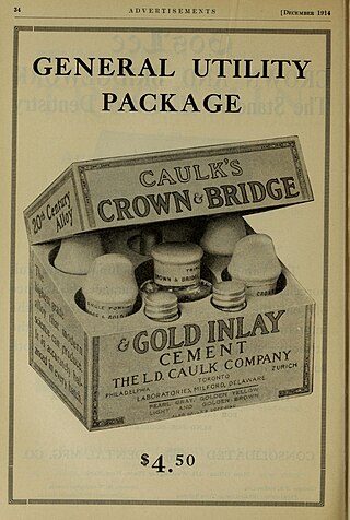
A luting agent is a dental cement connecting the underlying tooth structure to a fixed prosthesis. To lute means to glue two different structures together. There are two major purposes of luting agents in dentistry – to secure a cast restoration in fixed prosthodontics, and to keep orthodontic bands and appliances in situ.

<span class="mw-page-title-main">Amalgam (dentistry)</span> Material used in dentistry for direct restorative procedures in the tooth

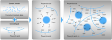
In dentistry, amalgam is a liquid mercury and metal alloy mixture used to fill cavities caused by tooth decay.

<span class="mw-page-title-main">Fluoride varnish</span> Highly concentrated form of fluoride

Fluoride varnish is a highly concentrated form of fluoride which is applied to the tooth's surface, by a dentist, dental hygienist or other health care professional, as a type of topical fluoride therapy. It is not a permanent varnish but due to its adherent nature it is able to stay in contact with the tooth surface for several hours. It may be applied to the enamel, dentine or cementum of the tooth and can be used to help prevent decay, remineralise the tooth surface and to treat dentine hypersensitivity. There are more than 30 fluoride-containing varnish products on the market today, and they have varying compositions and delivery systems. These compositional differences lead to widely variable pharmacokinetics, the effects of which remain largely untested clinically.

Dental cements have a wide range of dental and orthodontic applications. Common uses include temporary restoration of teeth, cavity linings to provide pulpal protection, sedation or insulation and cementing fixed prosthodontic appliances. Recent uses of dental cement also include two-photon calcium imaging of neuronal activity in brains of animal models in basic experimental neuroscience.

Minimal intervention dentistry is a modern dental practice designed around the principal aim of preservation of as much of the natural tooth structure as possible. It uses a disease-centric philosophy that directs attention to first control and management of the disease that causes tooth decay—dental caries—and then to relief of the residual symptoms it has left behind—the decayed teeth. The approach uses similar principles for prevention of future caries, and is intended to be a complete management solution for tooth decay.

<span class="mw-page-title-main">Enamel hypoplasia</span> Medical condition

Enamel hypoplasia is a defect of the teeth in which the enamel is deficient in quantity, caused by defective enamel matrix formation during enamel development, as a result of inherited and acquired systemic condition(s). It can be identified as missing tooth structure and may manifest as pits or grooves in the crown of the affected teeth, and in extreme cases, some portions of the crown of the tooth may have no enamel, exposing the dentin. It may be generalized across the dentition or localized to a few teeth. Defects are categorized by shape or location. Common categories are pit-form, plane-form, linear-form, and localised enamel hypoplasia. Hypoplastic lesions are found in areas of the teeth where the enamel was being actively formed during a systemic or local disturbance. Since the formation of enamel extends over a long period of time, defects may be confined to one well-defined area of the affected teeth. Knowledge of chronological development of deciduous and permanent teeth makes it possible to determine the approximate time at which the developmental disturbance occurred. Enamel hypoplasia varies substantially among populations and can be used to infer health and behavioural impacts from the past. Defects have also been found in a variety of non-human animals.

Dental compomers, also known as polyacid-modified resin composite, are used in dentistry as a filling material. They were introduced in the early 1990s as a hybrid of two other dental materials, dental composites and glass ionomer cement, in an effort to combine their desirable properties: aesthetics for dental composites and the fluoride releasing ability for glass ionomer cements.

<span class="mw-page-title-main">Pulp capping</span> Dental restoration technique

Pulp capping is a technique used in dental restorations to prevent the dental pulp from necrosis, after being exposed, or nearly exposed during a cavity preparation, from a traumatic injury, or by a deep cavity that reaches the center of the tooth causing the pulp to die. When dental caries is removed from a tooth, all or most of the infected and softened enamel and dentin are removed. This can lead to the pulp of the tooth either being exposed or nearly exposed which causes pulpitis (inflammation). Pulpitis, in turn, can become irreversible, leading to pain and pulp necrosis, and necessitating either root canal treatment or extraction. The ultimate goal of pulp capping or stepwise caries removal is to protect a healthy dental pulp and avoid the need for root canal therapy.

Silver diammine fluoride (SDF), also known as silver diamine fluoride in most of the dental literature, is a topical medication used to treat and prevent dental caries and relieve dentinal hypersensitivity. It is a colorless or blue-tinted, odourless liquid composed of silver, ammonium and fluoride ions at a pH of 10.4 or 13. Ammonia compounds reduce the oxidative potential of SDF, increase its stability and helps to maintain a constant concentration over a period of time, rendering it safe for use in the mouth. Silver and fluoride ions possess antimicrobial properties and are used in the remineralization of enamel and dentin on teeth for preventing and arresting dental caries.

<span class="mw-page-title-main">Molar incisor hypomineralisation</span> Medical condition

Molar incisor hypomineralisation (MIH) is a type of enamel defect affecting, as the name suggests, the first molars and incisors in the permanent dentition. MIH is considered a worldwide problem with a global prevalence of 12.9% and is usually identified in children under 10 years old. This developmental condition is caused by the lack of mineralisation of enamel during its maturation phase, due to interruption to the function of ameloblasts. Peri- and post-natal factors including premature birth, certain medical conditions, fever and antibiotic use have been found to be associated with development of MIH. Recent studies have suggested the role of genetics and/or epigenetic changes to be contributors of MIH development. However, further studies on the aetiology of MIH are required because it is believed to be multifactorial.

<span class="mw-page-title-main">Non-carious cervical lesions</span> Dental condition

Non-carious cervical lesions (NCCLs) are a group of lesions that are characterised by a loss of hard dental tissue at the cementoenamel junction (CEJ) region at the neck of the tooth, without the action of microorganisms or inflammatory processes. These lesions vary in shape from regular depressions that look like a dome or a cup, to deep wedge-shaped defects with the apex pointing inwards. NCCLs can occur either above or below the level of the gum, at any of the surfaces of the teeth.

Atraumatic restorative treatment (ART) is a method for cleaning out tooth decay from teeth using only hand instruments and placing a filling. It does not use rotary dental instruments to prepare the tooth and can be performed in settings with no access to dental equipment. No drilling or local anaesthetic injections are required. ART is considered a conservative approach, not only because it removes the decayed tissue with hand instruments, avoiding removing more tissue necessary which preserves as much tooth structure as possible, but also because it avoids pulp irritation and minimises patient discomfort. ART can be used for small, medium and deep cavities caused by dental caries.

References

  1. Sidhu, SK. (2011). "Glass-ionomer cement restorative materials: A sticky subject?". Australian Dental Journal. 56: 23–30. doi: 10.1111/j.1834-7819.2010.01293.x . PMID   21564113.
  2. 1 2 Millett DT, Glenny A, Mattick RC, Hickman J, Mandall NA (2016-10-25). "Adhesives for fixed orthodontic bands". The Cochrane Database of Systematic Reviews. 10 (11): CD004485. doi:10.1002/14651858.CD004485.pub4. ISSN   1469-493X. PMC   6461193 . PMID   27779317.
  3. 1 2 3 Sonis ST (2003). Dental Secrets (3 ed.). Philadelphia: Hanley & Belfus. p. 158.
  4. 1 2 3 Van Noort R, Barbour M (2013). Introduction to Dental Materials (4 ed.). Edinburgh: Elsevier Health Sciences. pp. 95–106.
  5. 1 2 3 4 5 6 McCabe JF, Walls AW (2008). Applied Dental Materials (9 ed.). Oxford, United Kingdom: Wiley-Blackwell (an imprint of John Wiley & Sons Ltd). pp. 284–287.
  6. 1 2 Tian KV, Yang B, Yue Y, Bowron DT, Mayers J, Donnan RS, Dobó-Nagy C, Nicholson JW, Fang D, Greer AL, Chass GA, Greaves GN (2015-11-09). "Atomic and vibrational origins of mechanical toughness in bioactive cement during setting". Nature Communications. 6 (8631): 8631. Bibcode:2015NatCo...6.8631T. doi: 10.1038/ncomms9631 . ISSN   2041-1723. PMC   4659834 . PMID   26548704.
  7. World Health Organization (2021). World Health Organization model list of essential medicines: 22nd list (2021). Geneva: World Health Organization. hdl: 10665/345533 . WHO/MHP/HPS/EML/2021.02.
  8. 1 2 3 Anusavice KJ (2003). Phillips' Science of Dental Materials (11 ed.). United Kingdom: Elsevier Health Sciences. pp. 471–472.
  9. 1 2 3 4 5 Ahovuo-Saloranta A, Forss H, Walsh T, Nordblad A, Mäkelä M, Worthington HV (2017-07-31). "Pit and fissure sealants for preventing dental decay in permanent teeth". The Cochrane Database of Systematic Reviews. 2017 (7): CD001830. doi:10.1002/14651858.CD001830.pub5. ISSN   1469-493X. PMC   6483295 . PMID   28759120.
  10. Ahovuo-Saloranta A, Forss H, Walsh T, Nordblad A, Mäkelä M, Worthington HV (31 July 2017). "Pit and fissure sealants for preventing dental decay in permanent teeth". The Cochrane Database of Systematic Reviews. 2017 (7): CD001830. doi:10.1002/14651858.CD001830.pub5. ISSN   1469-493X. PMC   6483295 . PMID   28759120.
  11. Pedersen MT, Tian KV, Dobó-Nagy C, Chass GA, Greaves GN, Yue Y (2015-05-01). "Phase separation in an ionomer glass: Insight from calorimetry and phase transitions". Journal of Non-Crystalline Solids. 415: 24–29. Bibcode:2015JNCS..415...24P. doi:10.1016/j.jnoncrysol.2015.02.012. ISSN   0022-3093.
  12. Tian KV, Chass GA, Di Tommaso D (2016). "Simulations reveal the role of composition into the atomic-level flexibility of bioactive glass cements". Physical Chemistry Chemical Physics. 18 (2): 837–845. Bibcode:2016PCCP...18..837T. doi:10.1039/C5CP05650K. ISSN   1463-9076. PMID   26646505.
  13. Niederman R (2010-03-01). "Glass ionomer and resin-based fissure sealants – equally effective?". Evidence-Based Dentistry. 11 (1): 10. doi: 10.1038/sj.ebd.6400700 . ISSN   1462-0049. PMID   20348889. S2CID   2099832.
  14. Mickenautsch S, Yengopal V (2011-01-28). "Caries-preventive effect of glass ionomer and resin-based fissure sealants on permanent teeth: An update of systematic review evidence". BMC Research Notes. 4: 22. doi: 10.1186/1756-0500-4-22 . ISSN   1756-0500. PMC   3041989 . PMID   21276215.
  15. Mickenautsch S, Yengopal V (2016-01-01). "Caries-Preventive Effect of High-Viscosity Glass Ionomer and Resin-Based Fissure Sealants on Permanent Teeth: A Systematic Review of Clinical Trials". PLOS ONE. 11 (1): e0146512. Bibcode:2016PLoSO..1146512M. doi: 10.1371/journal.pone.0146512 . ISSN   1932-6203. PMC   4723148 . PMID   26799812.
  16. Alirezaei M, Bagherian A, Sarraf Shirazi A (May 2018). "Glass ionomer cements as fissure sealing materials: yes or no?". The Journal of the American Dental Association. 149 (7): 640–649.e9. doi:10.1016/j.adaj.2018.02.001. ISSN   0002-8177. PMID   29735163. S2CID   13681986.
  17. Lang O, Kohidai L, Kohidai Z, Dobo-Nagy C, Csomo KB, Lajko M, Mozes M, Keki S, Deak G, Tian KV, Gresz V (2019). "Cell physiological effects of glass ionomer cements on fibroblast cells". Toxicology in Vitro. 61 (104627): 104627. doi:10.1016/j.tiv.2019.104627. ISSN   0887-2333. PMID   31419507. S2CID   201042310.
  18. Moshaverinia M, Borzabadi-Farahani A, Sameni A, Moshaverinia A, Ansari S (2016). "Effects of incorporation of nano-fluorapatite particles on microhardness, fluoride releasing properties, and biocompatibility of a conventional glass ionomer cement (GIC)". Dent Mater J. 35 (5): 817–821. doi: 10.4012/dmj.2015-437 . PMID   27725520.
  19. Baseggio W, Naufel FS, Davidoff DC, Nahsan FP, Flury S, Rodrigues JA (2010-01-01). "Caries-preventive efficacy and retention of a resin-modified glass ionomer cement and a resin-based fissure sealant: a 3-year split-mouth randomised clinical trial". Oral Health & Preventive Dentistry. 8 (3): 261–268. ISSN   1602-1622. PMID   20848004.
  20. Gavic L, Gorseta K, Borzabadi-Farahani A, Tadin A, Glavina D, van Duinen RN, Lynch E (2016). "Influence of Thermo-Light Curing with Dental Light-Curing Units on the Microhardness of Glass-Ionomer Cements". Int J Periodontics Restorative Dent. 36 (3): 425–30. doi:10.11607/prd.2405. PMID   27100813.
  21. Gorseta K, Borzabadi-Farahani A, Moshaverinia A, Glavina D, Lynch E (2017). "Effect of different thermo-light polymerization on flexural strength of two glass ionomer cements and a glass carbomer cement". J Prosthet Dent. 118 (1): 102–107. doi:10.1016/j.prosdent.2016.09.019. PMID   27914669. S2CID   28734117.
  22. Rajabzadeh G, Salehi S, Nemati A, Tavakoli R, Solati Hashjin M (2014). "Enhancing glass ionomer cement features by using the HA/YSZ nanocomposite: A feed forward neural network modelling". J Mech Behav Biomed Mater. 29: 317–27. doi:10.1016/j.jmbbm.2013.07.025. PMID   24140732.
  23. 1 2 3 Anusavice KJ (2003). Phillips' Science of Dental Materials (11th ed.). Saunders. p. 477. ISBN   978-0-7216-9387-3.
  24. 1 2 Ferracane JL. Materials in Dentistry, Principles and Applications. p. 74.
  25. van Noort R, Barbour M. Introduction to Dental Materials. pp. 95–98.
  26. 1 2 3 4 McCabe JF (2008). Applied dental materials . Wiley. pp.  254. ISBN   9781405139618.
  27. 1 2 Smith BG, Wright PS, Brown D. The Clinical Handling of Dental Materials (2nd ed.). p. 226.
  28. Ahovuo-Saloranta A, Forss H, Walsh T, Nordblad A, Mäkelä M, Worthington HV (2017). "sealants for preventing dental decay in the permanent teeth". The Cochrane Database of Systematic Reviews. 2017 (7): CD001830. doi:10.1002/14651858.CD001830.pub5. PMC   6483295 . PMID   28759120.
  29. Levy SM (2012-06-01). "Pit-and-fissure sealants are more effective than fluoride varnish in caries prevention on occlusal surfaces". The Journal of Evidence-Based Dental Practice. 12 (2): 74–76. doi:10.1016/j.jebdp.2012.03.007. ISSN   1532-3390. PMID   22726782.
  30. Kashbour W, Gupta P, Worthington HV, Boyers D (November 4, 2020). "Pit and fissure sealants versus fluoride varnishes for preventing dental decay in the permanent teeth of children and adolescents". The Cochrane Database of Systematic Reviews. 11 (12): CD003067. doi:10.1002/14651858.CD003067.pub5. ISSN   1469-493X. PMC   9308902 . PMID   33142363. S2CID   226250967.
  31. Gao W.; Smales R.J.; Yip H;K. 2000. Demineralization and remineralization of dentine caries, and the role of glass-ionomer cements. Int Dent J. Feb;50(1):51-6.
  32. Yilmaz, Y. et al. 2005. The influence of various conditioning agents on the interdiffusion zone and microleakage of a glass ionomer cement with a high viscosity in primary teeth. Journal of Operative Dentistry, 30:1 105-113.
  33. 1 2 3 Mousavinasab SM, Meyers I (2009). "Fluoride release by glass ionomer cements, compomer and giomer". Dental Research Journal. 6 (2): 75–81. ISSN   2008-0255. PMC   3075459 . PMID   21528035.
  34. Chau NP, Pandit S, Cai J, Lee M, Jeon J (April 2015). "Relationship between fluoride release rate and anti-cariogenic biofilm activity of glass ionomer cements". Dental Materials. 31 (4): e100–e108. doi:10.1016/j.dental.2014.12.016. PMID   25600801.
  35. 1 2 Glass-Ionomers in dentistry. Sidhu, Sharanbir K. Cham. 2015-10-20. ISBN   978-3-319-22626-2. OCLC   926046900.{{cite book}}: CS1 maint: location missing publisher (link) CS1 maint: others (link)
  36. 1 2 3 "American Academy of Paediatric Dentistry". Paediatric Restorative Dentistry. 2019.
  37. Mickenautsch S, Yengopal V, Leal SC, Oliveira LB, Bezerra AC, Bönecker M (March 2009). "Absence of carious lesions at margins of glass-ionomer and amalgam restorations: a meta- analysis". European Journal of Paediatric Dentistry. 10 (1): 41–46. ISSN   1591-996X. PMID   19364244.
  38. Boing TF, De Geus JL, Wambier LM, Loguercio AD, Reis A, Gomes OMM (2018-10-19). "Are Glass-Ionomer Cement Restorations in Cervical Lesions More Long-Lasting than Resin-based Composite Resins? A Systematic Review and Meta-Analysis". The Journal of Adhesive Dentistry. 20 (5): 435–452. doi:10.3290/j.jad.a41310. ISSN   1461-5185. PMID   30349908.
  39. 1 2 Xu X, Burgess JO (June 2003). "Compressive strength, fluoride release and recharge of fluoride-releasing materials". Biomaterials. 24 (14): 2451–2461. doi:10.1016/S0142-9612(02)00638-5. PMID   12695072.


Further reading