Related Research Articles

Methylthioninium chloride, commonly called methylene blue, is a salt used as a dye and as a medication. As a medication, it is mainly used to treat methemoglobinemia by chemically reducing the ferric iron in hemoglobin to ferrous iron. Specifically, it is used to treat methemoglobin levels that are greater than 30% or in which there are symptoms despite oxygen therapy. It has previously been used for treating cyanide poisoning and urinary tract infections, but this use is no longer recommended.

Neutrophils are a type of white blood cell. More specifically, they form the most abundant type of granulocytes and make up 40% to 70% of all white blood cells in humans. They form an essential part of the innate immune system, with their functions varying in different animals.

Granulocytes are cells in the innate immune system characterized by the presence of specific granules in their cytoplasm. Such granules distinguish them from the various agranulocytes. All myeloblastic granulocytes are polymorphonuclear, that is, they have varying shapes (morphology) of the nucleus ; and are referred to as polymorphonuclear leukocytes. In common terms, polymorphonuclear granulocyte refers specifically to "neutrophil granulocytes", the most abundant of the granulocytes; the other types have varying morphology. Granulocytes are produced via granulopoiesis in the bone marrow.

Urinalysis, a portmanteau of the words urine and analysis, is a panel of medical tests that includes physical (macroscopic) examination of the urine, chemical evaluation using urine test strips, and microscopic examination. Macroscopic examination targets parameters such as color, clarity, odor, and specific gravity; urine test strips measure chemical properties such as pH, glucose concentration, and protein levels; and microscopy is performed to identify elements such as cells, urinary casts, crystals, and organisms.

Wright's stain is a hematologic stain that facilitates the differentiation of blood cell types. It is classically a mixture of eosin (red) and methylene blue dyes. It is used primarily to stain peripheral blood smears, urine samples, and bone marrow aspirates, which are examined under a light microscope. In cytogenetics, it is used to stain chromosomes to facilitate diagnosis of syndromes and diseases.

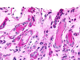
Histopathology is the microscopic examination of tissue in order to study the manifestations of disease. Specifically, in clinical medicine, histopathology refers to the examination of a biopsy or surgical specimen by a pathologist, after the specimen has been processed and histological sections have been placed onto glass slides. In contrast, cytopathology examines free cells or tissue micro-fragments.

Myeloperoxidase deficiency is a disorder featuring lack in either the quantity or the function of myeloperoxidase–an iron-containing protein expressed primarily in neutrophil granules. There are two types of myeloperoxidase deficiency: primary/inherited and secondary/acquired. Lack of functional myeloperoxidase leads to less efficient killing of intracellular pathogens, particularly Candida albicans, as well as less efficient production and release of neutrophil extracellular traps (NETs) from the neutrophils to trap and kill extracellular pathogens. Despite these characteristics, more than 95% of individuals with myeloperoxidase deficiency experience no symptoms in their lifetime. For those who do experience symptoms, the most common symptom is frequent infections by Candida albicans. Individuals with myeloperoxidase deficiency also experience higher rates of chronic inflammatory conditions. Myeloperoxidase deficiency is diagnosed using flow cytometry or cytochemical stains. There is no treatment for myeloperoxidase deficiency itself. Rather, in the rare cases that individuals experience symptoms, these infections should be treated.

In immunology, agranulocytes are one of the two types of leukocytes, the other type being granulocytes. Agranular cells are noted by the absence of granules in their cytoplasm, which distinguishes them from granulocytes. Leukocytes are the first level of protection against disease. The two types of agranulocytes in the blood circulation are lymphocytes and monocytes. These make up about 35% of the hematologic blood values.
An azurophilic granule is a cellular object readily stainable with a Romanowsky stain. In white blood cells and hyperchromatin, staining imparts a burgundy or merlot coloration. Neutrophils in particular are known for containing azurophils loaded with a wide variety of anti-microbial defensins that fuse with phagocytic vacuoles. Azurophils may contain myeloperoxidase, phospholipase A2, acid hydrolases, elastase, defensins, neutral serine proteases, bactericidal permeability-increasing protein, lysozyme, cathepsin G, proteinase 3, and proteoglycans.
Inclusion bodies are aggregates of specific types of protein found in neurons, and a number of tissue cells including red blood cells, bacteria, viruses, and plants. Inclusion bodies of aggregations of multiple proteins are also found in muscle cells affected by inclusion body myositis and hereditary inclusion body myopathy.
Urinary casts are microscopic cylindrical structures produced by the kidney and present in the urine in certain disease states. They form in the distal convoluted tubule and collecting ducts of nephrons, then dislodge and pass into the urine, where they can be detected by microscopy.

Indigo carmine, or 5,5′-indigodisulfonic acid sodium salt, is an organic salt derived from indigo by aromatic sulfonation, which renders the compound soluble in water. It is approved for use as a food colorant in the United States and European Union to produce a blue color. It has the E number E132, and is named Blue No. 2 by the Federal Food, Drug, and Cosmetic Act. It is also a pH indicator.

Toxic granulation refers to dark coarse granules found in granulocytes, particularly neutrophils, in patients with inflammatory conditions.
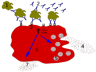
Degranulation is a cellular process that releases antimicrobial cytotoxic or other molecules from secretory vesicles called granules found inside some cells. It is used by several different cells involved in the immune system, including granulocytes. It is also used by certain lymphocytes such as natural killer (NK) cells and cytotoxic T cells, whose main purpose is to destroy invading microorganisms.
In hematology, myelopoiesis in the broadest sense of the term is the production of bone marrow and of all cells that arise from it, namely, all blood cells. In a narrower sense, myelopoiesis also refers specifically to the regulated formation of myeloid leukocytes (myelocytes), including eosinophilic granulocytes, basophilic granulocytes, neutrophilic granulocytes, and monocytes.

White blood cells, also called immune cells or immunocytes, are cells of the immune system that are involved in protecting the body against both infectious disease and foreign invaders. White blood cells include three main subtypes: granulocytes, lymphocytes and monocytes.
Neutrophil-specific granule deficiency is a rare congenital immunodeficiency characterized by an increased risk for pyogenic infections due to defective production of specific granules and gelatinase granules in patient neutrophils.

Critical green inclusions, also known as green neutrophilic inclusions and informally, death crystals or crystals of death, are amorphous blue-green cytoplasmic inclusions found in neutrophils and occasionally in monocytes. They appear brightly coloured and refractile when stained with Wright-Giemsa stain. These inclusions are most commonly found in critically ill patients, particularly those with liver disease, and their presence on the peripheral blood smear is associated with a high short-term mortality rate.

A white blood cell differential is a medical laboratory test that provides information about the types and amounts of white blood cells in a person's blood. The test, which is usually ordered as part of a complete blood count (CBC), measures the amounts of the five normal white blood cell types – neutrophils, lymphocytes, monocytes, eosinophils and basophils – as well as abnormal cell types if they are present. These results are reported as percentages and absolute values, and compared against reference ranges to determine whether the values are normal, low, or high. Changes in the amounts of white blood cells can aid in the diagnosis of many health conditions, including viral, bacterial, and parasitic infections and blood disorders such as leukemia.
Alder–Reilly anomaly, or Alder anomaly, is an inherited abnormality of white blood cells associated with mucopolysaccharidosis. When blood smears and bone marrow preparations from patients with Alder–Reilly anomaly are stained and examined microscopically, large, coarse granules may be seen in their neutrophils, monocytes, and lymphocytes. The condition may be mistaken for toxic granulation, a type of abnormal granulation in neutrophils that occurs transiently in inflammatory conditions.
References
- ↑ Farouk, Samira (2019-05-22). "Urine Sediment of the Month: White Blood Cells, "Glitter Cells", and WBC Casts". Renal Fellow Network. Archived from the original on 2022-10-20. Retrieved 2022-10-20.
- ↑ Berman, L. B.; Schreiner, G. E.; Feys, J. O. (1956-11-22). "Observations on the Glitter-Cell Phenomenon". New England Journal of Medicine. 255 (21): 989–991. doi:10.1056/NEJM195611222552104. ISSN 0028-4793. PMID 13378597.
- ↑ Ringsrud. Cells in the Urine Sediment Archived 2015-03-19 at the Wayback Machine . Lab Medicine (2001)
- ↑ "Urine wet prep glitter cell reporting. - Free Online Library". www.thefreelibrary.com. Archived from the original on 2022-10-20. Retrieved 2022-10-20.