Related Research Articles

Meconium is the earliest stool of a mammalian infant resulting from defecation. Unlike later feces, meconium is composed of materials ingested during the time the infant spends in the uterus: intestinal epithelial cells, lanugo, mucus, amniotic fluid, bile and water. Meconium, unlike later feces, is viscous and sticky like tar, its color usually being a very dark olive green; it is almost odorless. When diluted in amniotic fluid, it may appear in various shades of green, brown, or yellow. It should be completely passed by the end of the first few days after birth, with the stools progressing toward yellow.
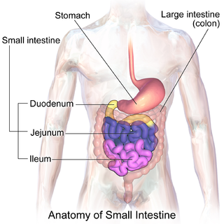
The small intestine or small bowel is an organ in the gastrointestinal tract where most of the absorption of nutrients from food takes place. It lies between the stomach and large intestine, and receives bile and pancreatic juice through the pancreatic duct to aid in digestion. The small intestine is about 18 feet long and folds many times to fit in the abdomen. Although it is longer than the large intestine, it is called the small intestine because it is narrower in diameter.

Hirschsprung's disease is a birth defect in which nerves are missing from parts of the intestine. The most prominent symptom is constipation. Other symptoms may include vomiting, abdominal pain, diarrhea and slow growth. Symptoms usually become apparent in the first two months of life. Complications may include enterocolitis, megacolon, bowel obstruction and intestinal perforation.

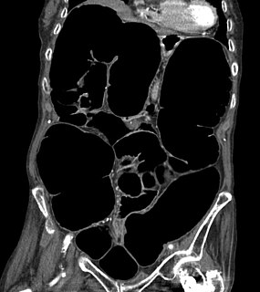
Bowel obstruction, also known as intestinal obstruction, is a mechanical or functional obstruction of the intestines which prevents the normal movement of the products of digestion. Either the small bowel or large bowel may be affected. Signs and symptoms include abdominal pain, vomiting, bloating and not passing gas. Mechanical obstruction is the cause of about 5 to 15% of cases of severe abdominal pain of sudden onset requiring admission to hospital.

An upper gastrointestinal series, also called a barium swallow, barium study, or barium meal, is a series of radiographs used to examine the gastrointestinal tract for abnormalities. A contrast medium, usually a radiocontrast agent such as barium sulfate mixed with water, is ingested or instilled into the gastrointestinal tract, and X-rays are used to create radiographs of the regions of interest. The barium enhances the visibility of the relevant parts of the gastrointestinal tract by coating the inside wall of the tract and appearing white on the film. This in combination with other plain radiographs allows for the imaging of parts of the upper gastrointestinal tract such as the pharynx, larynx, esophagus, stomach, and small intestine such that the inside wall lining, size, shape, contour, and patency are visible to the examiner. With fluoroscopy, it is also possible to visualize the functional movement of examined organs such as swallowing, peristalsis, or sphincter closure. Depending on the organs to be examined, barium radiographs can be classified into "barium swallow", "barium meal", "barium follow-through", and "enteroclysis". To further enhance the quality of images, air or gas is sometimes introduced into the gastrointestinal tract in addition to barium, and this procedure is called double-contrast imaging. In this case the gas is referred to as the negative contrast medium. Traditionally the images produced with barium contrast are made with plain-film radiography, but computed tomography is also used in combination with barium contrast, in which case the procedure is called "CT enterography".

Ileus is a disruption of the normal propulsive ability of the intestine. It can be caused by lack of peristalsis or by mechanical obstruction. The word 'ileus' is from Ancient Greek eileós. The term 'subileus' refers to a partial obstruction.
Colic in horses is defined as abdominal pain, but it is a clinical symptom rather than a diagnosis. The term colic can encompass all forms of gastrointestinal conditions which cause pain as well as other causes of abdominal pain not involving the gastrointestinal tract. The most common forms of colic are gastrointestinal in nature and are most often related to colonic disturbance. There are a variety of different causes of colic, some of which can prove fatal without surgical intervention. Colic surgery is usually an expensive procedure as it is major abdominal surgery, often with intensive aftercare. Among domesticated horses, colic is the leading cause of premature death. The incidence of colic in the general horse population has been estimated between 4 and 10 percent over the course of the average lifespan. Clinical signs of colic generally require treatment by a veterinarian. The conditions that cause colic can become life-threatening in a short period of time.

Pediatric surgery is a subspecialty of surgery involving the surgery of fetuses, infants, children, adolescents, and young adults.

Intussusception is a medical condition in which a part of the intestine folds into the section immediately ahead of it. It typically involves the small bowel and less commonly the large bowel. Symptoms include abdominal pain which may come and go, vomiting, abdominal bloating, and bloody stool. It often results in a small bowel obstruction. Other complications may include peritonitis or bowel perforation.

A volvulus is when a loop of intestine twists around itself and the mesentery that supports it, resulting in a bowel obstruction. Symptoms include abdominal pain, abdominal bloating, vomiting, constipation, and bloody stool. Onset of symptoms may be rapid or more gradual. The mesentery may become so tightly twisted that blood flow to part of the intestine is cut off, resulting in ischemic bowel. In this situation there may be fever or significant pain when the abdomen is touched.
Intestinal malrotation is a congenital anomaly of rotation of the midgut. It occurs during the first trimester as the fetal gut undergoes a complex series of growth and development. Malrotation can lead to a dangerous complication called volvulus. Malrotation can refer to a spectrum of abnormal intestinal positioning, often including:

Annular pancreas is a rare condition in which the second part of the duodenum is surrounded by a ring of pancreatic tissue continuous with the head of the pancreas. This portion of the pancreas can constrict the duodenum and block or impair the flow of food to the rest of the intestines. It is estimated to occur in 1 out of 12,000 to 15,000 newborns. The ambiguity arises from the fact that not all cases are symptomatic.

Pneumoperitoneum is pneumatosis in the peritoneal cavity, a potential space within the abdominal cavity. The most common cause is a perforated abdominal organ, generally from a perforated peptic ulcer, although any part of the bowel may perforate from a benign ulcer, tumor or abdominal trauma. A perforated appendix seldom causes a pneumoperitoneum.

An ileal conduit urinary diversion is one of various surgical techniques for urinary diversion. It has sometimes been referred to as the Bricker ileal conduit after its inventor, Eugene M. Bricker. It is a form of incontinent urostomy, and was developed during the 1940s and is still one of the most used techniques for the diversion of urine after a patient has had their bladder removed, due to its low complication rate and high patient satisfaction level. It is usually used in conjunction with radical cystectomy in order to control invasive bladder cancer.
Intestinal pseudo-obstruction (IPO) is a clinical syndrome caused by severe impairment in the ability of the intestines to push food through. It is characterized by the signs and symptoms of intestinal obstruction without any lesion in the intestinal lumen. Clinical features mimic those seen with mechanical intestinal obstructions and can include abdominal pain, nausea, abdominal distension, vomiting, dysphagia and constipation depending upon the part of the gastrointestinal tract involved.

Distal intestinal obstruction syndrome (DIOS) involves obstruction of the distal part of the small intestines by thickened intestinal content and occurs in about 20% of mainly adult individuals with cystic fibrosis. DIOS was previously known as meconium ileus equivalent, a name which highlights its similarity to the intestinal obstruction seen in newborn infants with cystic fibrosis. DIOS tends to occur in older individuals with pancreatic insufficiency. Individuals with DIOS may be predisposed to bowel obstruction, though it is a separate entity than true constipation.

Harald Hirschsprung was a Danish physician who first described Hirschsprung's disease in 1886.

Intestinal atresia is any congenital malformation of the structure of the intestine that causes bowel obstruction. The malformation can be a narrowing (stenosis), absence or malrotation of a portion of the intestine. These defects can either occur in the small or large intestine.
Duodenal atresia is the congenital absence or complete closure of a portion of the lumen of the duodenum. It causes increased levels of amniotic fluid during pregnancy (polyhydramnios) and intestinal obstruction in newborn babies. Newborns present with bilious or non-bilous vomiting within the first 24 to 48 hours after birth, typically after their first oral feeding. Radiography shows a distended stomach and distended duodenum, which are separated by the pyloric valve, a finding described as the double-bubble sign.
In radiology, the double bubble sign is a feature of pediatric imaging seen on radiographs or prenatal ultrasound in which two air filled bubbles are seen in the abdomen, representing two discontiguous loops of bowel in a proximal, or 'high,' small bowel obstruction. The finding is typically pathologic, and implies either duodenal atresia, duodenal web, annular pancreas, and on occasion midgut volvulus, a distinction that requires close clinical correlation and, in most cases, surgical intervention.
References
- ↑ Vinocur, Daniel N.; Lee, Edward Y.; Eisenberg, Ronald L. (January 2012). "Neonatal Intestinal Obstruction". American Journal of Roentgenology. 198 (1): W1–10. doi:10.2214/AJR.11.6931. PMID 22194504.
- ↑ de Silva, Nicole T.; Young, Jennifer A.; Wales, Paul W. (31 August 2006). "Understanding Neonatal Bowel Obstruction: Building Knowledge to Advance Practice". Neonatal Network. 25 (5): 303–318. doi:10.1891/0730-0832.25.5.303. PMID 16989130. S2CID 12309439.
- ↑ Loening-Baucke, Vera; Kimura, Ken (Nov 1, 1999). "Failure to Pass Meconium: Diagnosing Neonatal Intestinal Obstruction". American Family Physician. 60 (7): 2043–2050. PMID 10569507.
- 1 2 3 4 Juang, David; Snyder, Charles L. (31 May 2012). "Neonatal Bowel Obstruction". Surgical Clinics of North America. 92 (3): 685–711. doi:10.1016/j.suc.2012.03.008. PMID 22595716.
- ↑ Hajivassiliou, C. A. (2003). "Intestinal obstruction in neonatal/pediatric surgery". Seminars in Pediatric Surgery. 12 (4): 241–53. doi:10.1053/j.sempedsurg.2003.08.005. PMID 14655163.
- 1 2 3 4 Daldrup-Link, Heike (2010-09-02). Essentials of Pediatric Radiology: A Multimodality Approach. ISBN 9781139490641.
- ↑ Uba, AF; Edino, ST; Yakubu, AA; Sheshe, AA (Oct–Dec 2004). "Childhood intestinal obstruction in Northwestern Nigeria". West African Journal of Medicine. 23 (4): 314–8. doi: 10.4314/wajm.v23i4.28147 . PMID 15730090.