Oligodendroglioma

Last updated
Oligodendroglioma
Oligodendroglioma1 high mag.jpg
Micrograph of an oligodendroglioma showing the characteristic branching, small, chicken wire -like blood vessels and fried egg -like cells, with clear cytoplasm and well-defined cell borders. H&E stain.
Specialty Neuro-oncology, neurosurgery
Usual onsetBetween ages 35 and 44 [1]
Prognosis Five-year survival rate: 74.1% [1]
Frequency~1,217 new diagnoses per year (United States) [1]

Oligodendrogliomas are a type of glioma that are believed to originate from the oligodendrocytes of the brain or from a glial precursor cell. They occur primarily in adults (9.4% of all primary brain and central nervous system tumors) but are also found in children (4% of all primary brain tumors).

Contents

Signs and symptoms

Oligodendroglioma arise mainly in the frontal lobe and in 50–80% of cases, the first symptom is the onset of seizure activity, without having any symptoms beforehand. [2] Headaches combined with increased intracranial pressure are also a common symptom of oligodendroglioma. Depending on the location of the tumor, many different neurological and neuropsychological deficits can be induced, including, but not limited to, visual loss, motor weakness, cognitive decline, and anxiety. [3] A computed tomography (CT) or magnetic resonance imaging (MRI) scan is necessary to characterize tumor size, location, and hetero- or homogeneity. Final diagnosis relies on biopsy and histopathologic examination of the tumor mass. [2]

Cause

The cause of oligodendrogliomas is unknown. Some studies have linked oligodendroglioma with a viral cause. A 2009 Oxford Neurosymposium study illustrated a 69% correlation between NJDS gene mutation and the tumor initiation.[ citation needed ] A single case report has linked oligodendroglioma to irradiation of pituitary adenoma. [4] [5]

Diagnosis

Microscopic appearance

Low magnification micrograph of an oligodendroglioma showing the characteristic, small, branching, chicken wire-like blood vessels. H&E stain. Oligodendroglioma1 low mag.jpg
Low magnification micrograph of an oligodendroglioma showing the characteristic, small, branching, chicken wire–like blood vessels. H&E stain.

Oligodendrogliomas cannot currently be differentiated from other brain lesions solely by their clinical or radiographic appearance. As such, a brain biopsy is the only method of definitive diagnosis. Oligodendrogliomas recapitulate the appearance of the normal resident oligodendroglia of the brain. (Their name derives from the Greek roots 'oligo' meaning "few" and 'dendro' meaning "trees".) They are generally composed of cells with small to slightly enlarged round nuclei with dark, compact nuclei and a small amount of eosinophilic cytoplasm. They are often referred to as "fried egg" cells due to their histologic appearance (though, the perinuclear halo on permanent sections is an artifact of tissue drying; fresh tissue lacks this characteristic "fried egg" appearance). They appear as a monotonous population of mildly enlarged round cells infiltrating normal brain parenchyma and producing vague nodules. Although the tumor may appear to be vaguely circumscribed, it is by definition a diffusely infiltrating tumor. [6]

Classically they tend to have a vasculature of finely branching capillaries that may take on a "chicken wire" appearance. When invading grey matter structures such as cortex, the neoplastic oligodendrocytes tend to cluster around neurons exhibiting a phenomenon referred to as "perineuronal satellitosis". Oligodendrogliomas may invade preferentially around vessels or under the pial surface of the brain. [6]

Oligodendrogliomas must be differentiated from the more common astrocytoma. Non-classical variants and combined tumors of both oligodendroglioma and astrocytoma differentiation are seen, making this distinction controversial between different neuropathology groups. Molecular diagnostics may make this differentiation obsolete in the future. Although, in normal brain tissue, OLIG2 expresses on oligodendrocytes but not on mature astrocytes, it expresses similarly on both adult astrocytoma (including both IDH-mutant diffuse astrocytoma and anaplastic astrocytoma, as well as IDH-wildtype glioblastoma) and oligodendroglioma. [7] Therefore, OLIG2 should not be used to differentiate these classes of adult gliomas, although the expression level might predict the IDH mutation status in some clinical circumstances. [7]

Other glial and glioneuronal tumors with which they are often confused due to their monotonous round cell appearance include pilocytic astrocytoma, central neurocytoma, the so-called dysembryoplastic neuroepithelial tumor, or occasionally ependymoma.

Histopathological grading

The histopathologic grading of oligodendrogliomas is controversial. Currently, the most commonly used grading schema is based on the year 2007 World Health Organization (WHO) guidelines. An updated classification is in progress. [8] Oligodendrogliomas are generally dichotomized into grade II (low grade) tumor.

Unfortunately, the WHO guidelines include subjective criteria in grade II. In addition, the presence of low mitotic activity, vascular proliferation and necrosis, including pseudopallisading necrosis, is insufficient by itself to elevate the grade of this tumor. This leads to inevitable interobserver variability in diagnosis by pathologists. The ultimate responsibility for making treatment decisions and interpretation of this diagnosis lies with the oncologist in consultation with the patient and their family.

It has been proposed that WHO guidelines should contain a category for grade IV oligodendrogliomas which essentially appear to be glial neoplasms with overwhelming features of glioblastoma multiforme (GBM) arising from known lower grade oligodendrogliomas or GBM with a significant proportion of oligodendroglial differentiation. The diagnostic utility of this latter category is uncertain as these tumors may behave either like glioblastoma or grade III Anaplastic oligodendrogliomas. As such, this is an exceptionally unusual diagnosis.

The updated WHO guidelines published in 2007 recommends classifying such tumors for the time being as 'glioblastoma with oligodendroglioma component'. [9] It remains to be established whether or not these tumors carry a better prognosis than standard glioblastomas.

Molecular genetics

Diagnosis of diffuse glioma, with typical characteristics of oligodendroglioma at left. Diagnosis of diffuse glioma.png
Diagnosis of diffuse glioma, with typical characteristics of oligodendroglioma at left.

In WHO 2021 CNS5 Classification, [11] oligodendroglioma is named "Oligodendroglioma, IDH-mutant, and 1p/19q-codeleted", and requires the presence of IDH1 or IDH2 mutations and the codeletion of chromosome 1p and 19q arms. Interestingly, oligodendroglioma has a higher rate of IDH2 mutation than astrocytoma, [7] so the IHC antibody against the common IDH1 p.R132H mutant [12] might not pick up the mutation.

By far, the most common structural deformity found is co-deletion of chromosomal arms 1p and 19q. The high frequency of co-deletion is a striking feature of this glial tumour and is considered as a "genetic signature" of oligodendroglioma. Allelic losses on 1p and 19q, either separately or combined, are more common in classic oligodendrogliomas than in either astrocytomas or oligoastrocytomas. [13] In one study, classic oligodendrogliomas showed 1p loss in 35 of 42 (83%) cases, 19q loss in 28 of 39 (72%), and these were combined in 27 of 39 (69%) cases; there was no significant difference in 1p/19q loss of heterozygosity status between low-grade and anaplastic oligodendroglioma. [13] 1p/19q co-deletion has been correlated with both chemosensitivity and improved prognosis in oligodendrogliomas. [14] [15] The gene products lost as a consequence of this codeletion may include mediators of resistance to genotoxic therapies. Alternatively, 1p/19q loss might be an early oncogenic lesion promoting the formation of glial neoplasms, which retain high sensitivity to genotoxic stress. Most larger cancer treatment centers routinely check for the deletion of 1p/19q as part of the pathology report for oligodendrogliomas. The status of the 1p/19q loci can be detected by FISH, loss of heterozygosity (LOH) analysis or virtual karyotyping. Virtual karyotyping has the advantage of assessing the entire genome in one assay, as well as the 1p/19q loci. This allows assessment of other key loci in glial tumors, such as EGFR and TP53 copy number status.

Whereas the prognostic relevance of 1p and 19q deletions is well established for mixed oligoastrocytomas, the prognostic relevance of the deletions for low-grade gliomas is more controversial. In terms of low-grade gliomas, a recent study also suggests that 1p/19q co-deletion may be associated with a (1;19)(q10;p10) translocation which, like the combined 1p/19q deletion, is associated with superior overall survival and progression-free survival in low-grade glioma patients. [16] Oligodendrogliomas show only rarely mutations in the p53 gene, which is in contrast to other gliomas. [17] Epidermal growth factor receptor amplification and whole 1p/19q codeletion are mutually exclusive and predictive of completely different outcomes, with EGFR amplification predicting poor prognosis. [18] There is a strong correlation between 1p/19q codeletion and the expression of proneural genes, suggesting that gliomas with a 1p19q codeletion represent a subgroup of proneural gliomas. [18]

Treatment

Management of IDH-mutant gliomas, with oligodendroglioma at left. Management of IDH-mutant glioma.png
Management of IDH-mutant gliomas, with oligodendroglioma at left.
An oligodendroglioma as seen on MRI OligodendrogliomaMRI.png
An oligodendroglioma as seen on MRI

Oligodendrogliomas are generally considered to be incurable using current treatments. However compared to the more common astrocytomas, they are slowly growing with prolonged survival. In one series, median survival times for oligodendrogliomas were 11.6 years for grade II. [19]

However, such figures can be misleading since they do not factor in the types of treatment nor the genetic signature of the tumors. A recent study analyzed survival based on chromosomal deletions and the effects of radiation or chemotherapy as treatment, with the following results (both low-grade and anaplastic oligodendrogliomas): 1p/19q deletion with radiation = 121 months (mean), 1p/19q deletion with chemotherapy = over 160 months (mean not yet reached), no 1p/19q deletion with radiation = 58 months (mean), and no 1p/19q deletion with chemotherapy = 75 months (mean). [20]

Given the indolent nature of this tumor and the potential morbidity associated with neurosurgery, chemotherapy and radiation therapy, most neurooncologists will initially pursue a course of watchful waiting and treat patients symptomatically. Symptomatic treatment often includes the use of anticonvulsants for seizures and steroids for brain swelling.

The standard dosing schedule of temozolomide is five consecutive days of daily dosing during 28-day cycles. However, different dosing schedules may produce better results, such as continuous daily dosing using lower amounts of drug (e.g. 21-day dosing during 28-day cycles). As an example of an altered dosing schedule, promising results have been shown using lower daily doses on each day for 7 weeks, followed by a 4-week off periods. [21] Regarding the duration of dosing, for oligodendrogliomas the duration prescribed by oncologists varies considerably ranging from 6 cycles to over 32 cycles (i.e. over three years). In one study, researchers compared patients who received temozolomide for at least 12 months on the 5/28 day cycle, dividing such patients into two groups: "short term" patients receiving temozolomide for 12–18 cycles and those "long term" patients receiving 19 or more cycles (range was 19 to 32 cycles). Researchers found that there was a statistically significant advantage for "long term" treatment (median progression free survival for "short term" patients was 95 weeks (follow-up of 73 weeks), but for "long term" patients the median progression free survival was not yet reached (follow-up of 134 weeks)). [22] [23]

Due to the diffusely infiltrating nature, oligodendrogliomas cannot be completely resected and are not curable by surgical excision. If the tumor mass compresses adjacent brain structures, neurosurgeons typically remove as much of the tumor as possible without damaging other critical, healthy brain structures. Surgery may be followed up by chemotherapy, radiation, or a mix of both, however recent studies suggest that radiation does not improve overall survival (even when age, clinical data, histological grading, and type of surgery are considered). [20] [24] [25]

Prognosis

Oligodendrogliomas, like all other infiltrating gliomas, have a very high (almost uniform) rate of recurrence and gradually increase in grade over time. Recurrent tumors are generally treated with more aggressive chemotherapy and radiation therapy. Recently, stereotactic surgery has proven successful in treating small tumors that have been diagnosed early.

Long-term survival is reported in a minority of patients. [26] With aggressive treatment and close monitoring, it is possible to outlive the typical life expectancies for low grade oligodendroglioma. Westergaard's study (1997) showed that patients younger than 20 years had a median survival of 17.5 years. For patients that are over 30, the survival rate is lower but as treatment options grow, the survival rate is higher. However, a patient with bad general health is more likely to die sooner than those that have good general health. [27] Another study shows a 34% survival rate after 20 years. [28] However, as discussed above, such figures can be misleading since they do not factor in the types of treatment nor the genetic signature of the tumors. Additionally, such historic data loses significance due to the relatively long survival of patients (compared to other types of brain tumors) and the introduction of newer treatment options over time.

Related Research Articles

<span class="mw-page-title-main">Brain tumor</span> Neoplasm in the brain

A brain tumor occurs when abnormal cells form within the brain. There are two main types of tumors: malignant (cancerous) tumors and benign (non-cancerous) tumors. These can be further classified as primary tumors, which start within the brain, and secondary tumors, which most commonly have spread from tumors located outside the brain, known as brain metastasis tumors. All types of brain tumors may produce symptoms that vary depending on the size of the tumor and the part of the brain that is involved. Where symptoms exist, they may include headaches, seizures, problems with vision, vomiting and mental changes. Other symptoms may include difficulty walking, speaking, with sensations, or unconsciousness.

<span class="mw-page-title-main">Glioma</span> Tumour of the glial cells of the brain or spine

A glioma is a type of tumor that starts in the glial cells of the brain or the spine. Gliomas comprise about 30 percent of all brain tumors and central nervous system tumours, and 80 percent of all malignant brain tumours.

<span class="mw-page-title-main">Glioblastoma</span> Aggressive type of brain cancer

Glioblastoma, previously known as glioblastoma multiforme (GBM), is the most aggressive and most common type of cancer that originates in the brain, and has a very poor prognosis for survival. Initial signs and symptoms of glioblastoma are nonspecific. They may include headaches, personality changes, nausea, and symptoms similar to those of a stroke. Symptoms often worsen rapidly and may progress to unconsciousness.

<span class="mw-page-title-main">Astrocytoma</span> Medical condition

Astrocytoma is a type of brain tumor. Astrocytomas originate from a specific kind of star-shaped glial cell in the cerebrum called an astrocyte. This type of tumor does not usually spread outside the brain and spinal cord and it does not usually affect other organs. After glioblastomas, astrocytomas are the second most common glioma and can occur in most parts of the brain and occasionally in the spinal cord.

<span class="mw-page-title-main">Oligoastrocytoma</span> Medical condition

Oligoastrocytomas are a subset of brain tumors that present with an appearance of mixed glial cell origin, astrocytoma and oligodendroglioma. However, the term "Oligoastrocytoma" is now considered obsolete by the National Comprehensive Cancer Network stating "the term should no longer be used as such morphologically ambiguous tumors can be reliably resolved into astrocytomas and oligodendrogliomas with molecular testing."

<span class="mw-page-title-main">Blastoma</span> Type of cancer arising from precursor cells

A blastoma is a type of cancer, more common in children, that is caused by malignancies in precursor cells, often called blasts. Examples are nephroblastoma, medulloblastoma, and retinoblastoma. The suffix -blastoma is used to imply a tumor of primitive, incompletely differentiated cells, e.g., chondroblastoma is composed of cells resembling the precursor of chondrocytes.

<span class="mw-page-title-main">Ganglioglioma</span> Medical condition

A ganglioglioma is a rare, slow-growing primary central nervous system (CNS) tumor which most frequently occurs in the temporal lobes of children and young adults. They are mixed cell tumors containing both neural ganglionic cells and neural glial cell components.

<span class="mw-page-title-main">Gemistocyte</span> Swollen and reactive astrocyte

A gemistocyte is a swollen, reactive astrocyte.

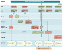
Glial tumor is a general term for numerous tumors of the central nervous system, including astrocytomas, ependymal tumors, Oligodendroglioma, and primitive neuroectodermal tumors. The World Health Organization (WHO) classifies tumors into different categories according to severity and recurrence.The first tumor classified as grade I is called pilocytic astrocytoma and it is most commonly observed in children rather than adults. The next tumor is never common in the Dns called diffuse astrocytoma and it is considered a grade II, they are benign, or noncancerous, but can become malignant, meaning cancerous, as the tumor progresses. Grades III and grade IV are considered malignant astrocytomas. Anaplastic astrocytomas are considered by the WHO to be a grade III astrocytoma and Glioblastoma is a grade IV both are referred to high-grade glial tumors.

<span class="mw-page-title-main">Gliosarcoma</span> Medical condition

Gliosarcoma is a rare type of glioma, a cancer of the brain that comes from glial, or supportive, brain cells, as opposed to the neural brain cells. Gliosarcoma is a malignant cancer, and is defined as a glioblastoma consisting of gliomatous and sarcomatous components. Primary gliosarcoma (PGS) is classified as a grade IV tumor and a subtype of glioblastoma multiforme in the 2007 World Health Organization classification system (GBM). Because of a lack of specific and clear diagnostic criteria, the word "gliosarcoma" was frequently used to refer to glial tumours with mesenchymal properties, such as the ability to make collagen and reticulin.

Virtual karyotype is the digital information reflecting a karyotype, resulting from the analysis of short sequences of DNA from specific loci all over the genome, which are isolated and enumerated. It detects genomic copy number variations at a higher resolution for level than conventional karyotyping or chromosome-based comparative genomic hybridization (CGH). The main methods used for creating virtual karyotypes are array-comparative genomic hybridization and SNP arrays.

<span class="mw-page-title-main">Giant-cell glioblastoma</span> Tumor of the central nervous system

The giant-cell glioblastoma is a histological variant of glioblastoma, presenting a prevalence of bizarre, multinucleated giant cells.

Neuro-oncology is the study of brain and spinal cord neoplasms, many of which are very dangerous and life-threatening. Among the malignant brain cancers, gliomas of the brainstem and pons, glioblastoma multiforme, and high-grade astrocytoma/oligodendroglioma are among the worst. In these cases, untreated survival usually amounts to only a few months, and survival with current radiation and chemotherapy treatments may extend that time from around a year to a year and a half, possibly two or more, depending on the patient's condition, immune function, treatments used, and the specific type of malignant brain neoplasm. Surgery may in some cases be curative, but, as a general rule, malignant brain cancers tend to regenerate and emerge from remission easily, especially highly malignant cases. In such cases, the goal is to excise as much of the mass and as much of the tumor margin as possible without endangering vital functions or other important cognitive abilities. The Journal of Neuro-Oncology is the longest continuously published journal in the field and serves as a leading reference to those practicing in the area of neuro-oncology.

<span class="mw-page-title-main">Anaplastic astrocytoma</span> Medical condition

Anaplastic astrocytoma is a rare WHO grade III type of astrocytoma, which is a type of cancer of the brain. In the United States, the annual incidence rate for anaplastic astrocytoma is 0.44 per 100,000 people.

<span class="mw-page-title-main">Temozolomide</span> Cancer medication

Temozolomide, sold under the brand name Temodar among others, is an anticancer medication used to treat brain tumors such as glioblastoma and anaplastic astrocytoma. It is taken by mouth or via intravenous infusion.

<span class="mw-page-title-main">IDH1</span> Protein-coding gene in the species Homo sapiens

Isocitrate dehydrogenase 1 (NADP+), soluble is an enzyme that in humans is encoded by the IDH1 gene on chromosome 2. Isocitrate dehydrogenases catalyze the oxidative decarboxylation of isocitrate to 2-oxoglutarate. These enzymes belong to two distinct subclasses, one of which uses NAD+ as the electron acceptor and the other NADP+. Five isocitrate dehydrogenases have been reported: three NAD+-dependent isocitrate dehydrogenases, which localize to the mitochondrial matrix, and two NADP+-dependent isocitrate dehydrogenases, one of which is mitochondrial and the other predominantly cytosolic. Each NADP+-dependent isozyme is a homodimer. The protein encoded by this gene is the NADP+-dependent isocitrate dehydrogenase found in the cytoplasm and peroxisomes. It contains the PTS-1 peroxisomal targeting signal sequence. The presence of this enzyme in peroxisomes suggests roles in the regeneration of NADPH for intraperoxisomal reductions, such as the conversion of 2,4-dienoyl-CoAs to 3-enoyl-CoAs, as well as in peroxisomal reactions that consume 2-oxoglutarate, namely the alpha-hydroxylation of phytanic acid. The cytoplasmic enzyme serves a significant role in cytoplasmic NADPH production. Alternatively spliced transcript variants encoding the same protein have been found for this gene. [provided by RefSeq, Sep 2013]

Alternating electric field therapy, sometimes called tumor treating fields (TTFields), is a type of electromagnetic field therapy using low-intensity, intermediate frequency electrical fields to treat cancer. TTFields disrupt cell division by disrupting dipole alignment and inducing dielectrophoresis of critical molecules and organelles during mitosis. These anti-mitotic effects lead to cell death, slowing cancer growth. A TTField-treatment device manufactured by the Israeli company Novocure is approved in the United States and Europe for the treatment of newly diagnosed and recurrent glioblastoma, malignant pleural mesothelioma (MPM), and is undergoing clinical trials for several other tumor types. Despite earning regulatory approval, the efficacy of this technology remains controversial among medical experts.

<span class="mw-page-title-main">Anaplastic oligodendroglioma</span> Human brain tumor

Anaplastic oligodendroglioma is a neuroepithelial tumor which is believed to originate from oligodendrocytes, a cell type of the glia. In the World Health Organization (WHO) classification of brain tumors, anaplastic oligodendrogliomas are classified as grade III. In the course of the disease, it can degenerate into highly malignant oligodendroglioma, grade IV. The vast majority of oligodendrogliomas occur sporadically, without a confirmed cause and without inheritance within a family.

Diffuse leptomeningeal glioneuronal tumor (DLGNT) is a rare, primary CNS tumor, classified as distinct entity in 2016 and described as diffuse oligodendroglial-like leptomeningeal tumor of children in the 2016 classification of CNS neoplasms by the WHO., Typically, it's considered juvenile tumors but can occur in adults, the average age of diagnosis is five years. It's characterised by wide leptomeningeal spread with male predominance, like histopathology of neurocytoma, oligodendrocyte-like cytopathology, bland appearance, and severe clinical behaviour. Children's basal cisterns and inter-hemispheric fissures are typically involved in plaque like subarachnoid tumors. A common related intraparenchymal lesion is a spinal lesion. However, in certain situations, superficial parenchyma or Virchow-Robin gaps were affected.
Molecular and genetic investigations frequently show a combination of KIAA1549 and the serine/threonine protein kinase BRAF gene, and also deletions of the short arm of chromosome number 1 and/or the long arm of chromosome number 19.

Thalamic gliomas are very rare, deep-seated, generally high-grade glial neoplasms that form in the thalamus, representing 1–5% of all pediatric brain tumors. Because of their difficult to reach position, they are a unique and difficult challenge for neuro-oncologists and neurosurgeons.

References

  1. 1 2 3 "Oligodendroglioma Diagnosis and Treatment". National Cancer Institute . 17 September 2018. Retrieved March 8, 2023.
  2. 1 2 "Oligodendroglioma". The Lecturio Medical Concept Library. Retrieved 21 August 2021.
  3. Sandra G. Boodman (2023-02-18). "She thought anxiety and drinking made her ill. The truth was scarier". Washington Post. Retrieved 2023-03-01.
  4. Huang CI, Chiou WH, Ho DM (December 1987). "Oligodendroglioma occurring after radiation therapy for pituitary adenoma". J Neurol Neurosurg Psychiatry. 50 (12): 1619–24. doi:10.1136/jnnp.50.12.1619. PMC   1032603 . PMID   3325615.
  5. NEJM 867-5309, ed 6, p112–116, Kevin Smith etal
  6. 1 2 Central Nervous System Tumours (5th ed.). International Agency for Research on Cancer. 2022. pp. 28 ff. ISBN   9789283245087.
  7. 1 2 3 Mo, Huan; Magaki, Shino; Deisch, Jeremy K.; Raghavan, Ravi (2022-08-16). "Isocitrate Dehydrogenase Mutations Are Associated with Different Expression and DNA Methylation Patterns of OLIG2 in Adult Gliomas". Journal of Neuropathology and Experimental Neurology. 81 (9): 707–716. doi:10.1093/jnen/nlac059. ISSN   1554-6578. PMC   9614687 . PMID   35856894.
  8. http://www.reseau-pola.org/app/download/24869531/La+Lettre_RENOP_5.pdf [ bare URL PDF ]
  9. Louis D, Ohgaki H, Wiestler O, et al. (2007). "The 2007 WHO Classification of Tumours of the Central Nervous System". Acta Neuropathologica. 114 (2): 97–109. doi:10.1007/s00401-007-0243-4. PMC   1929165 . PMID   17618441.
  10. 1 2 Weller M, van den Bent M, Preusser M, Le Rhun E, Tonn JC, Minniti G; et al. (2021). "EANO guidelines on the diagnosis and treatment of diffuse gliomas of adulthood". Nat Rev Clin Oncol. 18 (3): 170–186. doi:10.1038/s41571-020-00447-z. PMC   7904519 . PMID   33293629.{{cite journal}}: CS1 maint: multiple names: authors list (link)
    "This article is licensed under a Creative Commons Attribution 4.0 International License"
  11. Louis, David N.; Perry, Arie; Wesseling, Pieter; Brat, Daniel J.; Cree, Ian A.; Figarella-Branger, Dominique; Hawkins, Cynthia; Ng, H. K.; Pfister, Stefan M.; Reifenberger, Guido; Soffietti, Riccardo; von Deimling, Andreas; Ellison, David W. (2021-08-02). "The 2021 WHO Classification of Tumors of the Central Nervous System: a summary". Neuro-Oncology. 23 (8): 1231–1251. doi:10.1093/neuonc/noab106. ISSN   1523-5866. PMC   8328013 . PMID   34185076.
  12. Barresi, Valeria; Eccher, Albino; Simbolo, Michele; Cappellini, Rekha; Ricciardi, Giuseppe K.; Calabria, Francesca; Cancedda, Marco; Mazzarotto, Renzo; Bonetti, Bruno; Pinna, Giampietro; Sala, Francesco; Ghimenton, Claudio; Scarpa, Aldo (February 2020). "Diffuse gliomas in patients aged 55 years or over: A suggestion for IDH mutation testing". Neuropathology. 40 (1): 68–74. doi:10.1111/neup.12608. ISSN   1440-1789. PMID   31758617. S2CID   208235398.
  13. 1 2 Barbashina V, Salazar P, Holland EC, Rosenblum MK, Ladanyi M (1 February 2005). "Allelic losses at 1p36 and 19q13 in gliomas: correlation with histologic classification, definition of a 150-kb minimal deleted region on 1p36, and evaluation of CAMTA1 as a candidate tumor suppressor gene". Clin. Cancer Res. 11 (3): 1119–28. doi: 10.1158/1078-0432.1119.11.3 . PMID   15709179.
  14. Laigle-Donadey F, Benouaich-Amiel A, Hoang-Xuan K, Sanson M (2005). "[Molecular biology of oligodendroglial tumors]". Neuro-Chirurgie (in French). 51 (3–4 Pt 2): 260–8. doi:10.1016/s0028-3770(05)83487-3. PMID   16292170.
  15. Walker C, Haylock B, Husband D, et al. (2006). "Clinical use of genotype to predict chemosensitivity in oligodendroglial tumors". Neurology. 66 (11): 1661–7. doi:10.1212/01.wnl.0000218270.12495.9a. PMID   16769937. S2CID   39812093.
  16. Jenkins RB, Blair H, Ballman KV, et al. (October 2006). "A t(1;19)(q10;p10) mediates the combined deletions of 1p and 19q and predicts a better prognosis of patients with oligodendroglioma". Cancer Res. 66 (20): 9852–61. doi: 10.1158/0008-5472.CAN-06-1796 . PMID   17047046.
  17. Ohgaki H, Eibl RH, Wiestler OD, Yasargil MG, Newcomb EW, Kleihues P (15 November 1991). "p53 mutations in nonastrocytic human brain tumors". Cancer Res. 51 (22): 6202–5. PMID   1933879.
  18. 1 2 Ducray F, Idbaih A, de Reyniès A, et al. (2008). "Anaplastic oligodendrogliomas with 1p19q codeletion have a proneural gene expression profile". Mol. Cancer. 7: 41. doi: 10.1186/1476-4598-7-41 . PMC   2415112 . PMID   18492260.
  19. Ohgaki H, Kleihues P (June 2005). "Population-based studies on incidence, survival rates, and genetic alterations in astrocytic and oligodendroglial gliomas". J Neuropathol Exp Neurol. 64 (6): 479–89. doi: 10.1093/jnen/64.6.479 . PMID   15977639.
  20. 1 2 Hamlat A, Saikali S, Chaperon J, et al. (November 2005). "Oligodendroglioma: clinical study and survival analysis correlated with chromosomal anomalies". Neurosurg Focus. 19 (5): E15. doi: 10.3171/foc.2005.19.5.16 . PMID   16398465. S2CID   2416961.
  21. Kesari S, Schiff D, Drappatz J, et al. (January 2009). "Phase II Study of Protracted Daily Temozolomide for Low-Grade Gliomas in Adults". Clin. Cancer Res. 15 (1): 330–337. doi:10.1158/1078-0432.CCR-08-0888. PMID   19118062. S2CID   15174867.
  22. Coleman H, et al. (2002). "(Abstract) Impact of duration of temozolomide therapy on progression-free survival in recurrent malignant glioma". Neuro-Oncology. 4 (4): 308–381. doi: 10.1215/15228517-4-4-308 .
  23. Anderson K. and Lindsey W., "What is the optimal length of treatment with Temodar (temozolomide) for glioblastoma?", Online at http://virtualtrials.com/pdf/temodarlength.pdf
  24. Lassman, A. B. (20 May 2009). "Retrospective analysis of outcomes among more than 1,000 patients with newly diagnosed anaplastic oligodendroglial tumors". Journal of Clinical Oncology. 27 (15S): 2014. doi:10.1200/jco.2009.27.15_suppl.2014. ISSN   0732-183X.
  25. Sunyach MP, Jouvet A, Perol D, et al. (December 2007). "Role of exclusive chemotherapy as first line treatment in oligodendroglioma". J. Neurooncol. 85 (3): 319–28. doi:10.1007/s11060-007-9422-3. PMID   17568995. S2CID   27456056.
  26. Tatter SB (December 2002). "Recurrent malignant glioma in adults". Curr Treat Options Oncol. 3 (6): 509–24. doi:10.1007/s11864-002-0070-8. PMID   12392640. S2CID   9844062.
  27. Herbert H. Engelhard, M.D., Ph.D., Ana Stelea, M.D., and Arno Mundt, M.D., "Oligodendroglioma and Anaplastic Oligodendroglioma: Clinical Features, Treatment, and Prognosis", p. 449
  28. Feigenberg SJ, Amdur RJ, Morris CG, Mendenhall WM, Marcus RB, Friedman WA (2003). "Oligodendroglioma: does deferring treatment compromise outcome?". Am. J. Clin. Oncol. 26 (3): e60–6. doi:10.1097/01.COC.0000072507.25834.D6. PMID   12796617. S2CID   31560940.