Pilocytic astrocytoma

Last updated
Pilocytic astrocytoma
Other namesJuvenile pilocytic astrocytoma or Cystic cerebellar astrocytoma
Pilocytic astrocytoma - smear - very high mag.jpg
Micrograph of a pilocytic astrocytoma, showing characteristic bipolar cells with long pilocytic (hair-like) processes. Smear preparation. H&E stain.
Specialty Neuro-oncology, neurosurgery
Symptoms lack of appropriate weight gain/ weight loss
headaches
nausea
vomiting
irritability
torticollis
difficulty to coordinate movements
Usual onsetFirst 20 years of life
Diagnostic method MRI, CT scan

Pilocytic astrocytoma (and its variant pilomyxoid astrocytoma) is a brain tumor that occurs most commonly in children and young adults (in the first 20 years of life). They usually arise in the cerebellum, near the brainstem, in the hypothalamic region, or the optic chiasm, but they may occur in any area where astrocytes are present, including the cerebral hemispheres and the spinal cord. These tumors are usually slow growing and benign, corresponding to WHO malignancy grade 1. [1]

Contents

Signs and symptoms

Stereotactic MRI brain scan showing a recurrent postoperative brain stem cystic pilocytic astrocytoma. Pilocytic.jpg
Stereotactic MRI brain scan showing a recurrent postoperative brain stem cystic pilocytic astrocytoma.

Children affected by pilocytic astrocytoma can present with different symptoms that might include failure to thrive (lack of appropriate weight gain/ weight loss), headache, nausea, vomiting, irritability, torticollis (tilt neck or wry neck), difficulty to coordinate movements, and visual complaints (including nystagmus). The complaints may vary depending on the location and size of the neoplasm. The most common symptoms are associated with increased intracranial pressure due to the size of the tumor mass. [2]

Causes

Pilocytic astrocytoma can be associated with the genetic condition neurofibromatosis type 1 (NF1), and optic nerve gliomas are among the most frequently encountered tumors in patients with this disorder. The majority of pilocytic astrocytomas, however, arise sporadically – with no evidence of a link to an underlying hereditary predisposition or lifestyle factor. They are associated with genetic alterations in the MAPK/ERK pathway, most frequently a characteristic KIAA1549BRAF fusion gene. [3] [4]

Diagnosis

Pilocytic astrocytoma in the hypothalamic region 405615R-PA-HYPOTHALAMIC.jpg
Pilocytic astrocytoma in the hypothalamic region
Axial non-contrast CT in a nine-year-old girl showing a slightly hypodense mass in the tectum of the brainstem, compressing the aqueduct of Sylvius and causing obstructive hydrocephalus Tectal plate glioma CT.jpg
Axial non-contrast CT in a nine-year-old girl showing a slightly hypodense mass in the tectum of the brainstem, compressing the aqueduct of Sylvius and causing obstructive hydrocephalus
Sagittal T1-weighted MRI showing a well-circumscribed hypointense mass in the tectum (presumably a tectal plate glioma). These lesions are a distinct subset of pilocytic astrocytoma which present with hydrocephalus typically in 6 to 10 year-olds and are rarely progressive lesions. When imaging is characteristic, a biopsy is usually not performed because of the risks to adjacent structures, often shunting to relieve intracranial pressure is the only treatment required. Tectal plate glioma sag T1 MRI.jpg
Sagittal T1-weighted MRI showing a well-circumscribed hypointense mass in the tectum (presumably a tectal plate glioma). These lesions are a distinct subset of pilocytic astrocytoma which present with hydrocephalus typically in 6 to 10 year-olds and are rarely progressive lesions. When imaging is characteristic, a biopsy is usually not performed because of the risks to adjacent structures, often shunting to relieve intracranial pressure is the only treatment required.
T1-weighted coronal MRI image postcontrast showing heterogeneous contrast enhancement within the presumed tectal plate glioma T1-weighted coronal MRI image post contrast tectal plate glioma.jpg
T1-weighted coronal MRI image postcontrast showing heterogeneous contrast enhancement within the presumed tectal plate glioma

Usually – depending on the interview of the patient and after a clinical exam which includes a neurological exam and an ophthalmological exam – a CT scan and/or an MRI scan will be performed to confirm the presence of a tumor. They are usually easily distinguishable from normal brain structures using these imaging techniques. A special dye may be injected into a vein before these scans to provide contrast and make tumors easier to identify. Pilocytic astrocytomas are typically clearly visible on such scans, but it is often difficult to say based on imaging alone what type of tumor is present.

If a tumor is found, it will be necessary for a neurosurgeon to perform a biopsy of it. This involves the removal of a small amount of tumorous tissue, which is then sent to a (neuro)pathologist for examination and staging. [5] The biopsy may take place before surgical removal of the tumor, or the sample may be taken during surgery to remove the bulk of the tumor.

Microscopic appearance

Pilocytic astrocytomas are often cystic tumors, and, if solid, tend to be well-circumscribed.

Under the microscope, the tumor is seen to be composed of bipolar cells with long "hair-like" GFAP-positive processes, giving the designation "pilocytic" (that is, made up of cells that look like fibers when viewed under a microscope [6] ). Some pilocytic astrocytomas may be more fibrillary and dense in composition. The presence of Rosenthal fibers, [7] eosinophilic granular bodies, and microcysts can often be seen. Myxoid foci and oligodendroglioma-like cells may also be present, though these are not specific to pilocytic astrocytoma. Long-standing lesions may show hemosiderin-laden macrophages and calcifications.

Treatment

The most common form of treatment is having the tumor surgically removed. Complete removal of the tumor will generally allow functional survival for many years. [8] In particular for pilocytic astrocytomas (commonly indolent masses that may permit normal neurologic function), surgeons may decide to monitor the neoplasm's evolution and postpone surgical intervention for some time. However, total resection is often not possible. The location could prohibit access to the neoplasm and lead to incomplete or no resection at all. Left unattended, these tumors may eventually lead to further symptoms due to continued slow growth. Extremely rarely, they may also undergo malignant transformation.

If surgery is not possible, recommendations such as chemotherapy or radiation may be suggested. However, side effects from these treatments can be extensive and long term, resulting in some cases in life-long difficulties. [9] [10]

Side effects

After treatment, children with pilocytic astrocytoma may experience an improvement of symptoms related to the tumor itself depending on the location, but may also experience side effects related to the treatment:

Prognosis

In keeping with their assignment as WHO grade 1, pilocytic astrocytoma is not usually associated with recurrence after complete resection. The pilomyxoid astrocytoma variant may behave more aggressively than classic pilocytic astrocytoma, but this might also be associated with the younger age at presentation and their more frequent midline location.

In cases of progressive/recurrent disease or when maximal surgical removal has been achieved but some residual tumor remains, chemotherapy and/or radiation therapy may be considered by the medical team. [11]

Incidence

Regularly updated statistics about the incidence, epidemiology, and survival outcomes of brain tumors can be found in the annual reports of the Central Brain Tumor Registry of the United States (CBTRUS). [12] These figures suggest that an average of just over 1,000 pilocytic astrocytomas are diagnosed per year in the US, representing about 1% of all CNS tumors. In children, however, the proportion is much higher. Pilocytic astrocytoma is the single most common childhood brain tumor, accounting for almost 20% of brain tumors diagnosed in 0–14 year-olds, with a peak incidence between 5–14 years of age. [13]

Additional images

Related Research Articles

<span class="mw-page-title-main">Brain tumor</span> Neoplasm in the brain

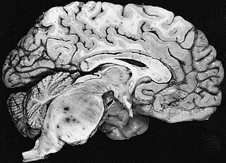
A brain tumor occurs when abnormal cells form within the brain. There are two main types of tumors: malignant tumors and benign (non-cancerous) tumors. These can be further classified as primary tumors, which start within the brain, and secondary tumors, which most commonly have spread from tumors located outside the brain, known as brain metastasis tumors. All types of brain tumors may produce symptoms that vary depending on the size of the tumor and the part of the brain that is involved. Where symptoms exist, they may include headaches, seizures, problems with vision, vomiting and mental changes. Other symptoms may include difficulty walking, speaking, with sensations, or unconsciousness.

<span class="mw-page-title-main">Glioma</span> Tumour of the glial cells of the brain or spine

A glioma is a type of tumor that starts in the glial cells of the brain or the spine. Gliomas comprise about 30 percent of all brain tumors and central nervous system tumours, and 80 percent of all malignant brain tumours.

Spinal tumors are neoplasms located in either the vertebral column or the spinal cord. There are three main types of spinal tumors classified based on their location: extradural and intradural. Extradural tumors are located outside the dura mater lining and are most commonly metastatic. Intradural tumors are located inside the dura mater lining and are further subdivided into intramedullary and extramedullary tumors. Intradural-intramedullary tumors are located within the dura and spinal cord parenchyma, while intradural-extramedullary tumors are located within the dura but outside the spinal cord parenchyma. The most common presenting symptom of spinal tumors is nocturnal back pain. Other common symptoms include muscle weakness, sensory loss, and difficulty walking. Loss of bowel and bladder control may occur during the later stages of the disease.

<span class="mw-page-title-main">Oligodendroglioma</span> Medical condition

Oligodendrogliomas are a type of glioma that are believed to originate from the oligodendrocytes of the brain or from a glial precursor cell. They occur primarily in adults but are also found in children.

<span class="mw-page-title-main">Ependymoma</span> Medical condition

An ependymoma is a tumor that arises from the ependyma, a tissue of the central nervous system. Usually, in pediatric cases the location is intracranial, while in adults it is spinal. The common location of intracranial ependymomas is the fourth ventricle. Rarely, ependymomas can occur in the pelvic cavity.

<span class="mw-page-title-main">Meningioma</span> Type of tumor

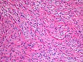
Meningioma, also known as meningeal tumor, is typically a slow-growing tumor that forms from the meninges, the membranous layers surrounding the brain and spinal cord. Symptoms depend on the location and occur as a result of the tumor pressing on nearby tissue. Many cases never produce symptoms. Occasionally seizures, dementia, trouble talking, vision problems, one sided weakness, or loss of bladder control may occur.

<span class="mw-page-title-main">Glioblastoma</span> Aggressive type of brain cancer

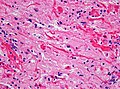
Glioblastoma, previously known as glioblastoma multiforme (GBM), is the most aggressive and most common type of cancer that originates in the brain, and has very poor prognosis for survival. Initial signs and symptoms of glioblastoma are nonspecific. They may include headaches, personality changes, nausea, and symptoms similar to those of a stroke. Symptoms often worsen rapidly and may progress to unconsciousness.

<span class="mw-page-title-main">Astrocytoma</span> Medical condition

Astrocytoma is a type of brain tumor. Astrocytomas originate from a specific kind of star-shaped glial cell in the cerebrum called an astrocyte. This type of tumor does not usually spread outside the brain and spinal cord and it does not usually affect other organs. After glioblastomas, astrocytomas are the second most common glioma and can occur in most parts of the brain and occasionally in the spinal cord.

<span class="mw-page-title-main">Oligoastrocytoma</span> Medical condition

Oligoastrocytomas are a subset of brain tumors that present with an appearance of mixed glial cell origin, astrocytoma and oligodendroglioma. However, the term "Oligoastrocytoma" is now considered obsolete by the National Comprehensive Cancer Network stating "the term should no longer be used as such morphologically ambiguous tumors can be reliably resolved into astrocytomas and oligodendrogliomas with molecular testing."

<span class="mw-page-title-main">Craniopharyngioma</span> Medical condition

A craniopharyngioma is a rare type of brain tumor derived from pituitary gland embryonic tissue that occurs most commonly in children, but also affects adults. It may present at any age, even in the prenatal and neonatal periods, but peak incidence rates are childhood-onset at 5–14 years and adult-onset at 50–74 years. People may present with bitemporal inferior quadrantanopia leading to bitemporal hemianopsia, as the tumor may compress the optic chiasm. It has a point prevalence around two per 1,000,000. Craniopharyngiomas are distinct from Rathke's cleft tumours and intrasellar arachnoid cysts.

A blastoma is a type of cancer, more common in children, that is caused by malignancies in precursor cells, often called blasts. Examples are nephroblastoma, medulloblastoma, and retinoblastoma. The suffix -blastoma is used to imply a tumor of primitive, incompletely differentiated cells, e.g., chondroblastoma is composed of cells resembling the precursor of chondrocytes.

<span class="mw-page-title-main">Gemistocyte</span> Swollen and reactive astrocyte

A gemistocyte is a swollen, reactive astrocyte.

<span class="mw-page-title-main">Brain metastasis</span> Cancer that has metastasized (spread) to the brain from another location in the body

A brain metastasis is a cancer that has metastasized (spread) to the brain from another location in the body and is therefore considered a secondary brain tumor. The metastasis typically shares a cancer cell type with the original site of the cancer. Metastasis is the most common cause of brain cancer, as primary tumors that originate in the brain are less common. The most common sites of primary cancer which metastasize to the brain are lung, breast, colon, kidney, and skin cancer. Brain metastases can occur in patients months or even years after their original cancer is treated. Brain metastases have a poor prognosis for cure, but modern treatments are allowing patients to live months and sometimes years after the diagnosis.

<span class="mw-page-title-main">Pleomorphic xanthoastrocytoma</span> Medical condition

Pleomorphic xanthoastrocytoma (PXA) is a brain tumor that occurs most frequently in children and teenagers. At Boston Children's Hospital, the average age at diagnosis is 12 years.

Neuro-oncology is the study of brain and spinal cord neoplasms, many of which are very dangerous and life-threatening. Among the malignant brain cancers, gliomas of the brainstem and pons, glioblastoma multiforme, and high-grade astrocytoma/oligodendroglioma are among the worst. In these cases, untreated survival usually amounts to only a few months, and survival with current radiation and chemotherapy treatments may extend that time from around a year to a year and a half, possibly two or more, depending on the patient's condition, immune function, treatments used, and the specific type of malignant brain neoplasm. Surgery may in some cases be curative, but, as a general rule, malignant brain cancers tend to regenerate and emerge from remission easily, especially highly malignant cases. In such cases, the goal is to excise as much of the mass and as much of the tumor margin as possible without endangering vital functions or other important cognitive abilities. The Journal of Neuro-Oncology is the longest continuously published journal in the field and serves as a leading reference to those practicing in the area of neuro-oncology.

<span class="mw-page-title-main">Fibrillary astrocytoma</span> Type of brain tumor

Fibrillary astrocytomas are a group of primary slow-growing brain tumors that typically occur in adults between the ages of 20 and 50.

<span class="mw-page-title-main">Anaplastic astrocytoma</span> Medical condition

Anaplastic astrocytoma is a rare WHO grade III type of astrocytoma, which is a type of cancer of the brain. In the United States, the annual incidence rate for anaplastic astrocytoma is 0.44 per 100,000 people.

<span class="mw-page-title-main">Subependymal giant cell astrocytoma</span> Medical condition

Subependymal giant cell astrocytoma is a low-grade astrocytic brain tumor (astrocytoma) that arises within the ventricles of the brain. It is most commonly associated with tuberous sclerosis complex (TSC). Although it is a low-grade tumor, its location can potentially obstruct the ventricles and lead to hydrocephalus.

<span class="mw-page-title-main">Astroblastoma</span> Medical condition

Astroblastoma is a rare glial tumor derived from the astroblast, a type of cell that closely resembles spongioblastoma and astrocytes. Astroblastoma cells are most likely found in the supratentorial region of the brain that houses the cerebrum, an area responsible for all voluntary movements in the body. It also occurs significantly in the frontal lobe, parietal lobe, and temporal lobe, areas where movement, language creation, memory perception, and environmental surroundings are expressed. These tumors can be present in major brain areas not associated with the main cerebral hemispheres, including the cerebellum, optic nerve, cauda equina, hypothalamus, and brain stem.

<span class="mw-page-title-main">Angiocentric glioma</span>

Angiocentric glioma (AG) refers to a rare neuroepithelial tumor when the superficial brain malignant cells enclose the brain vessels, commonly found in children and young adults. Initially identified in 2005 by Wang and his team from the University of Texas, AG was classified as Grade I by 2007 WHO Classification of Tumors of the Central Nervous System due to its benign clinical behavior, low proliferation index, and curative properties. AG primarily affects children and young adults at an average initial diagnosis age of 16 years old. Over 85% AG patients experience intractable seizures since childhood, especially partial epilepsy.

References

  1. Louis, David N.; Ohgaki, Hiroko; Wiestler, Otmar D.; Cavenee, Webster K. (2016). WHO classification of tumours of the central nervous system (Revised 4th ed.). Lyon. ISBN   978-92-832-4492-9.{{cite book}}: CS1 maint: location missing publisher (link)
  2. "Pilocytic astrocytoma". National Center For Advancing Translational Sciences. Retrieved 2 July 2021.
  3. Jones DT, Kocialkowski S, Liu L, Pearson DM, Bäcklund LM, Ichimura K, Collins VP (November 2008). "Tandem duplication producing a novel oncogenic BRAF fusion gene defines the majority of pilocytic astrocytomas". Cancer Research. 68 (21): 8673–8677. doi:10.1158/0008-5472.CAN-08-2097. PMC   2577184 . PMID   18974108.
  4. Sadighi Z, Slopis J (May 2013). "Pilocytic astrocytoma: a disease with evolving molecular heterogeneity". Journal of Child Neurology. 28 (5): 625–632. doi:10.1177/0883073813476141. PMID   23439714. S2CID   8437596.
  5. "College of American Pathologists".
  6. National Cancer Institute > Dictionary of Cancer Terms > pilocytic Retrieved on July 16, 2010
  7. Wippold FJ, Perry A, Lennerz J (May 2006). "Neuropathology for the neuroradiologist: Rosenthal fibers". AJNR. American Journal of Neuroradiology. 27 (5): 958–961. PMC   7975751 . PMID   16687524.
  8. Dodgshun AJ, Maixner WJ, Hansford JR, Sullivan MJ (May 2016). "Low rates of recurrence and slow progression of pediatric pilocytic astrocytoma after gross-total resection: justification for reducing surveillance imaging". Journal of Neurosurgery. Pediatrics. 17 (5): 569–572. doi: 10.3171/2015.9.PEDS15449 . PMID   26722760.
  9. Armstrong GT, Conklin HM, Huang S, Srivastava D, Sanford R, Ellison DW, et al. (February 2011). "Survival and long-term health and cognitive outcomes after low-grade glioma". Neuro-Oncology. 13 (2): 223–234. doi: 10.1093/neuonc/noq178 . PMC   3064628 . PMID   21177781.
  10. Ris MD, Leisenring WM, Goodman P, Di C, Noll J, Levy W, et al. (September 2019). "Neuropsychological and socioeconomic outcomes in adult survivors of pediatric low-grade glioma". Cancer. 125 (17): 3050–3058. doi: 10.1002/cncr.32186 . PMC   6690772 . PMID   31231797.
  11. "Astrocytoma". The Lecturio Medical Concept Library. Retrieved 1 July 2021.
  12. "Central Brain Tumor Registry of the United States". CBTRUS.
  13. Ostrom QT, Patil N, Cioffi G, Waite K, Kruchko C, Barnholtz-Sloan JS (October 2020). "CBTRUS Statistical Report: Primary Brain and Other Central Nervous System Tumors Diagnosed in the United States in 2013–2017". Neuro-Oncology. 22 (12 Suppl 2): iv1–iv96. doi:10.1093/neuonc/noaa200. PMC   7596247 . PMID   33123732.