Related Research Articles

Microscopy is the technical field of using microscopes to view objects and areas of objects that cannot be seen with the naked eye. There are three well-known branches of microscopy: optical, electron, and scanning probe microscopy, along with the emerging field of X-ray microscopy.

Spectroscopy is the field of study that measures and interprets electromagnetic spectra. In narrower contexts, spectroscopy is the precise study of color as generalized from visible light to all bands of the electromagnetic spectrum.
Fourier-transform spectroscopy is a measurement technique whereby spectra are collected based on measurements of the coherence of a radiative source, using time-domain or space-domain measurements of the radiation, electromagnetic or not. It can be applied to a variety of types of spectroscopy including optical spectroscopy, infrared spectroscopy, nuclear magnetic resonance (NMR) and magnetic resonance spectroscopic imaging (MRSI), mass spectrometry and electron spin resonance spectroscopy.

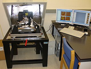
Atomic force microscopy (AFM) or scanning force microscopy (SFM) is a very-high-resolution type of scanning probe microscopy (SPM), with demonstrated resolution on the order of fractions of a nanometer, more than 1000 times better than the optical diffraction limit.

Absorption spectroscopy is spectroscopy that involves techniques that measure the absorption of electromagnetic radiation, as a function of frequency or wavelength, due to its interaction with a sample. The sample absorbs energy, i.e., photons, from the radiating field. The intensity of the absorption varies as a function of frequency, and this variation is the absorption spectrum. Absorption spectroscopy is performed across the electromagnetic spectrum.

A synchrotron light source is a source of electromagnetic radiation (EM) usually produced by a storage ring, for scientific and technical purposes. First observed in synchrotrons, synchrotron light is now produced by storage rings and other specialized particle accelerators, typically accelerating electrons. Once the high-energy electron beam has been generated, it is directed into auxiliary components such as bending magnets and insertion devices in storage rings and free electron lasers. These supply the strong magnetic fields perpendicular to the beam that are needed to stimulate the high energy electrons to emit photons.
In physics and physical chemistry, time-resolved spectroscopy is the study of dynamic processes in materials or chemical compounds by means of spectroscopic techniques. Most often, processes are studied after the illumination of a material occurs, but in principle, the technique can be applied to any process that leads to a change in properties of a material. With the help of pulsed lasers, it is possible to study processes that occur on time scales as short as 10−16 seconds. All time-resolved spectra are suitable to be analyzed using the two-dimensional correlation method for a correlation map between the peaks.
Laser-ultrasonics uses lasers to generate and detect ultrasonic waves. It is a non-contact technique used to measure materials thickness, detect flaws and carry out materials characterization. The basic components of a laser-ultrasonic system are a generation laser, a detection laser and a detector.
Ultrafast laser spectroscopy is a spectroscopic technique that uses ultrashort pulse lasers for the study of dynamics on extremely short time scales. Different methods are used to examine the dynamics of charge carriers, atoms, and molecules. Many different procedures have been developed spanning different time scales and photon energy ranges; some common methods are listed below.

Near-field scanning optical microscopy (NSOM) or scanning near-field optical microscopy (SNOM) is a microscopy technique for nanostructure investigation that breaks the far field resolution limit by exploiting the properties of evanescent waves. In SNOM, the excitation laser light is focused through an aperture with a diameter smaller than the excitation wavelength, resulting in an evanescent field on the far side of the aperture. When the sample is scanned at a small distance below the aperture, the optical resolution of transmitted or reflected light is limited only by the diameter of the aperture. In particular, lateral resolution of 6 nm and vertical resolution of 2–5 nm have been demonstrated.
Photothermal microspectroscopy (PTMS), alternatively known as photothermal temperature fluctuation (PTTF), is derived from two parent instrumental techniques: infrared spectroscopy and atomic force microscopy (AFM). In one particular type of AFM, known as scanning thermal microscopy (SThM), the imaging probe is a sub-miniature temperature sensor, which may be a thermocouple or a resistance thermometer. This same type of detector is employed in a PTMS instrument, enabling it to provide AFM/SThM images: However, the chief additional use of PTMS is to yield infrared spectra from sample regions below a micrometer, as outlined below.
Photothermal optical microscopy / "photothermal single particle microscopy" is a technique that is based on detection of non-fluorescent labels. It relies on absorption properties of labels, and can be realized on a conventional microscope using a resonant modulated heating beam, non-resonant probe beam and lock-in detection of photothermal signals from a single nanoparticle. It is the extension of the macroscopic photothermal spectroscopy to the nanoscopic domain. The high sensitivity and selectivity of photothermal microscopy allows even the detection of single molecules by their absorption. Similar to Fluorescence Correlation Spectroscopy (FCS), the photothermal signal may be recorded with respect to time to study the diffusion and advection characteristics of absorbing nanoparticles in a solution. This technique is called photothermal correlation spectroscopy (PhoCS).
The technique of vibrational analysis with scanning probe microscopy allows probing vibrational properties of materials at the submicrometer scale, and even of individual molecules. This is accomplished by integrating scanning probe microscopy (SPM) and vibrational spectroscopy. This combination allows for much higher spatial resolution than can be achieved with conventional Raman/FTIR instrumentation. The technique is also nondestructive, requires non-extensive sample preparation, and provides more contrast such as intensity contrast, polarization contrast and wavelength contrast, as well as providing specific chemical information and topography images simultaneously.
The photoacoustic effect or optoacoustic effect is the formation of sound waves following light absorption in a material sample. In order to obtain this effect the light intensity must vary, either periodically or as a single flash. The photoacoustic effect is quantified by measuring the formed sound with appropriate detectors, such as microphones or piezoelectric sensors. The time variation of the electric output from these detectors is the photoacoustic signal. These measurements are useful to determine certain properties of the studied sample. For example, in photoacoustic spectroscopy, the photoacoustic signal is used to obtain the actual absorption of light in either opaque or transparent objects. It is useful for substances in extremely low concentrations, because very strong pulses of light from a laser can be used to increase sensitivity and very narrow wavelengths can be used for specificity. Furthermore, photoacoustic measurements serve as a valuable research tool in the study of the heat evolved in photochemical reactions, particularly in the study of photosynthesis.
AFM-IR or infrared nanospectroscopy is one of a family of techniques that are derived from a combination of two parent instrumental techniques. AFM-IR combines the chemical analysis power of infrared spectroscopy and the high-spatial resolution of scanning probe microscopy (SPM). The term was first used to denote a method that combined a tuneable free electron laser with an atomic force microscope equipped with a sharp probe that measured the local absorption of infrared light by a sample with nanoscale spatial resolution.
The operation of a photon scanning tunneling microscope (PSTM) is analogous to the operation of an electron scanning tunneling microscope, with the primary distinction being that PSTM involves tunneling of photons instead of electrons from the sample surface to the probe tip. A beam of light is focused on a prism at an angle greater than the critical angle of the refractive medium in order to induce total internal reflection within the prism. Although the beam of light is not propagated through the surface of the refractive prism under total internal reflection, an evanescent field of light is still present at the surface.
Photo-reflectance is an optical technique for investigating the material and electronic properties of thin films. Photo-reflectance measures the change in reflectivity of a sample in response to the application of an amplitude modulated light beam. In general, a photo-reflectometer consists of an intensity modulated "pump" light beam used to modulate the reflectivity of the sample, a second "probe" light beam used to measure the reflectance of the sample, an optical system for directing the pump and probe beams to the sample, and for directing the reflected probe light onto a photodetector, and a signal processor to record the differential reflectance. The pump light is typically modulated at a known frequency so that a lock-in amplifier may be used to suppress unwanted noise, resulting in the ability to detect reflectance changes at the ppm level.
The collimated transmission method is a direct way of measuring the optical properties of materials. It is especially useful for sensing the optical properties of tissues to guide developments of both diagnostic and therapeutic techniques. These optical properties are described by the absorption coefficient μa, scattering coefficient μs, and anisotropy factor g.
Ultrafast scanning electron microscopy (UFSEM) combines two microscopic modalities, Pump-probe microscopy and Scanning electron microscope, to gather temporal and spatial resolution phenomena. The technique uses ultrashort laser pulses for pump excitation of the material and the sample response will be detected by an Everhart-Thornley detector. Acquiring data depends mainly on formation of images by raster scan mode after pumping with short laser pulse at different delay times. The characterization of the output image will be done through the temporal resolution aspect. Thus, the idea is to exploit the shorter DeBroglie wavelength in respect to the photons which has great impact to increase the resolution about 1 nm. That technique is an up-to-date approach to study the dynamic of charge on material surfaces.
References
- ↑ In the true mirage, however, a light ray is bent gradually until it is totally reflected by the very hot air near soil. Here, instead, there is no reflection, and the ray is bent in a smooth way by the refraction index gradient, which acts like a prism.
- ↑ Boccara, A.C.; Fournier, D.; Jackson, Warren; Amer, Nabil. (1980). "Sensitive photothermal deflection technique for measuring absorption in optically thin media". Optics Letters . 5 (9): 377–379. Bibcode:1980OptL....5..377B. doi:10.1364/OL.5.000377. PMID 19693234.
- 1 2 Jackson, W.B.; Amer, N.M.; Boccara, A.C.; Fournier, D. (1981-04-15). "Photothermal deflection spectroscopy and detection". Applied Optics . 20 (8): 1333–1344. Bibcode:1981ApOpt..20.1333J. doi:10.1364/AO.20.001333. PMID 20309309.
- ↑ Zhao Y, Liu L, Zhao X, Lu M (2016). "Enhanced photothermal lens using a photonic crystal surface". Applied Physics Letters. 109 (7): 071108. Bibcode:2016ApPhL.109g1108Z. doi:10.1063/1.4961376.
- J. A. Sell Photothermal Investigations of Solids and Fluids Academic Press, New York 1989
- D. P. Almond and P. M. Patel Photothermal Science and Techniques Chapman and Hall, London 1996
- S. E. Bialkowski Photothermal Spectroscopy Methods for Chemical Analysis John Wiley, New York 1996