Circular dichroism

Last updated

Circular dichroism (CD) is dichroism involving circularly polarized light, i.e., the differential absorption of left- and right-handed light. [1] [2] Left-hand circular (LHC) and right-hand circular (RHC) polarized light represent two possible spin angular momentum states for a photon, and so circular dichroism is also referred to as dichroism for spin angular momentum. [3] This phenomenon was discovered by Jean-Baptiste Biot, Augustin Fresnel, and Aimé Cotton in the first half of the 19th century. [4] Circular dichroism and circular birefringence are manifestations of optical activity. It is exhibited in the absorption bands of optically active chiral molecules. CD spectroscopy has a wide range of applications in many different fields. Most notably, UV CD is used to investigate the secondary structure of proteins. [5] UV/Vis CD is used to investigate charge-transfer transitions. [6] Near-infrared CD is used to investigate geometric and electronic structure by probing metal dd transitions. [2] Vibrational circular dichroism, which uses light from the infrared energy region, is used for structural studies of small organic molecules, and most recently proteins and DNA. [5]

Contents

Physical principles

Circular polarization of light

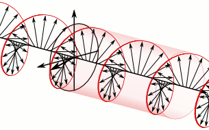
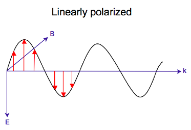
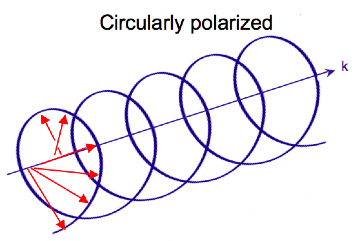
Electromagnetic radiation consists of an electric and magnetic field that oscillate perpendicular to one another and to the propagating direction, [7] a transverse wave. While linearly polarized light occurs when the electric field vector oscillates only in one plane, circularly polarized light occurs when the direction of the electric field vector rotates about its propagation direction while the vector retains constant magnitude. At a single point in space, the circularly polarized-vector will trace out a circle over one period of the wave frequency, hence the name. The two diagrams below show the electric field vectors of linearly and circularly polarized light, at one moment of time, for a range of positions; the plot of the circularly polarized electric vector forms a helix along the direction of propagation . For left circularly polarized light (LCP) with propagation towards the observer, the electric vector rotates counterclockwise. [2] For right circularly polarized light (RCP), the electric vector rotates clockwise.

Linearly pol.png Circularly pol.png

Interaction of circularly polarized light with matter

When circularly polarized light passes through an absorbing optically active medium, the speeds between right and left polarizations differ () as well as their wavelength() and the extent to which they are absorbed (). Circular dichroism is the difference . [5] The electric field of a light beam causes a linear displacement of charge when interacting with a molecule (electric dipole), whereas its magnetic field causes a circulation of charge (magnetic dipole). These two motions combined cause an excitation of an electron in a helical motion, which includes translation and rotation and their associated operators. The experimentally determined relationship between the rotational strength of a sample and the is given by

The rotational strength has also been determined theoretically,

We see from these two equations that in order to have non-zero , the electric and magnetic dipole moment operators ( and ) must transform as the same irreducible representation. and are the only point groups where this can occur, making only chiral molecules CD active.

Simply put, since circularly polarized light itself is "chiral", it interacts differently with chiral molecules. That is, the two types of circularly polarized light are absorbed to different extents. In a CD experiment, equal amounts of left and right circularly polarized light of a selected wavelength are alternately radiated into a (chiral) sample. One of the two polarizations is absorbed more than the other one, and this wavelength-dependent difference of absorption is measured, yielding the CD spectrum of the sample. Due to the interaction with the molecule, the electric field vector of the light traces out an elliptical path after passing through the sample.

It is important that the chirality of the molecule can be conformational rather than structural. That is, for instance, a protein molecule with a helical secondary structure can have a CD that changes with changes in the conformation.

Delta absorbance

By definition,

where (Delta Absorbance) is the difference between absorbance of left circularly polarized (LCP) and right circularly polarized (RCP) light (this is what is usually measured). is a function of wavelength, so for a measurement to be meaningful the wavelength at which it was performed must be known.

Molar circular dichroism

It can also be expressed, by applying Beer's law, as:

where

and are the molar extinction coefficients for LCP and RCP light,
is the molar concentration,
is the path length in centimeters (cm).

Then

is the molar circular dichroism. This intrinsic property is what is usually meant by the circular dichroism of the substance. Since is a function of wavelength, a molar circular dichroism value () must specify the wavelength at which it is valid.

Extrinsic effects on circular dichroism

In many practical applications of circular dichroism (CD), as discussed below, the measured CD is not simply an intrinsic property of the molecule, but rather depends on the molecular conformation. In such a case the CD may also be a function of temperature, concentration, and the chemical environment, including solvents. In this case the reported CD value must also specify these other relevant factors in order to be meaningful.

In ordered structures lacking two-fold rotational symmetry, optical activity, [8] [9] including differential transmission [10] (and reflection [11] ) of circularly polarized waves also depends on the propagation direction through the material. In this case, so-called extrinsic 3d chirality is associated with the mutual orientation of light beam and structure.

Molar ellipticity

Although is usually measured, for historical reasons most measurements are reported in degrees of ellipticity. Molar ellipticity is circular dichroism corrected for concentration. Molar circular dichroism and molar ellipticity, , are readily interconverted by the equation:

Elliptical polarized light (violet) is composed of unequal contributions of right (blue) and left (red) circular polarized light. Electric Vectors 1.png
Elliptical polarized light (violet) is composed of unequal contributions of right (blue) and left (red) circular polarized light.

This relationship is derived by defining the ellipticity of the polarization as:

where

and are the magnitudes of the electric field vectors of the right-circularly and left-circularly polarized light, respectively.

When equals (when there is no difference in the absorbance of right- and left-circular polarized light), is 0° and the light is linearly polarized. When either or is equal to zero (when there is complete absorbance of the circular polarized light in one direction), is 45° and the light is circularly polarized.

Generally, the circular dichroism effect is small, so is small and can be approximated as in radians. Since the intensity or irradiance, , of light is proportional to the square of the electric-field vector, the ellipticity becomes:

Then by substituting for I using Beer's law in natural logarithm form:

The ellipticity can now be written as:

Since , this expression can be approximated by expanding the exponentials in a Taylor series to first-order and then discarding terms of in comparison with unity and converting from radians to degrees:

The linear dependence of solute concentration and pathlength is removed by defining molar ellipticity as,

Then combining the last two expression with Beer's law, molar ellipticity becomes:

The units of molar ellipticity are historically (deg·cm2/dmol). To calculate molar ellipticity, the sample concentration (g/L), cell pathlength (cm), and the molecular weight (g/mol) must be known.

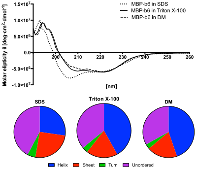
If the sample is a protein, the mean residue weight (average molecular weight of the amino acid residues it contains) is often used in place of the molecular weight, essentially treating the protein as a solution of amino acids. Using mean residue ellipticity facilitates comparing the CD of proteins of different molecular weight; use of this normalized CD is important in studies of protein structure.

Mean residue ellipticity

Methods for estimating secondary structure in polymers, proteins and polypeptides in particular, often require that the measured molar ellipticity spectrum be converted to a normalized value, specifically a value independent of the polymer length. Mean residue ellipticity is used for this purpose; it is simply the measured molar ellipticity of the molecule divided by the number of monomer units (residues) in the molecule.

Application to biological molecules

Upper panel: Circular dichroism spectroscopy in the ultraviolet wavelength region (UV-CD) of MBP-cytochrome b6 fusion protein in different detergent solutions. It shows that the protein in DM, as well as in Triton X-100 solution, recovered its structure. However the spectra obtained from SDS solution shows decreased ellipticity in the range between 200 and 210 nm, which indicates incomplete secondary structure recovery.
Lower panel: The content of secondary structures predicted from the CD spectra using the CDSSTR algorithm. The protein in SDS solution shows increased content of unordered structures and decreased helices content. Circular dichroism and structures calculation-protein in detergents.svg
Upper panel: Circular dichroism spectroscopy in the ultraviolet wavelength region (UV-CD) of MBP-cytochrome b6 fusion protein in different detergent solutions. It shows that the protein in DM, as well as in Triton X-100 solution, recovered its structure. However the spectra obtained from SDS solution shows decreased ellipticity in the range between 200 and 210 nm, which indicates incomplete secondary structure recovery.
Lower panel: The content of secondary structures predicted from the CD spectra using the CDSSTR algorithm. The protein in SDS solution shows increased content of unordered structures and decreased helices content.

In general, this phenomenon will be exhibited in absorption bands of any optically active molecule. As a consequence, circular dichroism is exhibited by biological molecules, because of their dextrorotary and levorotary components. Even more important is that a secondary structure will also impart a distinct CD to its respective molecules. Therefore, the alpha helix of proteins and the double helix of nucleic acids have CD spectral signatures representative of their structures. The capacity of CD to give a representative structural signature makes it a powerful tool in modern biochemistry with applications that can be found in virtually every field of study.

CD is closely related to the optical rotatory dispersion (ORD) technique, and is generally considered to be more advanced. CD is measured in or near the absorption bands of the molecule of interest, while ORD can be measured far from these bands. CD's advantage is apparent in the data analysis. Structural elements are more clearly distinguished since their recorded bands do not overlap extensively at particular wavelengths as they do in ORD. In principle, these two spectral measurements can be interconverted through an integral transform (Kramers–Kronig relation), if all the absorptions are included in the measurements.

The far-UV (ultraviolet) CD spectrum of proteins can reveal important characteristics of their secondary structure. CD spectra can be readily used to estimate the fraction of a molecule that is in the alpha-helix conformation, the beta-sheet conformation, the beta-turn conformation, or some other (e.g. random coil) conformation. [13] [14] [15] [16] These fractional assignments place important constraints on the possible secondary conformations that the protein can be in. CD cannot, in general, say where the alpha helices that are detected are located within the molecule or even completely predict how many there are. Despite this, CD is a valuable tool, especially for showing changes in conformation. It can, for instance, be used to study how the secondary structure of a molecule changes as a function of temperature or of the concentration of denaturing agents, e.g. Guanidinium chloride or urea. In this way it can reveal important thermodynamic information about the molecule (such as the enthalpy and Gibbs free energy of denaturation) that cannot otherwise be easily obtained. Anyone attempting to study a protein will find CD a valuable tool for verifying that the protein is in its native conformation before undertaking extensive and/or expensive experiments with it. Also, there are a number of other uses for CD spectroscopy in protein chemistry not related to alpha-helix fraction estimation. Moreover, CD spectroscopy has been used in bioinorganic interface studies. Specifically it has been used to analyze the differences in secondary structure of an engineered protein before and after titration with a reagent. [17]

The near-UV CD spectrum (>250 nm) of proteins provides information on the tertiary structure. The signals obtained in the 250–300 nm region are due to the absorption, dipole orientation and the nature of the surrounding environment of the phenylalanine, tyrosine, cysteine (or S-S disulfide bridges) and tryptophan amino acids. Unlike in far-UV CD, the near-UV CD spectrum cannot be assigned to any particular 3D structure. Rather, near-UV CD spectra provide structural information on the nature of the prosthetic groups in proteins, e.g., the heme groups in hemoglobin and cytochrome c.

Visible CD spectroscopy is a very powerful technique to study metal–protein interactions and can resolve individual d–d electronic transitions as separate bands. CD spectra in the visible light region are only produced when a metal ion is in a chiral environment, thus, free metal ions in solution are not detected. This has the advantage of only observing the protein-bound metal, so pH dependence and stoichiometries are readily obtained. Optical activity in transition metal ion complexes have been attributed to configurational, conformational and the vicinal effects. Klewpatinond and Viles (2007) have produced a set of empirical rules for predicting the appearance of visible CD spectra for Cu2+ and Ni2+ square-planar complexes involving histidine and main-chain coordination.

CD gives less specific structural information than X-ray crystallography and protein NMR spectroscopy, for example, which both give atomic resolution data. However, CD spectroscopy is a quick method that does not require large amounts of proteins or extensive data processing. Thus CD can be used to survey a large number of solvent conditions, varying temperature, pH, salinity, and the presence of various cofactors.

CD spectroscopy is usually used to study proteins in solution, and thus it complements methods that study the solid state. This is also a limitation, in that many proteins are embedded in membranes in their native state, and solutions containing membrane structures are often strongly scattering. CD is sometimes measured in thin films.

CD spectroscopy has also been done using semiconducting materials such as TiO2 to obtain large signals in the UV range of wavelengths, where the electronic transitions for biomolecules often occur. [18]

Experimental limitations

CD has also been studied in carbohydrates, but with limited success due to the experimental difficulties associated with measurement of CD spectra in the vacuum ultraviolet (VUV) region of the spectrum (100–200 nm), where the corresponding CD bands of unsubstituted carbohydrates lie. Substituted carbohydrates with bands above the VUV region have been successfully measured.

Measurement of CD is also complicated by the fact that typical aqueous buffer systems often absorb in the range where structural features exhibit differential absorption of circularly polarized light. Phosphate, sulfate, carbonate, and acetate buffers are generally incompatible with CD unless made extremely dilute e.g. in the 10–50 mM range. The TRIS buffer system should be completely avoided when performing far-UV CD. Borate and Onium compounds are often used to establish the appropriate pH range for CD experiments. Some experimenters have substituted fluoride for chloride ion because fluoride absorbs less in the far UV, and some have worked in pure water. Another, almost universal, technique is to minimize solvent absorption by using shorter path length cells when working in the far UV, 0.1 mm path lengths are not uncommon in this work.

In addition to measuring in aqueous systems, CD, particularly far-UV CD, can be measured in organic solvents e.g. ethanol, methanol, trifluoroethanol (TFE). The latter has the advantage to induce structure formation of proteins, inducing beta-sheets in some and alpha helices in others, which they would not show under normal aqueous conditions. Most common organic solvents such as acetonitrile, THF, chloroform, dichloromethane are however, incompatible with far-UV CD.

It may be of interest to note that the protein CD spectra used in secondary structure estimation are related to the π to π* orbital absorptions of the amide bonds linking the amino acids. These absorption bands lie partly in the so-called vacuum ultraviolet (wavelengths less than about 200 nm). The wavelength region of interest is actually inaccessible in air because of the strong absorption of light by oxygen at these wavelengths. In practice these spectra are measured not in vacuum but in an oxygen-free instrument (filled with pure nitrogen gas).

Once oxygen has been eliminated, perhaps the second most important technical factor in working below 200 nm is to design the rest of the optical system to have low losses in this region. Critical in this regard is the use of aluminized mirrors whose coatings have been optimized for low loss in this region of the spectrum.

The usual light source in these instruments is a high pressure, short-arc xenon lamp. Ordinary xenon arc lamps are unsuitable for use in the low UV. Instead, specially constructed lamps with envelopes made from high-purity synthetic fused silica must be used.

Light from synchrotron sources has a much higher flux at short wavelengths, and has been used to record CD down to 160 nm. In 2010 the CD spectrophotometer at the electron storage ring facility ISA at the University of Aarhus in Denmark was used to record solid state CD spectra down to 120 nm. [19] At the quantum mechanical level, the feature density of circular dichroism and optical rotation are identical. Optical rotary dispersion and circular dichroism share the same quantum information content.

See also

Related Research Articles

The Beer-Lambert law is commonly applied to chemical analysis measurements to determine the concentration of chemical species that absorb light. It is often referred to as Beer's law. In physics, the Bouguer–Lambert law is an empirical law which relates the extinction or attenuation of light to the properties of the material through which the light is travelling. It had its first use in astronomical extinction. The fundamental law of extinction is sometimes called the Beer-Bouguer-Lambert law or the Bouguer-Beer-Lambert law or merely the extinction law. The extinction law is also used in understanding attenuation in physical optics, for photons, neutrons, or rarefied gases. In mathematical physics, this law arises as a solution of the BGK equation.

In physics, the cross section is a measure of the probability that a specific process will take place when some kind of radiant excitation intersects a localized phenomenon. For example, the Rutherford cross-section is a measure of probability that an alpha particle will be deflected by a given angle during an interaction with an atomic nucleus. Cross section is typically denoted σ (sigma) and is expressed in units of area, more specifically in barns. In a way, it can be thought of as the size of the object that the excitation must hit in order for the process to occur, but more exactly, it is a parameter of a stochastic process.

<span class="mw-page-title-main">Refractive index</span> Ratio of the speed of light in vacuum to that in the medium

In optics, the refractive index of an optical medium is a dimensionless number that gives the indication of the light bending ability of that medium.

<span class="mw-page-title-main">Optical rotation</span> Concept in enantioselective synthesis

Optical rotation, also known as polarization rotation or circular birefringence, is the rotation of the orientation of the plane of polarization about the optical axis of linearly polarized light as it travels through certain materials. Circular birefringence and circular dichroism are the manifestations of optical activity. Optical activity occurs only in chiral materials, those lacking microscopic mirror symmetry. Unlike other sources of birefringence which alter a beam's state of polarization, optical activity can be observed in fluids. This can include gases or solutions of chiral molecules such as sugars, molecules with helical secondary structure such as some proteins, and also chiral liquid crystals. It can also be observed in chiral solids such as certain crystals with a rotation between adjacent crystal planes or metamaterials.

<span class="mw-page-title-main">Circular polarization</span> Polarization state

In electrodynamics, circular polarization of an electromagnetic wave is a polarization state in which, at each point, the electromagnetic field of the wave has a constant magnitude and is rotating at a constant rate in a plane perpendicular to the direction of the wave.

<span class="mw-page-title-main">Permittivity</span> Measure of the electric polarizability of a dielectric

In electromagnetism, the absolute permittivity, often simply called permittivity and denoted by the Greek letter ε (epsilon), is a measure of the electric polarizability of a dielectric. A material with high permittivity polarizes more in response to an applied electric field than a material with low permittivity, thereby storing more energy in the material. In electrostatics, the permittivity plays an important role in determining the capacitance of a capacitor.

<span class="mw-page-title-main">Bremsstrahlung</span> Electromagnetic radiation due to deceleration of charged particles

In particle physics, bremsstrahlung is electromagnetic radiation produced by the deceleration of a charged particle when deflected by another charged particle, typically an electron by an atomic nucleus. The moving particle loses kinetic energy, which is converted into radiation, thus satisfying the law of conservation of energy. The term is also used to refer to the process of producing the radiation. Bremsstrahlung has a continuous spectrum, which becomes more intense and whose peak intensity shifts toward higher frequencies as the change of the energy of the decelerated particles increases.

<span class="mw-page-title-main">Magnetic circular dichroism</span>

Magnetic circular dichroism (MCD) is the differential absorption of left and right circularly polarized (LCP and RCP) light, induced in a sample by a strong magnetic field oriented parallel to the direction of light propagation. MCD measurements can detect transitions which are too weak to be seen in conventional optical absorption spectra, and it can be used to distinguish between overlapping transitions. Paramagnetic systems are common analytes, as their near-degenerate magnetic sublevels provide strong MCD intensity that varies with both field strength and sample temperature. The MCD signal also provides insight into the symmetry of the electronic levels of the studied systems, such as metal ion sites.

Chebyshev filters are analog or digital filters that have a steeper roll-off than Butterworth filters, and have either passband ripple or stopband ripple. Chebyshev filters have the property that they minimize the error between the idealized and the actual filter characteristic over the operating frequency range of the filter, but they achieve this with ripples in the passband. This type of filter is named after Pafnuty Chebyshev because its mathematical characteristics are derived from Chebyshev polynomials. Type I Chebyshev filters are usually referred to as "Chebyshev filters", while type II filters are usually called "inverse Chebyshev filters". Because of the passband ripple inherent in Chebyshev filters, filters with a smoother response in the passband but a more irregular response in the stopband are preferred for certain applications.

Absorbance is defined as "the logarithm of the ratio of incident to transmitted radiant power through a sample ". Alternatively, for samples which scatter light, absorbance may be defined as "the negative logarithm of one minus absorptance, as measured on a uniform sample". The term is used in many technical areas to quantify the results of an experimental measurement. While the term has its origin in quantifying the absorption of light, it is often entangled with quantification of light which is “lost” to a detector system through other mechanisms. What these uses of the term tend to have in common is that they refer to a logarithm of the ratio of a quantity of light incident on a sample or material to that which is detected after the light has interacted with the sample.

<span class="mw-page-title-main">Arc length</span> Distance along a curve

Arc length is the distance between two points along a section of a curve.

<span class="mw-page-title-main">Voigt effect</span>

The Voigt effect is a magneto-optical phenomenon which rotates and elliptizes linearly polarised light sent into an optically active medium. Unlike many other magneto-optical effects such as the Kerr or Faraday effect which are linearly proportional to the magnetization, the Voigt effect is proportional to the square of the magnetization and can be seen experimentally at normal incidence. There are several denominations for this effect in the literature: the Cotton–Mouton effect, the Voigt effect, and magnetic-linear birefringence. This last denomination is closer in the physical sense, where the Voigt effect is a magnetic birefringence of the material with an index of refraction parallel and perpendicular ) to the magnetization vector or to the applied magnetic field.

The Newman–Penrose (NP) formalism is a set of notation developed by Ezra T. Newman and Roger Penrose for general relativity (GR). Their notation is an effort to treat general relativity in terms of spinor notation, which introduces complex forms of the usual variables used in GR. The NP formalism is itself a special case of the tetrad formalism, where the tensors of the theory are projected onto a complete vector basis at each point in spacetime. Usually this vector basis is chosen to reflect some symmetry of the spacetime, leading to simplified expressions for physical observables. In the case of the NP formalism, the vector basis chosen is a null tetrad: a set of four null vectors—two real, and a complex-conjugate pair. The two real members often asymptotically point radially inward and radially outward, and the formalism is well adapted to treatment of the propagation of radiation in curved spacetime. The Weyl scalars, derived from the Weyl tensor, are often used. In particular, it can be shown that one of these scalars— in the appropriate frame—encodes the outgoing gravitational radiation of an asymptotically flat system.

Photon polarization is the quantum mechanical description of the classical polarized sinusoidal plane electromagnetic wave. An individual photon can be described as having right or left circular polarization, or a superposition of the two. Equivalently, a photon can be described as having horizontal or vertical linear polarization, or a superposition of the two.

<span class="mw-page-title-main">Gravitational lensing formalism</span>

In general relativity, a point mass deflects a light ray with impact parameter by an angle approximately equal to

The Binet equation, derived by Jacques Philippe Marie Binet, provides the form of a central force given the shape of the orbital motion in plane polar coordinates. The equation can also be used to derive the shape of the orbit for a given force law, but this usually involves the solution to a second order nonlinear ordinary differential equation. A unique solution is impossible in the case of circular motion about the center of force.

The table of chords, created by the Greek astronomer, geometer, and geographer Ptolemy in Egypt during the 2nd century AD, is a trigonometric table in Book I, chapter 11 of Ptolemy's Almagest, a treatise on mathematical astronomy. It is essentially equivalent to a table of values of the sine function. It was the earliest trigonometric table extensive enough for many practical purposes, including those of astronomy. Since the 8th and 9th centuries, the sine and other trigonometric functions have been used in Islamic mathematics and astronomy, reforming the production of sine tables. Khwarizmi and Habash al-Hasib later produced a set of trigonometric tables.

<span class="mw-page-title-main">Differential optical absorption spectroscopy</span>

In atmospheric chemistry, differential optical absorption spectroscopy (DOAS) is used to measure concentrations of trace gases. When combined with basic optical spectrometers such as prisms or diffraction gratings and automated, ground-based observation platforms, it presents a cheap and powerful means for the measurement of trace gas species such as ozone and nitrogen dioxide. Typical setups allow for detection limits corresponding to optical depths of 0.0001 along lightpaths of up to typically 15 km and thus allow for the detection also of weak absorbers, such as water vapour, Nitrous acid, Formaldehyde, Tetraoxygen, Iodine oxide, Bromine oxide and Chlorine oxide.

<span class="mw-page-title-main">Two-photon circular dichroism</span>

Two-photon circular dichroism (TPCD), the nonlinear counterpart of electronic circular dichroism (ECD), is defined as the differences between the two-photon absorption (TPA) cross-sections obtained using left circular polarized light and right circular polarized light.

<span class="mw-page-title-main">Stochastic gradient Langevin dynamics</span>

Stochastic gradient Langevin dynamics (SGLD) is an optimization and sampling technique composed of characteristics from Stochastic gradient descent, a Robbins–Monro optimization algorithm, and Langevin dynamics, a mathematical extension of molecular dynamics models. Like stochastic gradient descent, SGLD is an iterative optimization algorithm which uses minibatching to create a stochastic gradient estimator, as used in SGD to optimize a differentiable objective function. Unlike traditional SGD, SGLD can be used for Bayesian learning as a sampling method. SGLD may be viewed as Langevin dynamics applied to posterior distributions, but the key difference is that the likelihood gradient terms are minibatched, like in SGD. SGLD, like Langevin dynamics, produces samples from a posterior distribution of parameters based on available data. First described by Welling and Teh in 2011, the method has applications in many contexts which require optimization, and is most notably applied in machine learning problems.

References

  1. P. Atkins; J. de Paula (2005). Elements of Physical Chemistry (4th ed.). Oxford University Press. ISBN   978-0-7167-7329-0.
  2. 1 2 3 Edward I. Solomon; A. B. P. Lever (3 February 2006). Inorganic electronic structure and spectroscopy. Wiley-Interscience. p. 78. ISBN   978-0-471-97124-5 . Retrieved 29 April 2011.
  3. Introduction to Quantum Theory 2ED David Park Sec 2.2 Pg32 "...the polarization of a beam of light is exactly the same kind of thing as the spin of a beam of electrons, the differences of terminology reflecting only the accidents of the historical order of discovery."
  4. Gerald D. Fasman (1996). Circular dichroism and the conformational analysis of biomolecules. Springer. pp. 3–. ISBN   978-0-306-45142-3 . Retrieved 29 April 2011.
  5. 1 2 3 Kōji Nakanishi; Nina Berova; Robert Woody (1994). Circular dichroism: principles and applications. VCH. p. 473. ISBN   978-1-56081-618-8 . Retrieved 29 April 2011.
  6. Solomon, Neidig; A. T. Wecksler; G. Schenk; T. R. Holman (2007). "Kinetic and Spectroscopic Studies of N694C Lipoxygenase: A Probe of the Substrate Activation Mechanism of a Non-Heme Ferric Enzyme". J. Am. Chem. Soc. 129 (24): 7531–7537. doi:10.1021/ja068503d. PMC   2896304 . PMID   17523638.
  7. Alison Rodger; Bengt Nordén (1997). Circular dichroism and linear dichroism. Oxford University Press. ISBN   978-0-19-855897-2 . Retrieved 29 April 2011.
  8. R. Williams (1968). "Optical Rotatory Effect in the Nematic Liquid Phase of p-Azoxyanisole". Physical Review Letters. 21 (6): 342. Bibcode:1968PhRvL..21..342W. doi:10.1103/PhysRevLett.21.342.
  9. R. Williams (1969). "Optical-rotary power and linear electro-optic effect in nematic liquid crystals of p-azoxyanisole". Journal of Chemical Physics. 50 (3): 1324. Bibcode:1969JChPh..50.1324W. doi:10.1063/1.1671194.
  10. Plum, E.; Fedotov, V. A.; Zheludev, N. I. (2008). "Optical activity in extrinsically chiral metamaterial" (PDF). Applied Physics Letters. 93 (19): 191911. arXiv: 0807.0523 . Bibcode:2008ApPhL..93s1911P. doi:10.1063/1.3021082. S2CID   117891131.
  11. Plum, E.; Fedotov, V. A.; Zheludev, N. I. (2016). "Specular optical activity of achiral metasurfaces" (PDF). Applied Physics Letters. 108 (14): 141905. Bibcode:2016ApPhL.108n1905P. doi:10.1063/1.4944775. hdl:10220/40854.
  12. Surma M.A.; Szczepaniak A.; Króliczewski J. (2014). "Comparative Studies on Detergent-Assisted Apocytochrome b6 Reconstitution into Liposomal Bilayers Monitored by Zetasizer Instruments". PLOS ONE. 9 (11): e111341. Bibcode:2014PLoSO...9k1341S. doi: 10.1371/journal.pone.0111341 . ISSN   1932-6203. PMC   4244035 . PMID   25423011.
  13. Hall V, Nash A, Rodger A (2014). "SSNN, a method for neural network protein secondary structure fitting using circular dichroism data" (PDF). Analytical Methods. 6 (17): 6721–26. doi:10.1039/C3AY41831F. Archived (PDF) from the original on 2022-10-09.
  14. Hall V, Nash A, Hines E, Rodger A (2013). "Elucidating protein secondary structure with circular dichroism and a neural network". Journal of Computational Chemistry. 34 (32): 2774–86. doi:10.1002/jcc.23456. PMID   24122928. S2CID   19685126.
  15. Whitmore L, Wallace BA (2008). "Protein secondary structure analyses from circular dichroism spectroscopy: methods and reference databases". Biopolymers. 89 (5): 392–400. doi:10.1002/bip.20853. PMID   17896349.
  16. Greenfield NJ (2006). "Using circular dichroism spectra to estimate protein secondary structure". Nature Protocols. 1 (6): 2876–90. doi:10.1038/nprot.2006.202. PMC   2728378 . PMID   17406547.
  17. Bioinorganic Interface: Mechanistic Studies of Protein-Directed Nanomaterial Synthesis. (2016, May 5). Retrieved March 1, 2019, from https://pubs.acs.org/doi/pdf/10.1021/acs.jpcc.6b02569
  18. Sarkar, Sumant, Ryan Behunin, and John G. Gibbs. "Shape-Dependent, Chiro-Optical Response of UV-Active, Nanohelix Metamaterials." Nano letters (2019). https://pubs.acs.org/doi/10.1021/acs.nanolett.9b03274
  19. U. Meierhenrich; J.J. Filippi; C. Meinert; J. H. Bredehöft; J. Takahashi; L. Nahon; N. C. Jones; S. V. Hoffmann (2010). "Circular Dichroism of Amino Acids in the Vacuum-Ultraviolet Region". Angew. Chem. Int. Ed. 49 (42): 7799–7802. doi:10.1002/anie.201003877. PMID   20845349.