Fiber diffraction

Last updated

Fiber diffraction is a subarea of scattering, an area in which molecular structure is determined from scattering data (usually of X-rays, electrons or neutrons). In fiber diffraction the scattering pattern does not change, as the sample is rotated about a unique axis (the fiber axis). Such uniaxial symmetry is frequent with filaments or fibers consisting of biological or man-made macromolecules. In crystallography fiber symmetry is an aggravation regarding the determination of crystal structure, because reflections are smeared and may overlap in the fiber diffraction pattern. Materials science considers fiber symmetry a simplification, because almost the complete obtainable structure information is in a single two-dimensional (2D) diffraction pattern exposed on photographic film or on a 2D detector. 2 instead of 3 co-ordinate directions suffice to describe fiber diffraction.

Contents

Ideal fiber diffraction pattern of a semi-crystalline material with amorphous halo and reflections on layer lines. High intensity is represented by dark color. The fiber axis is vertical FibDiffSketch.png
Ideal fiber diffraction pattern of a semi-crystalline material with amorphous halo and reflections on layer lines. High intensity is represented by dark color. The fiber axis is vertical

The ideal fiber pattern exhibits 4-quadrant symmetry. In the ideal pattern the fiber axis is called the meridian, the perpendicular direction is called equator. In case of fiber symmetry, many more reflections than in single-crystal diffraction show up in the 2D pattern. In fiber patterns these reflections clearly appear arranged along lines (layer lines) running almost parallel to the equator. Thus, in fiber diffraction the layer line concept of crystallography becomes palpable. Bent layer lines indicate that the pattern must be straightened. Reflections are labelled by the Miller index hkl, i.e. 3 digits. Reflections on the i-th layer line share l=i. Reflections on the meridian are 00l-reflections. In crystallography artificial fiber diffraction patterns are generated by rotating a single crystal about an axis (rotating crystal method).

Non-ideal fiber patterns are obtained in experiments. They only show mirror symmetry about the meridian. The reason is that the fiber axis and the incident beam (X-rays, electrons, neutrons) cannot be perfectly oriented perpendicular to each other. The corresponding geometric distortion has been extensively studied by Michael Polanyi introducing the concept of Polanyi's sphere (German: "Lagenkugel") intersecting Ewald's sphere. Later Rosalind Franklin and Raymond Gosling have carried out their own geometrical reasoning and presented an approximative equation for the fiber tilt angle β. Analysis starts by mapping the distorted 2D pattern on the representative plane of the fiber. This is the plane that contains the cylinder axis in reciprocal space. In crystallography first an approximation of the mapping into reciprocal space is computed that is refined iteratively. The digital method frequently called Fraser correction starts from the Franklin approximation for the tilt angle β. It eliminates fiber tilt, unwarps the detector image, and corrects the scattering intensity. The correct equation for the determination of β has been presented by Norbert Stribeck.

Historical role

Fibrous materials such as wool or cotton easily form aligned bundles, and were among the first biological macromolecules studied by X-ray diffraction, notably by William Astbury in the early 1930s. Fiber diffraction data led to several important advances in the development of structural biology, e.g., the original models of the α-helix and the Watson-Crick model of double-stranded DNA.

Fiber diffraction geometry

Fiber diffraction geometry changes as the fiber is tilted (tilt-angle b is between the blue rigid axis and the axis labelled s-space). Structure information is in reciprocal space (black axes), expanded on surfaces of Polanyi spheres. In the animation 1 Polanyi sphere with 1 reflection on it is monitored FibDiffTiltDemo.gif
Fiber diffraction geometry changes as the fiber is tilted (tilt-angle β is between the blue rigid axis and the axis labelled s-space). Structure information is in reciprocal space (black axes), expanded on surfaces of Polanyi spheres. In the animation 1 Polanyi sphere with 1 reflection on it is monitored

The animation shows the geometry of fiber diffraction. It is based on the notions proposed by Michael Polanyi. Reference direction is the primary beam (label: X-ray). If the fiber is tilted away from the perpendicular direction by an angle β, as well the information about its molecular structure in reciprocal space (trihedron labelled s-space) is tilted. In reciprocal space the Ewald sphere has its center in the sample. Its radius is 1/λ, with λ the wavelength of the incident radiation. On the surface of the Ewald sphere all the points of reciprocal space are found that are seen by the detector. These points are mapped on the pixels of the detector by central projection.

In s-space each reflection is found on its Polanyi-sphere. Intrinsically the ideal reflection is a point in s-space, but fiber symmetry turns it into a ring smeared out by rotation about the fiber direction. Two rings represent each reflection on the Polanyi sphere, because scattering is point symmetric with respect to the origin of s-space. Mapped onto the detector are only those points of the reflection in s-space that are both on the Ewald sphere and on the Polanyi sphere. These points form the reflection circle (blue ring). It does not change as the fiber is tilted. As with a slide projector the reflection circle is projected (red moving rays) on the detector (detector circle, blue ring). There up to 4 images (red spots) of the monitored reflection can show up. The position of the reflection images is a function of the orientation of the fiber in the primary beam (Polanyi equation). Inverted, from the positions of the reflection images the orientation of the fiber can be determined, if for the Miller index both and is valid. From the Polanyi representation of fiber diffraction geometry the relations of the fiber mapping are established by elementary and spherical geometry.

Pattern correction

A measured fiber pattern WaxsPPrawlfig.png
A measured fiber pattern
Fiber pattern of polypropylene mapped into (the representative plane of) reciprocal space WaxsPPmapl.png
Fiber pattern of polypropylene mapped into (the representative plane of) reciprocal space

The figure on the left shows a typical fiber pattern of polypropylene before mapping it into reciprocal space. The mirror axis in the pattern is rotated by the angle with respect to the vertical direction. This shortcoming is compensated by simple rotation of the picture. 4 straight arrows point at 4 reflection images of a chosen reference reflection. Their positions are used to determine the fiber tilt angle . The image has been recorded on a CCD detector. It shows the logarithmic intensitity in pseudo-color representation. Here bright colors represent high intensity.

After determination of the distance between sample and detector is computed using known crystallographic data of the reference reflection, a uniformly gridded map for the representative fiber plane in reciprocal space is constructed and the diffraction data are fed into this map. The figure on the right shows the result. Change of scattering intensity has been considered in the unwarping process. Because of the curvature of the surface of the Ewald sphere there remain white spots at the meridian, in which structure information is missing. Only in the center of the image and at an s-value related to the scattering angle there is structure information on the meridian. Of course, there is now 4-quadrant symmetry. This means that in the example pattern part of the missing information may be copied "from the lower half to the upper half" into the white areas. Thus, it frequently makes sense to tilt the fiber intentionally.

3D representation of the reciprocal space filled with scattering data from the polypropylene fiber WaxsPPmap3Dlz.png
3D representation of the reciprocal space filled with scattering data from the polypropylene fiber

The three-dimensional sketch demonstrates that in the example experiment the collected information on the molecular structure of the polypropylene fiber is almost complete. By rotation of the plane pattern about the meridian the scattering data collected in 4 s fill an almost spherical volume of s-space. In the example the 4-quadrant symmetry has not yet been considered to fill part of the white spots. For clarity a quarter of the sphere has been cut out, but keeping the equatorial plane itself.

Related Research Articles

<span class="mw-page-title-main">Crystallography</span> Scientific study of crystal structures

Crystallography is the experimental science of determining the arrangement of atoms in crystalline solids. Crystallography is a fundamental subject in the fields of materials science and solid-state physics. The word crystallography is derived from the Ancient Greek word κρύσταλλος, and γράφειν. In July 2012, the United Nations recognised the importance of the science of crystallography by proclaiming that 2014 would be the International Year of Crystallography.

<span class="mw-page-title-main">X-ray crystallography</span> Technique used for determining crystal structures and identifying mineral compounds

X-ray crystallography is the experimental science determining the atomic and molecular structure of a crystal, in which the crystalline structure causes a beam of incident X-rays to diffract into many specific directions. By measuring the angles and intensities of these diffracted beams, a crystallographer can produce a three-dimensional picture of the density of electrons within the crystal. From this electron density, the positions of the atoms in the crystal can be determined, as well as their chemical bonds, crystallographic disorder, and various other information.

<span class="mw-page-title-main">Crystal structure</span> Ordered arrangement of atoms, ions, or molecules in a crystalline material

In crystallography, crystal structure is a description of the ordered arrangement of atoms, ions, or molecules in a crystalline material. Ordered structures occur from the intrinsic nature of the constituent particles to form symmetric patterns that repeat along the principal directions of three-dimensional space in matter.

In X-ray crystallography, wide-angle X-ray scattering (WAXS) or wide-angle X-ray diffraction (WAXD) is the analysis of Bragg peaks scattered to wide angles, which are caused by sub-nanometer-sized structures. It is an X-ray-diffraction method and commonly used to determine a range of information about crystalline materials. The term WAXS is commonly used in polymer sciences to differentiate it from SAXS but many scientists doing "WAXS" would describe the measurements as Bragg/X-ray/powder diffraction or crystallography.

<span class="mw-page-title-main">Electron diffraction</span> Bending of electron beams due to electrostatic interactions with matter

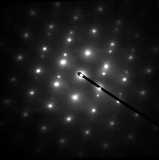
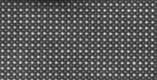
Electron diffraction is a generic term for phenomena associated with changes in the direction of electron beams due to elastic interactions with atoms. It occurs due to elastic scattering, when there is no change in the energy of the electrons. The negatively charged electrons are scattered due to Coulomb forces when they interact with both the positively charged atomic core and the negatively charged electrons around the atoms. The resulting map of the directions of the electrons far from the sample is called a diffraction pattern, see for instance Figure 1. Beyond patterns showing the directions of electrons, electron diffraction also plays a major role in the contrast of images in electron microscopes.

In many areas of science, Bragg's law, Wulff–Bragg's condition, or Laue–Bragg interference are a special case of Laue diffraction, giving the angles for coherent scattering of waves from a large crystal lattice. It describes how the superposition of wave fronts scattered by lattice planes leads to a strict relation between the wavelength and scattering angle. This law was initially formulated for X-rays, but it also applies to all types of matter waves including neutron and electron waves if there are a large number of atoms, as well as visible light with artificial periodic microscale lattices.

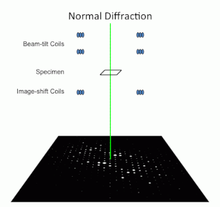
Reflection high-energy electron diffraction (RHEED) is a technique used to characterize the surface of crystalline materials. RHEED systems gather information only from the surface layer of the sample, which distinguishes RHEED from other materials characterization methods that also rely on diffraction of high-energy electrons. Transmission electron microscopy, another common electron diffraction method samples mainly the bulk of the sample due to the geometry of the system, although in special cases it can provide surface information. Low-energy electron diffraction (LEED) is also surface sensitive, but LEED achieves surface sensitivity through the use of low energy electrons.

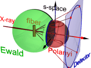
The Ewald sphere is a geometric construction used in electron, neutron, and x-ray diffraction which shows the relationship between:

<span class="mw-page-title-main">Powder diffraction</span> Experimental method in X-ray diffraction

Powder diffraction is a scientific technique using X-ray, neutron, or electron diffraction on powder or microcrystalline samples for structural characterization of materials. An instrument dedicated to performing such powder measurements is called a powder diffractometer.

X-ray crystal truncation rod scattering is a powerful method in surface science, based on analysis of surface X-ray diffraction (SXRD) patterns from a crystalline surface.

<span class="mw-page-title-main">Low-energy electron diffraction</span> Technique for determining surface structures

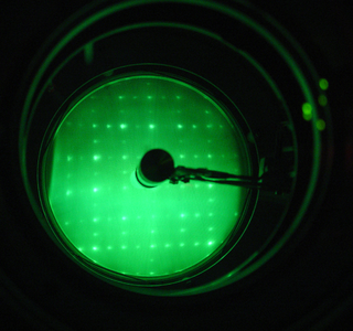
Low-energy electron diffraction (LEED) is a technique for the determination of the surface structure of single-crystalline materials by bombardment with a collimated beam of low-energy electrons (30–200 eV) and observation of diffracted electrons as spots on a fluorescent screen.

In condensed matter physics and crystallography, the static structure factor is a mathematical description of how a material scatters incident radiation. The structure factor is a critical tool in the interpretation of scattering patterns obtained in X-ray, electron and neutron diffraction experiments.

Diffraction topography is a imaging technique based on Bragg diffraction. Diffraction topographic images ("topographies") record the intensity profile of a beam of X-rays diffracted by a crystal. A topography thus represents a two-dimensional spatial intensity mapping of reflected X-rays, i.e. the spatial fine structure of a Laue reflection. This intensity mapping reflects the distribution of scattering power inside the crystal; topographs therefore reveal the irregularities in a non-ideal crystal lattice. X-ray diffraction topography is one variant of X-ray imaging, making use of diffraction contrast rather than absorption contrast which is usually used in radiography and computed tomography (CT). Topography is exploited to a lesser extends with neutrons, and has similarities to dark field imaging in the electron microscope community.

The International Union of Crystallography (IUCr) is an organisation devoted to the international promotion and coordination of the science of crystallography. The IUCr is a member of the International Council for Science (ICSU).

Helium atom scattering (HAS) is a surface analysis technique used in materials science. It provides information about the surface structure and lattice dynamics of a material by measuring the diffracted atoms from a monochromatic helium beam incident on the sample.

Single-wavelength anomalous diffraction (SAD) is a technique used in X-ray crystallography that facilitates the determination of the structure of proteins or other biological macromolecules by allowing the solution of the phase problem. In contrast to multi-wavelength anomalous diffraction (MAD), SAD uses a single dataset at a single appropriate wavelength.

A crystallographic database is a database specifically designed to store information about the structure of molecules and crystals. Crystals are solids having, in all three dimensions of space, a regularly repeating arrangement of atoms, ions, or molecules. They are characterized by symmetry, morphology, and directionally dependent physical properties. A crystal structure describes the arrangement of atoms, ions, or molecules in a crystal.

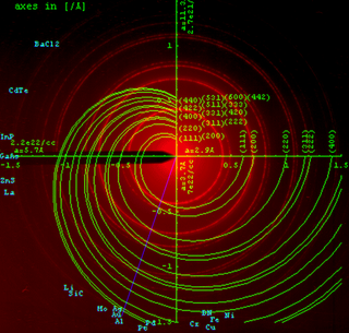
A Bernal chart in crystallography, is a chart used for indexing X-ray diffraction photographs from single crystals. From such a chart may be read the axial and radial cylindrical coordinates of that point in reciprocal space which corresponds to any particular X-ray reflection.

<span class="mw-page-title-main">Zone axis</span> High symmetry orientation of a crystal

Zone axis, a term sometimes used to refer to "high-symmetry" orientations in a crystal, most generally refers to any direction referenced to the direct lattice of a crystal in three dimensions. It is therefore indexed with direct lattice indices, instead of with Miller indices.

<span class="mw-page-title-main">Precession electron diffraction</span> Averaging technique for electron diffraction

Precession electron diffraction (PED) is a specialized method to collect electron diffraction patterns in a transmission electron microscope (TEM). By rotating (precessing) a tilted incident electron beam around the central axis of the microscope, a PED pattern is formed by integration over a collection of diffraction conditions. This produces a quasi-kinematical diffraction pattern that is more suitable as input into direct methods algorithms to determine the crystal structure of the sample.

References

Text books