Electron crystallography

Last updated

Electron crystallography is a subset of methods in electron diffraction focusing just upon detailed determination of the positions of atoms in solids using a transmission electron microscope (TEM). It can involve the use of high-resolution transmission electron microscopy images, electron diffraction patterns including convergent-beam electron diffraction or combinations of these. It has been successful in determining some bulk structures, and also surface structures. [1] [2] Two related methods are low-energy electron diffraction which has solved the structure of many surfaces, and reflection high-energy electron diffraction which is used to monitor surfaces often during growth.

Contents

History

The general approach dates back to the work in 1924 of Louis de Broglie in his PhD thesis Recherches sur la théorie des quanta [3] where he introduced the concept of electrons as matter waves. The wave nature was experimentally confirmed for electron beams in the work of two groups, the first the Davisson–Germer experiment, [4] [5] [6] [7] the other by George Paget Thomson and Alexander Reid. [8] Alexander Reid, who was Thomson's graduate student, performed the first experiments, [9] but he died soon after in a motorcycle accident. [10] These experiments were rapidly followed by the first non-relativistic diffraction model for electrons by Hans Bethe [11] based upon the Schrödinger equation, [12] which is very close to how electron diffraction is now described. Significantly, Clinton Davisson and Lester Germer noticed [6] [7] that their results could not be interpreted using a Bragg's law approach as the positions were systematically different; the approach of Hans Bethe [11] which includes both multiple scattering and the refraction due to the average potential yielded more accurate results. Very quickly there were multiple advances, for instance Seishi Kikuchi's observations of lines that can be used for crystallographic indexing due to combined elastic and inelastic scattering, [13] gas electron diffraction developed by Herman Mark and Raymond Weil, [14] [15] diffraction in liquids by Louis Maxwell, [16] and the first electron microscopes developed by Max Knoll and Ernst Ruska. [17] [18]

Despite early successes such as the determination of the positions of hydrogen atoms in NH4Cl crystals by W. E. Laschkarew and I. D. Usykin in 1933, [19] boric acid by John M. Cowley in 1953 [20] and orthoboric acid by William Houlder Zachariasen in 1954, [21] electron diffraction for many years was a qualitative technique used to check samples within electron microscopes. John M Cowley explains in a 1968 paper: [22]

Thus was founded the belief, amounting in some cases almost to an article of faith, and persisting even to the present day, that it is impossible to interpret the intensities of electron diffraction patterns to gain structural information.

This has slowly changed. One key step was the development in 1936 by Walther Kossel and Gottfried Möllenstedt of convergent beam electron diffraction (CBED), [23] This approach was extended by Peter Goodman and Gunter Lehmpfuhl, [24] then mainly by the groups of John Steeds [25] [26] [27] and Michiyoshi Tanaka [28] [29] who showed how to use CBED patterns to determine point groups and space groups. This was combined with other transmission electron microscopy approaches, typically where both local microstructure and atomic structure was of importance.

A second key set of work was that by the group of Boris Vainshtein who demonstrated solving the structure of many differnt materials such as clays and micas using powder diffraction patterns, a success attributed to the samples being relatively thin. [30] (Since the advent of precession electron diffraction [31] it has become clear that averaging over many different electron beam directions and thicknesses significantly reduces dynamical diffraction effects, [32] [33] so was probably also important.)

More complete crystallographic analysis of intensity data was slow to develop. One of the key steps was the demonstration in 1976 by Douglas L. Dorset and Herbert A. Hauptman that conventional direct methods could with care be used. [34] Another was the demonstration in 1986 that a Patterson function could be powerful in the seminal solution of the silicon (111) 7x7 reconstructed surface by Kunio Takanayagi using ultra-high vacuum electron diffraction. [35] [36] More complete analyses were the demonstration that classical inversion methods could be used for surfaces in 1997 by Dorset and Laurence D. Marks, and in 1998 by Jon Gjønnes who combined three-dimensional electron diffraction with precession electron diffraction and direct methods to solve an intermetallic, also using dynamical refinements. [37]

At the same time as approaches to invert diffraction data using electrons were established, the resolution of electron microscopes became good enough that images could be combined with diffraction information. At first resolution was poor, with in 1956 James Menter publishing the first electron microscope images showing the lattice structure of a material at 1.2nm resolution. [38] In 1968 Aaron Klug and David DeRosier used electron microscopy to visualise the structure of the tail of bacteriophage T4, a common virus, a key step in the use of electrons for macromolecular structure determination. [39] The first quantitative matching of atomic scale images and dynamical simulations was published in 1972 by J. G. Allpress, E. A. Hewat, A. F. Moodie and J. V. Sanders. [40] In the early 1980's the resolution of electron microscopes was now sufficient to resolve the atomic structure of materials, for instance with the 600 kV instrument led by Vernon Cosslett, [41] so combinations of high-resolution transmission electron microscopy and diffraction became standard across many areas of science. [42] Most of the research published using these approaches is described as electron microscopy, without the addition of the term electron crystallography.

Comparison with X-ray crystallography

It can complement X-ray crystallography for studies of very small crystals (<0.1 micrometers), both inorganic, organic, and proteins, such as membrane proteins, that cannot easily form the large 3-dimensional crystals required for that process. Protein structures are usually determined from either 2-dimensional crystals (sheets or helices), polyhedrons such as viral capsids, or dispersed individual proteins. Electrons can be used in these situations, whereas X-rays cannot, because electrons interact more strongly with atoms than X-rays do. Thus, X-rays will travel through a thin 2-dimensional crystal without diffracting significantly, whereas electrons can be used to form an image. Conversely, the strong interaction between electrons and protons makes thick (e.g. 3-dimensional > 1 micrometer) crystals impervious to electrons, which only penetrate short distances.

One of the main difficulties in X-ray crystallography is determining phases in the diffraction pattern. Because of the complexity of X-ray lenses, it is difficult to form an image of the crystal being diffracted, and hence phase information is lost. Fortunately, electron microscopes can resolve atomic structure in real space and the crystallographic structure factor phase information can be experimentally determined from an image's Fourier transform. The Fourier transform of an atomic resolution image is similar, but different, to a diffraction pattern—with reciprocal lattice spots reflecting the symmetry and spacing of a crystal. [43] Aaron Klug was the first to realize that the phase information could be read out directly from the Fourier transform of an electron microscopy image that had been scanned into a computer, already in 1968. For this, and his studies on virus structures and transfer-RNA, Klug received the Nobel Prize for chemistry in 1982.

Radiation damage

A common problem to X-ray crystallography and electron crystallography is radiation damage, by which especially organic molecules and proteins are damaged as they are being imaged, limiting the resolution that can be obtained. This is especially troublesome in the setting of electron crystallography, where that radiation damage is focused on far fewer atoms. One technique used to limit radiation damage is electron cryomicroscopy, in which the samples undergo cryofixation and imaging takes place at liquid nitrogen or even liquid helium temperatures. Because of this problem, X-ray crystallography has been much more successful in determining the structure of proteins that are especially vulnerable to radiation damage. Radiation damage was recently investigated using MicroED [44] [45] of thin 3D crystals in a frozen hydrated state.

Protein structures determined by electron crystallography

The first electron crystallographic protein structure to achieve atomic resolution was bacteriorhodopsin, determined by Richard Henderson and coworkers at the Medical Research Council Laboratory of Molecular Biology in 1990. [46] However, already in 1975 Unwin and Henderson had determined the first membrane protein structure at intermediate resolution (7 Ångström), showing for the first time the internal structure of a membrane protein, with its alpha-helices standing perpendicular to the plane of the membrane. Since then, several other high-resolution structures have been determined by electron crystallography, including the light-harvesting complex, [47] the nicotinic acetylcholine receptor, [48] and the bacterial flagellum. [49] The highest resolution protein structure solved by electron crystallography of 2D crystals is that of the water channel aquaporin-0. [50] In 2012, Jan Pieter Abrahams and coworkers extended electron crystallography to 3D protein nanocrystals [51] by rotation electron diffraction (RED). [52]

Electron microscopy image of an inorganic tantalum oxide, with its Fourier transform, inset. Notice how the appearance changes from the upper thin region to the thicker lower region. The unit cell of this compound is about 15 by 25 Angstrom. It is outlined at the centre of the figure, inside the result from image processing, where the symmetry has been taken into account. The black dots show clearly all the tantalum atoms. The diffraction extends to 6 orders along the 15 A direction and 10 orders in the perpendicular direction. Thus the resolution of the EM image is 2.5 A (15/6 or 25/10). This calculated Fourier transform contain both amplitudes (as seen) and phases (not displayed). Tantalum oxide EM image.jpg
Electron microscopy image of an inorganic tantalum oxide, with its Fourier transform, inset. Notice how the appearance changes from the upper thin region to the thicker lower region. The unit cell of this compound is about 15 by 25 Ångström. It is outlined at the centre of the figure, inside the result from image processing, where the symmetry has been taken into account. The black dots show clearly all the tantalum atoms. The diffraction extends to 6 orders along the 15 Å direction and 10 orders in the perpendicular direction. Thus the resolution of the EM image is 2.5 Å (15/6 or 25/10). This calculated Fourier transform contain both amplitudes (as seen) and phases (not displayed).
Electron diffraction pattern of the same crystal of inorganic tantalum oxide shown above. Notice that there are many more diffraction spots here than in the diffractogram calculated from the EM image above. The diffraction extends to 12 orders along the 15 A direction and 20 orders in the perpendicular direction. Thus the resolution of the ED pattern is 1.25 A (15/12 or 25/20). ED patterns do not contain phase information, but the clear differences between intensities of the diffraction spots can be used in crystal structure determination. Tant-ED.jpg
Electron diffraction pattern of the same crystal of inorganic tantalum oxide shown above. Notice that there are many more diffraction spots here than in the diffractogram calculated from the EM image above. The diffraction extends to 12 orders along the 15 Å direction and 20 orders in the perpendicular direction. Thus the resolution of the ED pattern is 1.25 Å (15/12 or 25/20). ED patterns do not contain phase information, but the clear differences between intensities of the diffraction spots can be used in crystal structure determination.

Application to inorganic materials

Electron crystallographic studies on inorganic crystals using high-resolution electron microscopy (HREM) images were first performed by Aaron Klug in 1978 [53] and by Sven Hovmöller and coworkers in 1984. [54] HREM images were used because they allow to select (by computer software) only the very thin regions close to the edge of the crystal for structure analysis (see also crystallographic image processing). This is of crucial importance since in the thicker parts of the crystal the exit-wave function (which carries the information about the intensity and position of the projected atom columns) is no longer linearly related to the projected crystal structure. Moreover, not only do the HREM images change their appearance with increasing crystal thickness, they are also very sensitive to the chosen setting of the defocus Δf of the objective lens (see the HREM images of GaN for example). To cope with this complexity methods based upon the Cowley-Moodie multislice algorithm [55] [56] and non-linear imaging theory [57] have been developed to simulate images; this only became possible [58] once the FFT method was developed. [59]

In addition to electron microscopy images, it is also possible to use electron diffraction (ED) patterns for crystal structure determination. [60] [61] The utmost care must be taken to record such ED patterns from the thinnest areas in order to keep most of the structure related intensity differences between the reflections (quasi-kinematical diffraction conditions). Just as with X-ray diffraction patterns, the important crystallographic structure factor phases are lost in electron diffraction patterns and must be uncovered by special crystallographic methods such as direct methods, maximum likelihood or (more recently) by the charge-flipping method. On the other hand, ED patterns of inorganic crystals have often a high resolution (= interplanar spacings with high Miller indices) much below 1 Ångström. This is comparable to the point resolution of the best electron microscopes. Under favourable conditions it is possible to use ED patterns from a single orientation to determine the complete crystal structure. [62] Alternatively a hybrid approach can be used which uses HRTEM images for solving and intensities from ED for refining the crystal structure. [63] [64]

Recent progress for structure analysis by ED was made by introducing the Vincent-Midgley [65] precession technique for recording electron diffraction patterns. [66] The thereby obtained intensities are usually much closer to the kinematical intensities, [67] [68] so that even structures can be determined that are out of range when processing conventional (selected area) electron diffraction data. [69] [70]

Crystal structures determined via electron crystallography can be checked for their quality by using first-principles calculations within density functional theory (DFT). This approach has been used to assist in solving surface structures [71] and for the validation of several metal-rich structures which were only accessible by HRTEM and ED, respectively. [72] [73]

Recently, two very complicated zeolite structures have been determined by electron crystallography combined with X-ray powder diffraction. [74] [75] These are more complex than the most complex zeolite structures determined by X-ray crystallography.

Related Research Articles

<span class="mw-page-title-main">Crystallography</span> Scientific study of crystal structures

Crystallography is the experimental science of determining the arrangement of atoms in crystalline solids. Crystallography is a fundamental subject in the fields of materials science and solid-state physics. The word crystallography is derived from the Ancient Greek word κρύσταλλος, and γράφειν. In July 2012, the United Nations recognised the importance of the science of crystallography by proclaiming that 2014 the International Year of Crystallography.

<span class="mw-page-title-main">X-ray crystallography</span> Technique used for determining crystal structures and identifying mineral compounds

X-ray crystallography is the experimental science of determining the atomic and molecular structure of a crystal, in which the crystalline structure causes a beam of incident X-rays to diffract in specific directions. By measuring the angles and intensities of the X-ray diffraction, a crystallographer can produce a three-dimensional picture of the density of electrons within the crystal and the positions of the atoms, as well as their chemical bonds, crystallographic disorder, and other information.

<span class="mw-page-title-main">Electron diffraction</span> Bending of electron beams due to electrostatic interactions with matter

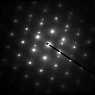
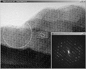
Electron diffraction is a generic term for phenomena associated with changes in the direction of electron beams due to elastic interactions with atoms. It occurs due to elastic scattering, when there is no change in the energy of the electrons. The negatively charged electrons are scattered due to Coulomb forces when they interact with both the positively charged atomic core and the negatively charged electrons around the atoms. The resulting map of the directions of the electrons far from the sample is called a diffraction pattern, see for instance Figure 1. Beyond patterns showing the directions of electrons, electron diffraction also plays a major role in the contrast of images in electron microscopes.

In physics, the phase problem is the problem of loss of information concerning the phase that can occur when making a physical measurement. The name comes from the field of X-ray crystallography, where the phase problem has to be solved for the determination of a structure from diffraction data. The phase problem is also met in the fields of imaging and signal processing. Various approaches of phase retrieval have been developed over the years.

<span class="mw-page-title-main">John M. Cowley</span> Australian physicist (1923–2004)

John Maxwell Cowley was an American Regents Professor at Arizona State University. The John M. Cowley Center for High-Resolution Electron Microscopy at Arizona State is named in his honor.

John Cowley was an extraordinarily productive scientist over more than five decades. He made pioneering contributions in the fields of electron microscopy, diffraction and crystallography, all of which brought him widespread recognition. He received the highest awards of the International Union of Crystallography, the Electron Microscopy Society of America and the American Crystallographic Society, and he was honored by election to Fellowship of the Australian Academy of Science, The Royal Society of London, and the American Physical Society. His monograph Diffraction Physics remains the standard reference in the field. His ideas, enthusiasm and basic understanding of electron optics and diffraction phenomena provided a valued source of leadership to many generations of students and co-workers, and he was universally admired by his peers and colleagues as a great and inspiring scientist.

A crystallographic database is a database specifically designed to store information about the structure of molecules and crystals. Crystals are solids having, in all three dimensions of space, a regularly repeating arrangement of atoms, ions, or molecules. They are characterized by symmetry, morphology, and directionally dependent physical properties. A crystal structure describes the arrangement of atoms, ions, or molecules in a crystal.

Eleanor Joy Dodson FRS is an Australian-born biologist who specialises in the computational modelling of protein crystallography. She holds a chair in the Department of Chemistry at the University of York. She is the widow of the scientist Guy Dodson.

<span class="mw-page-title-main">Crystallographic image processing</span>

Crystallographic image processing (CIP) is traditionally understood as being a set of key steps in the determination of the atomic structure of crystalline matter from high-resolution electron microscopy (HREM) images obtained in a transmission electron microscope (TEM) that is run in the parallel illumination mode. The term was created in the research group of Sven Hovmöller at Stockholm University during the early 1980s and became rapidly a label for the "3D crystal structure from 2D transmission/projection images" approach. Since the late 1990s, analogous and complementary image processing techniques that are directed towards the achieving of goals with are either complementary or entirely beyond the scope of the original inception of CIP have been developed independently by members of the computational symmetry/geometry, scanning transmission electron microscopy, scanning probe microscopy communities, and applied crystallography communities.

<span class="mw-page-title-main">Structure validation</span> Process of evaluating 3-dimensional atomic models of biomacromolecules

Macromolecular structure validation is the process of evaluating reliability for 3-dimensional atomic models of large biological molecules such as proteins and nucleic acids. These models, which provide 3D coordinates for each atom in the molecule, come from structural biology experiments such as x-ray crystallography or nuclear magnetic resonance (NMR). The validation has three aspects: 1) checking on the validity of the thousands to millions of measurements in the experiment; 2) checking how consistent the atomic model is with those experimental data; and 3) checking consistency of the model with known physical and chemical properties.

<span class="mw-page-title-main">Randy Read</span> Canadian-British scientist (1957–)

Randy John Read is a Wellcome Trust Principal Research Fellow and professor of protein crystallography at the University of Cambridge.

<span class="mw-page-title-main">Precession electron diffraction</span> Averaging technique for electron diffraction

Precession electron diffraction (PED) is a specialized method to collect electron diffraction patterns in a transmission electron microscope (TEM). By rotating (precessing) a tilted incident electron beam around the central axis of the microscope, a PED pattern is formed by integration over a collection of diffraction conditions. This produces a quasi-kinematical diffraction pattern that is more suitable as input into direct methods algorithms to determine the crystal structure of the sample.

In crystallography, direct methods is a set of techniques used for structure determination using diffraction data and a priori information. It is a solution to the crystallographic phase problem, where phase information is lost during a diffraction measurement. Direct methods provides a method of estimating the phase information by establishing statistical relationships between the recorded amplitude information and phases of strong reflections.

<span class="mw-page-title-main">Cryogenic electron microscopy</span> Form of transmission electron microscopy (TEM)

Cryogenic electron microscopy (cryo-EM) is a cryomicroscopy technique applied on samples cooled to cryogenic temperatures. For biological specimens, the structure is preserved by embedding in an environment of vitreous ice. An aqueous sample solution is applied to a grid-mesh and plunge-frozen in liquid ethane or a mixture of liquid ethane and propane. While development of the technique began in the 1970s, recent advances in detector technology and software algorithms have allowed for the determination of biomolecular structures at near-atomic resolution. This has attracted wide attention to the approach as an alternative to X-ray crystallography or NMR spectroscopy for macromolecular structure determination without the need for crystallization.

Microcrystal electron diffraction, or MicroED, is a CryoEM method that was developed by the Gonen laboratory in late 2013 at the Janelia Research Campus of the Howard Hughes Medical Institute. MicroED is a form of electron crystallography where thin 3D crystals are used for structure determination by electron diffraction. Prior to this demonstration, macromolecular (protein) electron crystallography was mainly used on 2D crystals, for example. The method is one of several modern versions of approaches to determine atomic structures using electron diffraction first demonstrated for the positions of hydrogen atoms in NH4Cl crystals by W. E. Laschkarew and I. D. Usykin in 1933, which has since been used for surfaces, via precession electron diffraction, with much of the early work described in the work of Boris Vainshtein and Douglas L. Dorset.

This is a timeline of crystallography.

Joanne Etheridge is an Australian physicist. She is Director of the Monash Centre for Electron Microscopy and Professor in the Department of Materials Science and Engineering at Monash University.

<span class="mw-page-title-main">Convergent beam electron diffraction</span> Convergent beam electron diffraction technique

Convergent beam electron diffraction (CBED) is an electron diffraction technique where a convergent or divergent beam of electrons is used to study materials.

<span class="mw-page-title-main">CrysTBox</span> Free crystallographic software

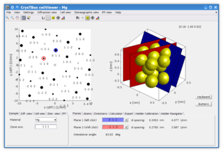
CrysTBox is a suite of computer tools designed to accelerate material research based on transmission electron microscope images via highly accurate automated analysis and interactive visualization. Relying on artificial intelligence and computer vision, CrysTBox makes routine crystallographic analyses simpler, faster and more accurate compared to human evaluators. The high level of automation together with sub-pixel precision and interactive visualization makes the quantitative crystallographic analysis accessible even for non-crystallographers allowing for an interdisciplinary research. Simultaneously, experienced material scientists can take advantage of advanced functionalities for comprehensive analyses.

4D scanning transmission electron microscopy is a subset of scanning transmission electron microscopy (STEM) which utilizes a pixelated electron detector to capture a convergent beam electron diffraction (CBED) pattern at each scan location. This technique captures a 2 dimensional reciprocal space image associated with each scan point as the beam rasters across a 2 dimensional region in real space, hence the name 4D STEM. Its development was enabled by evolution in STEM detectors and improvements computational power. The technique has applications in visual diffraction imaging, phase orientation and strain mapping, phase contrast analysis, among others.

Dark-field X-ray microscopy is an imaging technique used for multiscale structural characterisation. It is capable of mapping deeply embedded structural elements with nm-resolution using synchrotron X-ray diffraction-based imaging. The technique works by using scattered X-rays to create a high degree of contrast, and by measuring the intensity and spatial distribution of the diffracted beams, it is possible to obtain a three-dimensional map of the sample's structure, orientation, and local strain.

References

  1. Takayanagi, K.; Tanishiro, Y.; Takahashi, M.; Takahashi, S. (1985-05-01). "Structural analysis of Si(111)‐7×7 by UHV‐transmission electron diffraction and microscopy". Journal of Vacuum Science & Technology A. 3 (3): 1502–1506. Bibcode:1985JVSTA...3.1502T. doi:10.1116/1.573160. ISSN   0734-2101.
  2. Erdman, Natasha; Poeppelmeier, Kenneth R.; Asta, Mark; Warschkow, Oliver; Ellis, Donald E.; Marks, Laurence D. (2002). "The structure and chemistry of the TiO2-rich surface of SrTiO3 (001)". Nature. 419 (6902): 55–58. Bibcode:2002Natur.419...55E. doi:10.1038/nature01010. ISSN   0028-0836. PMID   12214229. S2CID   4384784.
  3. de Broglie, Louis Victor. "On the Theory of Quanta" (PDF). Foundation of Louis de Broglie (English translation by A.F. Kracklauer, 2004. ed.). Retrieved 25 February 2023.
  4. Davisson, C.; Germer, L. H. (1927). "The Scattering of Electrons by a Single Crystal of Nickel". Nature. 119 (2998): 558–560. Bibcode:1927Natur.119..558D. doi:10.1038/119558a0. ISSN   0028-0836. S2CID   4104602.
  5. Davisson, C.; Germer, L. H. (1927). "Diffraction of Electrons by a Crystal of Nickel". Physical Review. 30 (6): 705–740. Bibcode:1927PhRv...30..705D. doi: 10.1103/physrev.30.705 . ISSN   0031-899X.
  6. 1 2 Davisson, C. J.; Germer, L. H. (1928). "Reflection of Electrons by a Crystal of Nickel". Proceedings of the National Academy of Sciences. 14 (4): 317–322. Bibcode:1928PNAS...14..317D. doi: 10.1073/pnas.14.4.317 . ISSN   0027-8424. PMC   1085484 . PMID   16587341.
  7. 1 2 Davisson, C. J.; Germer, L. H. (1928). "Reflection and Refraction of Electrons by a Crystal of Nickel". Proceedings of the National Academy of Sciences. 14 (8): 619–627. Bibcode:1928PNAS...14..619D. doi: 10.1073/pnas.14.8.619 . ISSN   0027-8424. PMC   1085652 . PMID   16587378.
  8. Thomson, G. P.; Reid, A. (1927). "Diffraction of Cathode Rays by a Thin Film". Nature. 119 (3007): 890. Bibcode:1927Natur.119Q.890T. doi: 10.1038/119890a0 . ISSN   0028-0836. S2CID   4122313.
  9. Reid, Alexander (1928). "The diffraction of cathode rays by thin celluloid films". Proceedings of the Royal Society of London. Series A, Containing Papers of a Mathematical and Physical Character. 119 (783): 663–667. Bibcode:1928RSPSA.119..663R. doi: 10.1098/rspa.1928.0121 . ISSN   0950-1207. S2CID   98311959.
  10. Navarro, Jaume (2010). "Electron diffraction chez Thomson: early responses to quantum physics in Britain". The British Journal for the History of Science. 43 (2): 245–275. doi:10.1017/S0007087410000026. ISSN   0007-0874. S2CID   171025814.
  11. 1 2 Bethe, H. (1928). "Theorie der Beugung von Elektronen an Kristallen". Annalen der Physik (in German). 392 (17): 55–129. Bibcode:1928AnP...392...55B. doi:10.1002/andp.19283921704.
  12. Schrödinger, E. (1926). "An Undulatory Theory of the Mechanics of Atoms and Molecules". Physical Review. 28 (6): 1049–1070. Bibcode:1926PhRv...28.1049S. doi:10.1103/PhysRev.28.1049. ISSN   0031-899X.
  13. Kikuchi, Seishi (1928). "Diffraction of cathode rays by mica". Proceedings of the Imperial Academy. 4 (6): 271–274. doi: 10.2183/pjab1912.4.271 . S2CID   4121059 via Google Scholar.
  14. Mark, Herman; Wierl, Raymond (1930). "Neuere Ergebnisse der Elektronenbeugung". Die Naturwissenschaften. 18 (36): 778–786. Bibcode:1930NW.....18..778M. doi:10.1007/bf01497860. ISSN   0028-1042. S2CID   9815364.
  15. Mark, Herman; Wiel, Raymond (1930). "Die ermittlung von molekülstrukturen durch beugung von elektronen an einem dampfstrahl". Zeitschrift für Elektrochemie und angewandte physikalische Chemie. 36 (9): 675–676. doi:10.1002/bbpc.19300360921. S2CID   178706417.
  16. Maxwell, Louis R. (1933). "Electron Diffraction by Liquids". Physical Review. 44 (2): 73–76. Bibcode:1933PhRv...44...73M. doi:10.1103/PhysRev.44.73. ISSN   0031-899X.
  17. Knoll, M.; Ruska, E. (1932). "Beitrag zur geometrischen Elektronenoptik. I". Annalen der Physik. 404 (5): 607–640. Bibcode:1932AnP...404..607K. doi:10.1002/andp.19324040506. ISSN   0003-3804.
  18. Knoll, M.; Ruska, E. (1932). "Das Elektronenmikroskop". Zeitschrift für Physik (in German). 78 (5–6): 318–339. Bibcode:1932ZPhy...78..318K. doi:10.1007/BF01342199. ISSN   1434-6001. S2CID   186239132.
  19. Laschkarew, W. E.; Usyskin, I. D. (1933). "Die Bestimmung der Lage der Wasserstoffionen im NH4Cl-Kristallgitter durch Elektronenbeugung". Zeitschrift für Physik (in German). 85 (9–10): 618–630. Bibcode:1933ZPhy...85..618L. doi:10.1007/BF01331003. ISSN   1434-6001. S2CID   123199621.
  20. Cowley, J. M. (1953). "Structure analysis of single crystals by electron diffraction. II. Disordered boric acid structure". Acta Crystallographica. 6 (6): 522–529. Bibcode:1953AcCry...6..522C. doi: 10.1107/S0365110X53001423 . ISSN   0365-110X. S2CID   94391285.
  21. Zachariasen, W. H. (1954). "The precise structure of orthoboric acid". Acta Crystallographica. 7 (4): 305–310. Bibcode:1954AcCry...7..305Z. doi: 10.1107/S0365110X54000886 . ISSN   0365-110X.
  22. Cowley, J.M. (1968). "Crystal structure determination by electron diffraction". Progress in Materials Science. 13: 267–321. doi:10.1016/0079-6425(68)90023-6.
  23. Kossel, W.; Möllenstedt, G. (1939). "Elektroneninterferenzen im konvergenten Bündel". Annalen der Physik. 428 (2): 113–140. doi:10.1002/andp.19394280204. ISSN   0003-3804.
  24. Goodman, P.; Lehmpfuhl, G. (1968). "Observation of the breakdown of Friedel's law in electron diffraction and symmetry determination from zero-layer interactions". Acta Crystallographica Section A. 24 (3): 339–347. Bibcode:1968AcCrA..24..339G. doi:10.1107/S0567739468000677.
  25. Buxton, B. F.; Eades, J. A.; Steeds, John Wickham; Rackham, G. M.; Frank, Frederick Charles (1976). "The symmetry of electron diffraction zone axis patterns". Philosophical Transactions of the Royal Society of London. Series A, Mathematical and Physical Sciences. 281 (1301): 171–194. Bibcode:1976RSPTA.281..171B. doi:10.1098/rsta.1976.0024. S2CID   122890943.
  26. Steeds, J. W.; Vincent, R. (1983). "Use of high-symmetry zone axes in electron diffraction in determining crystal point and space groups". Journal of Applied Crystallography. 16 (3): 317–324. Bibcode:1983JApCr..16..317S. doi:10.1107/S002188988301050X. ISSN   0021-8898.
  27. Bird, D. M. (1989). "Theory of zone axis electron diffraction". Journal of Electron Microscopy Technique. 13 (2): 77–97. doi:10.1002/jemt.1060130202. ISSN   0741-0581. PMID   2681572.
  28. Tanaka, M.; Saito, R.; Sekii, H. (1983). "Point-group determination by convergent-beam electron diffraction". Acta Crystallographica Section A. 39 (3): 357–368. Bibcode:1983AcCrA..39..357T. doi:10.1107/S010876738300080X. ISSN   0108-7673.
  29. Tanaka, M.; Saito, R.; Watanabe, D. (1980). "Symmetry determination of the room-temperature form of LnNbO 4 (Ln = La,Nd) by convergent-beam electron diffraction". Acta Crystallographica Section A. 36 (3): 350–352. Bibcode:1980AcCrA..36..350T. doi:10.1107/S0567739480000800. ISSN   0567-7394. S2CID   98184340.
  30. Vaĭnshteĭn, B. K. (1964). Structure analysis by electron diffraction. Oxford: Pergamon Press. ISBN   978-0-08-010241-2. OCLC   681437461.
  31. Vincent, R.; Midgley, P.A. (1994). "Double conical beam-rocking system for measurement of integrated electron diffraction intensities". Ultramicroscopy. 53 (3): 271–282. doi:10.1016/0304-3991(94)90039-6. ISSN   0304-3991.
  32. Own, C. S.: PhD thesis, System Design and Verification of the Precession Electron Diffraction Technique, Northwestern University, 2005,http://www.numis.northwestern.edu/Research/Current/precession.shtml
  33. Own, C. S.; Marks, L. D.; Sinkler, W. (2006). "Precession electron diffraction 1: multislice simulation". Acta Crystallographica Section A Foundations of Crystallography. 62 (6): 434–443. doi:10.1107/S0108767306032892. ISSN   0108-7673.
  34. Dorset, Douglas L.; Hauptman, Herbert A. (1976). "Direct phase determination for quasi-kinematical electron diffraction intensity data from organic microcrystals". Ultramicroscopy. 1 (3–4): 195–201. doi:10.1016/0304-3991(76)90034-6. ISSN   0304-3991.
  35. Takayanagi, K.; Tanishiro, Y.; Takahashi, M.; Takahashi, S. (1985). "Structural analysis of Si(111)-7×7 by UHV-transmission electron diffraction and microscopy". Journal of Vacuum Science & Technology A: Vacuum, Surfaces, and Films. 3 (3): 1502–1506. Bibcode:1985JVSTA...3.1502T. doi:10.1116/1.573160. ISSN   0734-2101.
  36. Takayanagi, Kunio; Tanishiro, Yasumasa; Takahashi, Shigeki; Takahashi, Masaetsu (1985). "Structure analysis of Si(111)-7 × 7 reconstructed surface by transmission electron diffraction". Surface Science. 164 (2–3): 367–392. doi:10.1016/0039-6028(85)90753-8. ISSN   0039-6028.
  37. Gjønnes, J.; Hansen, V.; Berg, B. S.; Runde, P.; Cheng, Y. F.; Gjønnes, K.; Dorset, D. L.; Gilmore, C. J. (1998-05-01). "Structure Model for the Phase AlmFe Derived from Three-Dimensional Electron Diffraction Intensity Data Collected by a Precession Technique. Comparison with Convergent-Beam Diffraction". Acta Crystallographica Section A Foundations of Crystallography. 54 (3): 306–319. doi:10.1107/S0108767397017030.
  38. "The direct study by electron microscopy of crystal lattices and their imperfections". Proceedings of the Royal Society of London. Series A. Mathematical and Physical Sciences. 236 (1204): 119–135. 1956. doi:10.1098/rspa.1956.0117. ISSN   0080-4630.
  39. De Rosier, D. J.; Klug, A. (1968). "Reconstruction of Three Dimensional Structures from Electron Micrographs". Nature. 217 (5124): 130–134. Bibcode:1968Natur.217..130D. doi:10.1038/217130a0. PMID   23610788.
  40. Allpress, J. G.; Hewat, E. A.; Moodie, A. F.; Sanders, J. V. (1972). "n -Beam lattice images. I. Experimental and computed images from W 4 Nb 26 O 77". Acta Crystallographica Section A. 28 (6): 528–536. doi:10.1107/S0567739472001433. ISSN   0567-7394.
  41. "Principles and performance of a 600 kV high resolution electron microscope". Proceedings of the Royal Society of London. A. Mathematical and Physical Sciences. 370 (1740): 1–16. 1980-03-12. doi:10.1098/rspa.1980.0018. ISSN   0080-4630.
  42. Buseck, Peter; Cowley, John M; Eyring, Leyroy (1992). High-resolution Transmission Electron Microscopy and Associated Techniques. Oxford University Press.
  43. R Hovden; Y Jiang; HL Xin; LF Kourkoutis (2015). "Periodic Artifact Reduction in Fourier Transforms of Full Field Atomic Resolution Images". Microscopy and Microanalysis. 21 (2): 436–441. arXiv: 2210.09024 . Bibcode:2015MiMic..21..436H. doi:10.1017/S1431927614014639. PMID   25597865. S2CID   22435248.
  44. Nannenga, Brent L; Shi, Dan; Leslie, Andrew G W; Gonen, Tamir (2014-08-03). "High-resolution structure determination by continuous-rotation data collection in MicroED". Nature Methods. 11 (9): 927–930. doi:10.1038/nmeth.3043. ISSN   1548-7091. PMC   4149488 . PMID   25086503.
  45. Hattne, Johan; Shi, Dan; Glynn, Calina; Zee, Chih-Te; Gallagher-Jones, Marcus; Martynowycz, Michael W.; Rodriguez, Jose A.; Gonen, Tamir (2018). "Analysis of Global and Site-Specific Radiation Damage in Cryo-EM". Structure. 26 (5): 759–766.e4. doi:10.1016/j.str.2018.03.021. ISSN   0969-2126. PMC   6333475 . PMID   29706530.
  46. Henderson, R.; Baldwin, J.M.; Ceska, T.A.; Zemlin, F; Beckmann, E.; Downing, K.H. (June 1990). "Model for the structure of bacteriorhodopsin based on high-resolution electron cryo-microscopy". J Mol Biol. 213 (4): 899–929. doi:10.1016/S0022-2836(05)80271-2. PMID   2359127.
  47. Kühlbrandt, Werner; Wang, Da Neng; Fujiyoshi, Yoshinori (February 1994). "Atomic model of plant light-harvesting complex by electron crystallography". Nature. 367 (6464): 614–21. Bibcode:1994Natur.367..614K. doi:10.1038/367614a0. PMID   8107845. S2CID   4357116.
  48. Miyazawa, Atsuo; Fujiyoshi, Yoshinori; Unwin, Nigel (June 2003). "Structure and gating mechanism of the acetylcholine receptor pore". Nature. 423 (6943): 949–55. Bibcode:2003Natur.423..949M. doi:10.1038/nature01748. PMID   12827192. S2CID   205209809.
  49. Yonekura, Koji; Maki-Yonekura, Saori; Namba, Keiichi (August 2003). "Complete atomic model of the bacterial flagellar filament by electron cryomicroscopy". Nature. 424 (6949): 643–50. Bibcode:2003Natur.424..643Y. doi:10.1038/nature01830. PMID   12904785. S2CID   4301660.
  50. Gonen, Tamir; Cheng, Yifan; Sliz, Piotr; Hiroaki, Yoko; Fujiyoshi, Yoshinori; Harrison, Stephen C.; Walz, Thomas (2005). "Lipid–protein interactions in double-layered two-dimensional AQP0 crystals". Nature. 438 (7068): 633–638. Bibcode:2005Natur.438..633G. doi:10.1038/nature04321. ISSN   0028-0836. PMC   1350984 . PMID   16319884.
  51. Nederlof, I.; van Genderen, E.; Li, Y.-W.; Abrahams, J. P. (2013-07-01). "A Medipix quantum area detector allows rotation electron diffraction data collection from submicrometre three-dimensional protein crystals". Acta Crystallographica Section D: Biological Crystallography. 69 (7): 1223–1230. doi:10.1107/S0907444913009700. ISSN   0907-4449. PMC   3689525 . PMID   23793148.
  52. Zhang, Daliang; Oleynikov, Peter; Hovmöller, Sven; Zou, Xiaodong (March 2010). "Collecting 3D electron diffraction data by the rotation method". Zeitschrift für Kristallographie. 225 (2–3): 94–102. Bibcode:2010ZK....225...94Z. doi: 10.1524/zkri.2010.1202 . ISSN   0044-2968. S2CID   55751260.
  53. Klug, A (1978/79) Image Analysis and Reconstruction in the Electron Microscopy of Biological Macromolecules Chemica Scripta vol 14, p. 245-256.
  54. Hovmöller, Sven; Sjögren, Agneta; Farrants, George; Sundberg, Margareta; Marinder, Bengt-Olov (1984). "Accurate atomic positions from electron microscopy". Nature. 311 (5983): 238. Bibcode:1984Natur.311..238H. doi:10.1038/311238a0.
  55. Cowley, J. M.; Moodie, A. F. (1957-10-01). "The scattering of electrons by atoms and crystals. I. A new theoretical approach". Acta Crystallographica. 10 (10): 609–619. doi:10.1107/S0365110X57002194. ISSN   0365-110X.
  56. Ishizuka, Kazuo (2004). "FFT Multislice Method—The Silver Anniversary". Microscopy and Microanalysis. 10 (1): 34–40. Bibcode:2004MiMic..10...34I. doi:10.1017/S1431927604040292. ISSN   1431-9276. PMID   15306065. S2CID   8016041.
  57. Ishizuka, Kazuo (1980). "Contrast transfer of crystal images in TEM". Ultramicroscopy. 5 (1–3): 55–65. doi:10.1016/0304-3991(80)90011-X.
  58. Goodman, P.; Moodie, A. F. (1974-03-01). "Numerical evaluations of N -beam wave functions in electron scattering by the multi-slice method". Acta Crystallographica A. 30 (2): 280–290. Bibcode:1974AcCrA..30..280G. doi:10.1107/S056773947400057X. ISSN   0567-7394.
  59. Cooley, James W.; Tukey, John W. (1965). "An algorithm for the machine calculation of complex Fourier series". Mathematics of Computation. 19 (90): 297–301. doi: 10.1090/S0025-5718-1965-0178586-1 . ISSN   0025-5718.
  60. B. K. Vainshtein (1964), Structure Analysis by Electron Diffraction, Pergamon Press Oxford
  61. D. L. Dorset (1995), Structural Electron Crystallography, Plenum Publishing Corporation ISBN   0-306-45049-6
  62. Weirich, TE; Zou, X; Ramlau, R; Simon, A; Cascarano, GL; Giacovazzo, C; Hovmöller, S (2000). "Structures of nanometre-size crystals determined from selected-area electron diffraction data". Acta Crystallographica A. 56 (Pt 1): 29–35. doi:10.1107/S0108767399009605. PMID   10874414.
  63. Zandbergen, H. W. (1997). "Structure Determination of Mg5Si6 Particles in Al by Dynamic Electron Diffraction Studies". Science. 277 (5330): 1221–1225. doi:10.1126/science.277.5330.1221.
  64. Weirich, Thomas E.; Ramlau, Reiner; Simon, Arndt; Hovmöller, Sven; Zou, Xiaodong (1996). "A crystal structure determined with 0.02 Å accuracy by electron microscopy". Nature. 382 (6587): 144. Bibcode:1996Natur.382..144W. doi:10.1038/382144a0. S2CID   4327149.
  65. Vincent, R.; Midgley, P. A. (1994-03-01). "Double conical beam-rocking system for measurement of integrated electron diffraction intensities". Ultramicroscopy. 53 (3): 271–282. doi:10.1016/0304-3991(94)90039-6. ISSN   0304-3991.
  66. Precession Electron Diffraction
  67. Marks, L.D.; Sinkler, W. (2003). "Sufficient Conditions for Direct Methods with Swift Electrons". Microscopy and Microanalysis. 9 (5): 399–410. Bibcode:2003MiMic...9..399M. doi:10.1017/S1431927603030332. ISSN   1431-9276. PMID   19771696. S2CID   20112743.
  68. Own, C. S.; Marks, L. D.; Sinkler, W. (2006-11-01). "Precession electron diffraction 1: multislice simulation". Acta Crystallographica A. 62 (6): 434–443. doi:10.1107/S0108767306032892. ISSN   0108-7673. PMID   17057352.
  69. Gemmi, M; Zou, X; Hovmöller, S; Migliori, A; Vennström, M; Andersson, Y (2003). "Structure of Ti2P solved by three-dimensional electron diffraction data collected with the precession technique and high-resolution electron microscopy". Acta Crystallographica. 59 (Pt 2): 117–26. doi:10.1107/S0108767302022559. PMID   12604849.
  70. Weirich, T; Portillo, J; Cox, G; Hibst, H; Nicolopoulos, S (2006). "Ab initio determination of the framework structure of the heavy-metal oxide CsxNb2.54W2.46O14 from 100kV precession electron diffraction data". Ultramicroscopy. 106 (3): 164–75. doi:10.1016/j.ultramic.2005.07.002. PMID   16137828.
  71. Erdman, Natasha; Poeppelmeier, Kenneth R.; Asta, Mark; Warschkow, Oliver; Ellis, Donald E.; Marks, Laurence D. (2002). "The structure and chemistry of the TiO2-rich surface of SrTiO3 (001)". Nature. 419 (6902): 55–58. Bibcode:2002Natur.419...55E. doi:10.1038/nature01010. ISSN   0028-0836. PMID   12214229. S2CID   4384784.
  72. Albe, K; Weirich, TE (2003). "Structure and stability of alpha- and beta-Ti2Se. Electron diffraction versus density-functional theory calculations". Acta Crystallographica A. 59 (Pt 1): 18–21. doi:10.1107/S0108767302018275. PMID   12496457.
  73. Weirich, TE (2004). "First-principles calculations as a tool for structure validation in electron crystallography". Acta Crystallographica A. 60 (Pt 1): 75–81. Bibcode:2004AcCrA..60...75W. doi:10.1107/S0108767303025042. PMID   14691330.
  74. Gramm, Fabian; Baerlocher, Christian; McCusker, Lynne B.; Warrender, Stewart J.; Wright, Paul A.; Han, Bada; Hong, Suk Bong; Liu, Zheng; et al. (2006). "Complex zeolite structure solved by combining powder diffraction and electron microscopy". Nature. 444 (7115): 79–81. Bibcode:2006Natur.444...79G. doi:10.1038/nature05200. PMID   17080087. S2CID   4396820.
  75. Baerlocher, C.; Gramm, F.; Massuger, L.; McCusker, L. B.; He, Z.; Hovmoller, S.; Zou, X. (2007). "Structure of the Polycrystalline Zeolite Catalyst IM-5 Solved by Enhanced Charge Flipping". Science. 315 (5815): 1113–6. Bibcode:2007Sci...315.1113B. doi:10.1126/science.1137920. PMID   17322057. S2CID   19509220.

Further reading