Radioactivity in the life sciences

Last updated

Radioactivity is generally used in life sciences for highly sensitive and direct measurements of biological phenomena, and for visualizing the location of biomolecules radiolabelled with a radioisotope.

Contents

All atoms exist as stable or unstable isotopes and the latter decay at a given half-life ranging from attoseconds to billions of years; radioisotopes useful to biological and experimental systems have half-lives ranging from minutes to months. In the case of the hydrogen isotope tritium (half-life = 12.3 years) and carbon-14 (half-life = 5,730 years), these isotopes derive their importance from all organic life containing hydrogen and carbon and therefore can be used to study countless living processes, reactions, and phenomena. Most short lived isotopes are produced in cyclotrons, linear particle accelerators, or nuclear reactors and their relatively short half-lives give them high maximum theoretical specific activities which is useful for detection in biological systems.

DOTA linked to the monoclonal antibody tacatuzumab and chelating yttrium-90 Y-90 tacatuzumab tetraxetan structure.svg
DOTA linked to the monoclonal antibody tacatuzumab and chelating yttrium-90
Whole-body PET scan using F-FDG showing intestinal tumors and non-specific accumulation in bladder PET-MIPS-anim.gif
Whole-body PET scan using F-FDG showing intestinal tumors and non-specific accumulation in bladder

Radiolabeling is a technique used to track the passage of a molecule that incorporates a radioisotope through a reaction, metabolic pathway, cell, tissue, organism, or biological system. The reactant is 'labeled' by replacing specific atoms by their isotope. Replacing an atom with its own radioisotope is an intrinsic label that does not alter the structure of the molecule. Alternatively, molecules can be radiolabeled by chemical reactions that introduce an atom, moiety, or functional group that contains a radionuclide. For example, radio-iodination of peptides and proteins with biologically useful iodine isotopes is easily done by an oxidation reaction that replaces the hydroxyl group with iodine on tyrosine and histadine residues. Another example is to use chelators such DOTA that can be chemically coupled to a protein; the chelator in turn traps radiometals thus radiolabeling the protein. This has been used for introducing Yttrium-90 onto a monoclonal antibody for therapeutic purposes and for introducing Gallium-68 onto the peptide Octreotide for diagnostic imaging by PET imaging. [1] (See DOTA uses.)

Radiolabeling is not necessary for some applications. For some purposes, soluble ionic salts can be used directly without further modification (e.g., gallium-67, gallium-68, and radioiodine isotopes). These uses rely on the chemical and biological properties of the radioisotope itself, to localize it within the organism or biological system.

Molecular imaging is the biomedical field that employs radiotracers to visualize and quantify biological processes using positron emission tomography (PET) and single-photon emission computed tomography (SPECT) imaging. Again, a key feature of using radioactivity in life science applications is that it is a quantitative technique, so PET/SPECT not only reveals where a radiolabelled molecule is but how much is there.

Radiobiology (also known as radiation biology) is a field of clinical and basic medical sciences that involves the study of the action of radioactivity on biological systems. The controlled action of deleterious radioactivity on living systems is the basis of radiation therapy.

Examples of biologically useful radionuclides

Hydrogen

Tritium (hydrogen-3) is a very low beta energy emitter that can be used to label proteins, nucleic acids, drugs and almost any organic biomolecule. The maximum theoretical specific activity of tritium is 28.8  kCi / mol (1,070  TBq /mol). [2] However, there is often more than one tritium atom per molecule: for example, tritiated UTP is sold by most suppliers with carbons 5 and 6 each bonded to a tritium atom.

For tritium detection, liquid scintillation counters have been classically employed, in which the energy of a tritium decay is transferred to a scintillant molecule in solution which in turn gives off photons whose intensity and spectrum can be measured by a photomultiplier array. The efficiency of this process is 4–50%, depending on the scintillation cocktail used. [3] [4] The measurements are typically expressed in counts per minute (CPM) or disintegrations per minute (DPM). Alternatively, a solid-state, tritium-specific phosphor screen can be used together with a phosphorimager to measure and simultaneously image the radiotracer. [5] Measurements/images are digital in nature and can be expressed in intensity or densitometry units within a region of interest (ROI).

Carbon

Carbon-14 has a long half-life of 5730±40 years. Its maximum specific activity is 0.0624 kCi/mol (2.31 TBq/mol). It is used in applications such as radiometric dating or drug tests. [6] Carbon-14 labeling is common in drug development to do ADME (absorption, distribution, metabolism and excretion) studies in animal models and in human toxicology and clinical trials. Since tritium exchange may occur in some radiolabeled compounds, this does not happen with carbon-14 and may thus be preferred.

Sodium

Sodium-22 and chlorine-36 are commonly used to study ion transporters. However, sodium-22 is hard to screen off and chlorine-36, with a half-life of 300,000 years, has low activity. [7]

Sulfur

Sulfur-35 is used to label proteins and nucleic acids. Cysteine is an amino acid containing a thiol group which can be labeled by sulfur-35. For nucleotides that do not contain a sulfur group, the oxygen on one of the phosphate groups can be substituted with a sulfur. This thiophosphate acts the same as a normal phosphate group, although there is a slight bias against it by most polymerases. The maximum theoretical specific activity is 1,494 kCi/mol (55.3 PBq/mol).

Phosphorus

Phosphorus-32 is widely used for labeling nucleic acids and phosphoproteins. It has the highest emission energy (1.7 MeV) of all common research radioisotopes. This is a major advantage in experiments for which sensitivity is a primary consideration, such as titrations of very strong interactions (i.e., very low dissociation constant), footprinting experiments, and detection of low-abundance phosphorylated species. Phosphorus-32 is also relatively inexpensive. Because of its high energy, however, its safe use requires a number of engineering controls (e.g., acrylic glass) and administrative controls. The half-life of phosphorus-32 is 14.2 days, and its maximum specific activity is 9,131 kCi/mol (337.8 PBq/mol).

Phosphorus-33 is used to label nucleotides. It is less energetic than phosphorus-32 and does not require protection with plexiglass. A disadvantage is its higher cost compared to phosphorus-32, as most of the bombarded phosphorus-31 will have acquired only one neutron, while only some will have acquired two or more. Its maximum specific activity is 5,118 kCi/mol (189.4 PBq/mol).

Iodine

Iodine-125 is commonly used for labeling proteins, usually at tyrosine residues. Unbound iodine is volatile and must be handled in a fume hood. Its maximum specific activity is 2,176 kCi/mol (80.5 PBq/mol).

A good example of the difference in energy of the various radionuclei is the detection window ranges used to detect them, which are generally proportional to the energy of the emission, but vary from machine to machine: in a Perkin elmer TriLux Beta scintillation counter , the hydrogen-3 energy range window is between channel 5–360; carbon-14, sulfur-35 and phosphorus-33 are in the window of 361–660; and phosphorus-32 is in the window of 661–1024.[ citation needed ]

Detection

Autoradiograph of a coronal brain tissue slice, with a radiolabeled GAD67 probe. Most intense signal is seen in subventricular zone. Autoradiography of a brain slice from an embryonal rat - PMID19190758 PLoS 0004371.png
Autoradiograph of a coronal brain tissue slice, with a radiolabeled GAD67 probe. Most intense signal is seen in subventricular zone.
Autoradiograph of Southern blot membrane Southern-Blot-Autoradiogramm.jpg
Autoradiograph of Southern blot membrane

Quantitative

In liquid scintillation counting, a small aliquot, filter or swab is added to scintillation fluid and the plate or vial is placed in a scintillation counter to measure the radioactive emissions. Manufacturers have incorporated solid scintillants into multi-well plates to eliminate the need for scintillation fluid and make this into a high-throughput technique.

A gamma counter is similar in format to scintillation counting but it detects gamma emissions directly and does not require a scintillant.

A Geiger counter is a quick and rough approximation of activity. Lower energy emitters such as tritium can not be detected.

Qualitative AND Quantitative

Autoradiography: A tissue section affixed to a microscope slide or a membrane such as a Northern blot or a hybridized slot blot can be placed against x-ray film or phosphor screens to acquire a photographic or digital image. The density of exposure, if calibrated, can supply exacting quantitative information.

Phosphor storage screen: The slide or membrane is placed against a phosphor screen which is then scanned in a phosphorimager. This is many times faster than film/emulsion techniques and outputs data in a digital form, thus it has largely replaced film/emulsion techniques.

Microscopy

Electron microscopy: The sample is not exposed to a beam of electrons but detectors picks up the expelled electrons from the radionuclei.

Micro-autoradiography: A tissue section, typically cryosectioned, is placed against a phosphor screen as above.

Quantitative Whole Body Autoradiography (QWBA): Larger than micro-autoradiography, whole animals, typically rodents, can be analyzed for biodistribution studies.

Scientific methods

Schild regression is a radioligand binding assay. It is used for DNA labelling (5' and 3'), leaving the nucleic acids intact.

Radioactivity concentration

A vial of radiolabel has a "total activity". Taking as an example γ32P ATP, from the catalogues of the two major suppliers, Perkin Elmer NEG502H500UC or GE AA0068-500UCI, in this case, the total activity is 500 μCi (other typical numbers are 250 μCi or 1 mCi). This is contained in a certain volume, depending on the radioactive concentration, such as 5 to 10 mCi/mL (185 to 370 TBq/m3); typical volumes include 50 or 25 μL.

Not all molecules in the solution have a P-32 on the last (i.e., gamma) phosphate: the "specific activity" gives the radioactivity concentration and depends on the radionuclei's half-life. If every molecule were labelled, the maximum theoretical specific activity is obtained that for P-32 is 9131 Ci/mmol. Due to pre-calibration and efficiency issues this number is never seen on a label; the values often found are 800, 3000 and 6000 Ci/mmol. With this number it is possible to calculate the total chemical concentration and the hot-to-cold ratio.

"Calibration date" is the date in which the vial's activity is the same as on the label. "Pre-calibration" is when the activity is calibrated in a future date to compensate for the decay occurred during shipping.

Comparison with fluorescence

Prior to the widespread use of fluorescence in the past three decades radioactivity was the most common label.

The primary advantage of fluorescence over radiotracers is that it does not require radiological controls and their associated expenses and safety measures. The decay of radioisotopes may limit the shelf life of a reagent, requiring its replacement and thus increasing expenses. Several fluorescent molecules can be used simultaneously (given that they do not overlap, cf. FRET), whereas with radioactivity two isotopes can be used (tritium and a low energy isotope, e.g. 33P due to different intensities) but require special equipment (a tritium screen and a regular phosphor-imaging screen, a specific dual channel detector, e.g. ).

Fluorescence is not necessary easier or more convenient to use because fluorescence requires specialized equipment of its own and because quenching makes absolute and/or reproducible quantification difficult.

The primary disadvantage of fluorescence versus radiotracers is a significant biological problem: chemically tagging a molecule with a fluorescent dye radically changes the structure of the molecule, which in turn can radically change the way that molecule interacts with other molecules. In contrast, intrinsic radiolabeling of a molecule can be done without altering its structure in any way. For example, substituting a H-3 for a hydrogen atom or C-14 for a carbon atom does not change the conformation, structure, or any other property of the molecule, it's just switching forms of the same atom. Thus an intrinsically radiolabeled molecule is identical to its unlabeled counterpart.

Measurement of biological phenomena by radiotracers is always direct. In contrast, many life science fluorescence applications are indirect, consisting of a fluorescent dye increasing, decreasing, or shifting in wavelength emission upon binding to the molecule of interest.

Safety

If good health physics controls are maintained in a laboratory where radionuclides are used, it is unlikely that the overall radiation dose received by workers will be of much significance. Nevertheless, the effects of low doses are mostly unknown so many regulations exist to avoid unnecessary risks, such as skin or internal exposure. Due to the low penetration power and many variables involved it is hard to convert a radioactive concentration to a dose. 1 μCi of P-32 on a square centimetre of skin (through a dead layer of a thickness of 70 μm) gives 7961 rads (79.61 grays) per hour . Similarly a mammogram gives an exposure of 300 mrem (3 mSv) on a larger volume (in the US, the average annual dose is 620 mrem or 6.2 mSv [8] ).

See also

Related Research Articles

<span class="mw-page-title-main">Positron emission tomography</span> Medical imaging technique

Positron emission tomography (PET) is a functional imaging technique that uses radioactive substances known as radiotracers to visualize and measure changes in metabolic processes, and in other physiological activities including blood flow, regional chemical composition, and absorption. Different tracers are used for various imaging purposes, depending on the target process within the body.

<span class="mw-page-title-main">Beta particle</span> Ionizing radiation

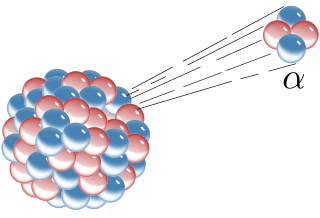
A beta particle, also called beta ray or beta radiation, is a high-energy, high-speed electron or positron emitted by the radioactive decay of an atomic nucleus during the process of beta decay. There are two forms of beta decay, β decay and β+ decay, which produce electrons and positrons respectively.

<span class="mw-page-title-main">Nuclear chemistry</span> Branch of chemistry dealing with radioactivity, transmutation and other nuclear processes

Nuclear chemistry is the sub-field of chemistry dealing with radioactivity, nuclear processes, and transformations in the nuclei of atoms, such as nuclear transmutation and nuclear properties.

A radioactive tracer, radiotracer, or radioactive label is a synthetic derivative of a natural compound in which one or more atoms have been replaced by a radionuclide. By virtue of its radioactive decay, it can be used to explore the mechanism of chemical reactions by tracing the path that the radioisotope follows from reactants to products. Radiolabeling or radiotracing is thus the radioactive form of isotopic labeling. In biological contexts, experiments that use radioisotope tracers are sometimes called radioisotope feeding experiments.

<span class="mw-page-title-main">Autoradiograph</span> Radiograph made by recording radiation emitted by samples on photographic plates

An autoradiograph is an image on an X-ray film or nuclear emulsion produced by the pattern of decay emissions from a distribution of a radioactive substance. Alternatively, the autoradiograph is also available as a digital image, due to the recent development of scintillation gas detectors or rare-earth phosphorimaging systems. The film or emulsion is apposed to the labeled tissue section to obtain the autoradiograph. The auto- prefix indicates that the radioactive substance is within the sample, as distinguished from the case of historadiography or microradiography, in which the sample is marked using an external source. Some autoradiographs can be examined microscopically for localization of silver grains in which the process is termed micro-autoradiography. For example, micro-autoradiography was used to examine whether atrazine was being metabolized by the hornwort plant or by epiphytic microorganisms in the biofilm layer surrounding the plant.

Liquid scintillation counting is the measurement of radioactive activity of a sample material which uses the technique of mixing the active material with a liquid scintillator, and counting the resultant photon emissions. The purpose is to allow more efficient counting due to the intimate contact of the activity with the scintillator. It is generally used for alpha particle or beta particle detection.

Phosphorus-32 (32P) is a radioactive isotope of phosphorus. The nucleus of phosphorus-32 contains 15 protons and 17 neutrons, one more neutron than the most common isotope of phosphorus, phosphorus-31. Phosphorus-32 only exists in small quantities on Earth as it has a short half-life of 14 days and so decays rapidly.

<span class="mw-page-title-main">Radioimmunoassay</span> Immunoassay that uses radiolabeled molecules

A radioimmunoassay (RIA) is an immunoassay that uses radiolabeled molecules in a stepwise formation of immune complexes. A RIA is a very sensitive in vitro assay technique used to measure concentrations of substances, usually measuring antigen concentrations by use of antibodies.

Isotopic labeling is a technique used to track the passage of an isotope through chemical reaction, metabolic pathway, or a biological cell. The reactant is 'labeled' by replacing one or more specific atoms with their isotopes. The reactant is then allowed to undergo the reaction. The position of the isotopes in the products is measured to determine the sequence the isotopic atom followed in the reaction or the cell's metabolic pathway. The nuclides used in isotopic labeling may be stable nuclides or radionuclides. In the latter case, the labeling is called radiolabeling.

<span class="mw-page-title-main">Neutron activation</span> Induction of radioactivity by neutron radiation

Neutron activation is the process in which neutron radiation induces radioactivity in materials, and occurs when atomic nuclei capture free neutrons, becoming heavier and entering excited states. The excited nucleus decays immediately by emitting gamma rays, or particles such as beta particles, alpha particles, fission products, and neutrons. Thus, the process of neutron capture, even after any intermediate decay, often results in the formation of an unstable activation product. Such radioactive nuclei can exhibit half-lives ranging from small fractions of a second to many years.

<span class="mw-page-title-main">Radiochemistry</span> Chemistry of radioactive materials

Radiochemistry is the chemistry of radioactive materials, where radioactive isotopes of elements are used to study the properties and chemical reactions of non-radioactive isotopes. Much of radiochemistry deals with the use of radioactivity to study ordinary chemical reactions. This is very different from radiation chemistry where the radiation levels are kept too low to influence the chemistry.

<span class="mw-page-title-main">Radioluminescence</span> Light produced in a material by bombardment with ionizing radiation

Radioluminescence is the phenomenon by which light is produced in a material by bombardment with ionizing radiation such as alpha particles, beta particles, or gamma rays. Radioluminescence is used as a low level light source for night illumination of instruments or signage. Radioluminescent paint is occasionally used for clock hands and instrument dials, enabling them to be read in the dark. Radioluminescence is also sometimes seen around high-power radiation sources, such as nuclear reactors and radioisotopes.

Various radionuclides emit beta particles, high-speed electrons or positrons, through radioactive decay of their atomic nucleus. These can be used in a range of different industrial, scientific, and medical applications. This article lists some common beta-emitting radionuclides of technological importance, and their properties.

An optoelectric nuclear battery is a type of nuclear battery in which nuclear energy is converted into light, which is then used to generate electrical energy. This is accomplished by letting the ionizing radiation emitted by the radioactive isotopes hit a luminescent material, which in turn emits photons that generate electricity upon striking a photovoltaic cell.

<span class="mw-page-title-main">Molecular imaging</span> Imaging molecules within living patients

Molecular imaging is a field of medical imaging that focuses on imaging molecules of medical interest within living patients. This is in contrast to conventional methods for obtaining molecular information from preserved tissue samples, such as histology. Molecules of interest may be either ones produced naturally by the body, or synthetic molecules produced in a laboratory and injected into a patient by a doctor. The most common example of molecular imaging used clinically today is to inject a contrast agent into a patient's bloodstream and to use an imaging modality to track its movement in the body. Molecular imaging originated from the field of radiology from a need to better understand fundamental molecular processes inside organisms in a noninvasive manner.

Krypton-85 (85Kr) is a radioisotope of krypton.

<span class="mw-page-title-main">Environmental radioactivity</span> Radioactivity naturally present within the Earth

Environmental radioactivity is produced by radioactive materials in the human environment. While some radioisotopes, such as strontium-90 (90Sr) and technetium-99 (99Tc), are only found on Earth as a result of human activity, and some, like potassium-40 (40K), are only present due to natural processes, a few isotopes, e.g. tritium (3H), result from both natural processes and human activities. The concentration and location of some natural isotopes, particularly uranium-238 (238U), can be affected by human activity.

Scintillation proximity assay (SPA) is an assay development and biochemical screening that permits the rapid and sensitive measurement of a broad range of biological processes in a homogeneous system. The type of beads that are involved in the SPA are microscopic in size and within the beads itself, there is a scintillant which emits light when it is stimulated. Stimulation occurs when radio-labelled molecules interact and bind to the surface of the bead. This interaction will trigger the bead to emit light, which can be detected using a photometer.

<span class="mw-page-title-main">Fluorescence in the life sciences</span> Scientific investigative technique

Fluorescence is used in the life sciences generally as a non-destructive way of tracking or analysing biological molecules. Some proteins or small molecules in cells are naturally fluorescent, which is called intrinsic fluorescence or autofluorescence. Alternatively, specific or general proteins, nucleic acids, lipids or small molecules can be "labelled" with an extrinsic fluorophore, a fluorescent dye which can be a small molecule, protein or quantum dot. Several techniques exist to exploit additional properties of fluorophores, such as fluorescence resonance energy transfer, where the energy is passed non-radiatively to a particular neighbouring dye, allowing proximity or protein activation to be detected; another is the change in properties, such as intensity, of certain dyes depending on their environment allowing their use in structural studies.

Radiofluorination is the process by which a radioactive isotope of fluorine is attached to a molecule and is preferably performed by nucleophilic substitution using nitro or halogens as leaving groups. Fluorine-18 is the most common isotope used for this procedure. This is due to its 97% positron emission and relatively long 109.8 min half-life. The half-life allows for a long enough time to be incorporated into the molecule and be used without causing exceedingly harmful effects. This process has many applications especially with the use of positron emission tomography (PET) as the aforementioned low positron energy is able to yield a high resolution in PET imaging.

References

  1. Breeman, W. A. P.; De Blois, E.; Sze Chan, H.; Konijnenberg, M.; Kwekkeboom, D. J.; Krenning, E. P. (2011). "68Ga-labeled DOTA-Peptides and 68Ga-labeled Radiopharmaceuticals for Positron Emission Tomography: Current Status of Research, Clinical Applications, and Future Perspectives". Seminars in Nuclear Medicine. 41 (4): 314–321. doi:10.1053/j.semnuclmed.2011.02.001. PMID   21624565.
  2. Voges, Rolf; Heys, J. Richard; Moenius, Thomas (2009). Preparation of compounds labeled with tritium and carbon-14. Chichester, U.K.: Wiley. p. 146. ISBN   978-0-470-51607-2 . Retrieved 11 September 2017.
  3. Jakonić, I; Nikolov, J; et al. (2014). "Study on quench effects in liquid scintillation counting during tritium measurements". Journal of Radioanalytical and Nuclear Chemistry. 302 (1): 253–259. doi:10.1007/s10967-014-3191-1. S2CID   95524987.
  4. "Scintillation Cocktails & Consumables - For every liquid scintillation counting application" (PDF). PerkinElmer. Archived from the original (PDF) on 27 March 2016. Retrieved 11 September 2017.
  5. "Storage Phosphor Screen BAS-IP" (PDF). GE Life Sciences. 2012. Archived from the original (PDF) on 11 September 2017. Retrieved 11 September 2017. Data file 29-0262-96 AA
  6. Radiolabeled Test Articles, AptoChem
  7. Biochemic methods. Sample for medicine Students. 2nd ed 2008, by Birgitte Lüttge. Aarhus University.
  8. NCRP. 160.{{cite journal}}: Missing or empty |title= (help)