Molecular imaging

Last updated
MolecularImagingTherapy.jpg

Molecular imaging is a field of medical imaging that focuses on imaging molecules of medical interest within living patients. This is in contrast to conventional methods for obtaining molecular information from preserved tissue samples, such as histology. Molecules of interest may be either ones produced naturally by the body, or synthetic molecules produced in a laboratory and injected into a patient by a doctor. The most common example of molecular imaging used clinically today is to inject a contrast agent (e.g., a microbubble, metal ion, or radioactive isotope) into a patient's bloodstream and to use an imaging modality (e.g., ultrasound, MRI, CT, PET) to track its movement in the body. Molecular imaging originated from the field of radiology from a need to better understand fundamental molecular processes inside organisms in a noninvasive manner.

Contents

The ultimate goal of molecular imaging is to be able to noninvasively monitor all of the biochemical processes occurring inside an organism in real time. Current research in molecular imaging involves cellular/molecular biology, chemistry, and medical physics, and is focused on: 1) developing imaging methods to detect previously undetectable types of molecules, 2) expanding the number and types of contrast agents available, and 3) developing functional contrast agents that provide information about the various activities that cells and tissues perform in both health and disease.

Overview

Molecular imaging emerged in the mid twentieth century as a discipline at the intersection of molecular biology and in vivo imaging. It enables the visualisation of the cellular function and the follow-up of the molecular process in living organisms without perturbing them. The multiple and numerous potentialities of this field are applicable to the diagnosis of diseases such as cancer, and neurological and cardiovascular diseases. This technique also contributes to improving the treatment of these disorders by optimizing the pre-clinical and clinical tests of new medication. They are also expected to have a major economic impact due to earlier and more precise diagnosis. Molecular and Functional Imaging has taken on a new direction since the description of the human genome. New paths in fundamental research, as well as in applied and industrial research, render the task of scientists more complex and increase the demands on them. Therefore, a tailor-made teaching program is in order.

Molecular imaging differs from traditional imaging in that probes known as biomarkers are used to help image particular targets or pathways. Biomarkers interact chemically with their surroundings and in turn alter the image according to molecular changes occurring within the area of interest. This process is markedly different from previous methods of imaging which primarily imaged differences in qualities such as density or water content. This ability to image fine molecular changes opens up an incredible number of exciting possibilities for medical application, including early detection and treatment of disease and basic pharmaceutical development. Furthermore, molecular imaging allows for quantitative tests, imparting a greater degree of objectivity to the study of these areas. One emerging technology is MALDI molecular imaging based on mass spectrometry.[ citation needed ]

Many areas of research are being conducted in the field of molecular imaging. Much research is currently centered on detecting what is known as a predisease state or molecular states that occur before typical symptoms of a disease are detected. Other important veins of research are the imaging of gene expression and the development of novel biomarkers. Organizations such as the SNMMI Center for Molecular Imaging Innovation and Translation (CMIIT) have formed to support research in this field. In Europe, other "networks of excellence" such as DiMI (Diagnostics in Molecular Imaging) or EMIL (European Molecular Imaging Laboratories) work on this new science, integrating activities and research in the field. In this way, a European Master Programme "EMMI" is being set up to train a new generation of professionals in molecular imaging.

Recently the term molecular imaging has been applied to a variety of microscopy and nanoscopy techniques including live-cell microscopy, Total Internal Reflection Fluorescence (TIRF)-microscopy, STimulated Emission Depletion (STED)-nanoscopy and Atomic Force Microscopy (AFM) as here images of molecules are the readout.

Imaging modalities

There are many different modalities that can be used for noninvasive molecular imaging. Each have their different strengths and weaknesses and some are more adept at imaging multiple targets than others.

Magnetic resonance imaging

Molecular MRI of a mouse brain presenting acute inflammation in the right hemisphere. Whereas unenhanced MRI failed to reveal any difference between right en left hemispheres, injection of a contrast-agent targeted to inflamed vessels allows to reveal inflammation specifically in the right hemisphere. Molecular MRI.jpg
Molecular MRI of a mouse brain presenting acute inflammation in the right hemisphere. Whereas unenhanced MRI failed to reveal any difference between right en left hemispheres, injection of a contrast-agent targeted to inflamed vessels allows to reveal inflammation specifically in the right hemisphere.

MRI has the advantages of having very high spatial resolution and is very adept at morphological imaging and functional imaging. MRI does have several disadvantages though. First, MRI has a sensitivity of around 10−3 mol/L to 10−5 mol/L which, compared to other types of imaging, can be very limiting. This problem stems from the fact that the difference between atoms in the high energy state and the low energy state is very small. For example, at 1.5 Tesla, a typical field strength for clinical MRI, the difference between high and low energy states is approximately 9 molecules per 2 million.[ citation needed ] Improvements to increase MR sensitivity include increasing magnetic field strength, and hyperpolarization via optical pumping, dynamic nuclear polarization or parahydrogen induced polarization. There are also a variety of signal amplification schemes based on chemical exchange that increase sensitivity. [1]

To achieve molecular imaging of disease biomarkers using MRI, targeted MRI contrast agents with high specificity and high relaxivity (sensitivity) are required. To date, many studies have been devoted to developing targeted-MRI contrast agents to achieve molecular imaging by MRI. Commonly, peptides, antibodies, or small ligands, and small protein domains, such as HER-2 affibodies, have been applied to achieve targeting. To enhance the sensitivity of the contrast agents, these targeting moieties are usually linked to high payload MRI contrast agents or MRI contrast agents with high relaxivities. [2] In particular, the recent development of micron-sized particles of iron oxide (MPIO) allowed to reach unprecedented levels of sensitivity to detect proteins expressed by arteries and veins. [3]

Optical imaging

Imaging of engineered E. coli Nissle 1917 in the mouse gut Imaging of engineered E. coli Nissle 1917 in the mouse gut.pdf
Imaging of engineered E. coli Nissle 1917 in the mouse gut

There are a number of approaches used for optical imaging. The various methods depend upon fluorescence, bioluminescence, absorption or reflectance as the source of contrast. [4]

Optical imaging's most valuable attribute is that it and ultrasound do not have strong safety concerns like the other medical imaging modalities.[ citation needed ]

The downside of optical imaging is the lack of penetration depth, especially when working at visible wavelengths. Depth of penetration is related to the absorption and scattering of light, which is primarily a function of the wavelength of the excitation source. Light is absorbed by endogenous chromophores found in living tissue (e.g. hemoglobin, melanin, and lipids). In general, light absorption and scattering decreases with increasing wavelength. Below ~700 nm (e.g. visible wavelengths), these effects result in shallow penetration depths of only a few millimeters. Thus, in the visible region of the spectrum, only superficial assessment of tissue features is possible. Above 900 nm, water absorption can interfere with signal-to-background ratio. Because the absorption coefficient of tissue is considerably lower in the near infrared (NIR) region (700-900 nm), light can penetrate more deeply, to depths of several centimeters. [5]

Near Infrared imaging

Fluorescent probes and labels are an important tool for optical imaging. Some researchers have applied NIR imaging in rat model of acute myocardial infarction (AMI), using a peptide probe that can binds to apoptotic and necrotic cells. [6] A number of near-infrared (NIR) fluorophores have been employed for in vivo imaging, including Kodak X-SIGHT Dyes and Conjugates, Pz 247, DyLight 750 and 800 Fluors, Cy 5.5 and 7 Fluors, Alexa Fluor 680 and 750 Dyes, IRDye 680 and 800CW Fluors. Quantum dots, with their photostability and bright emissions, have generated a great deal of interest; however, their size precludes efficient clearance from the circulatory and renal systems while exhibiting long-term toxicity.[ citation needed ].

Several studies have demonstrated the use of infrared dye-labeled probes in optical imaging.

  1. In a comparison of gamma scintigraphy and NIR imaging, a cyclopentapeptide dual-labeled with 111
    In
    and an NIR fluorophore was used to image αvβ3-integrin positive melanoma xenografts. [7]
  2. Near-infrared labeled RGD targeting αvβ3-integrin has been used in numerous studies to target a variety of cancers. [8]
  3. An NIR fluorophore has been conjugated to epidermal growth factor (EGF) for imaging of tumor progression. [9]
  4. An NIR fluorophore was compared to Cy5.5, suggesting that longer-wavelength dyes may produce more effective targeting agents for optical imaging. [10]
  5. Pamidronate has been labeled with an NIR fluorophore and used as a bone imaging agent to detect osteoblastic activity in a living animal. [11]
  6. An NIR fluorophore-labeled GPI, a potent inhibitor of PSMA (prostate specific membrane antigen). [12]
  7. Use of human serum albumin labeled with an NIR fluorophore as a tracking agent for mapping of sentinel lymph nodes. [13]
  8. 2-Deoxy-D-glucose labeled with an NIR fluorophore. [14]

It is important to note that addition of an NIR probe to any vector can alter the vector's biocompatibility and biodistribution. Therefore, it can not be unequivocally assumed that the conjugated vector will behave similarly to the native form.

Single photon emission computed tomography

SPECT image (bone tracer) of a mouse MIP Mouse02-spect.gif
SPECT image (bone tracer) of a mouse MIP

The development of computed tomography in the 1970s allowed mapping of the distribution of the radioisotopes in the organ or tissue, and led to the technique now called single photon emission computed tomography (SPECT).

The imaging agent used in SPECT emits gamma rays, as opposed to the positron emitters (such as 18
F
) used in PET. There are a range of radiotracers (such as 99m
Tc
, 111
In
, 123
I
, 201
Tl
) that can be used, depending on the specific application.

Xenon (133
Xe
) gas is one such radiotracer. It has been shown to be valuable for diagnostic inhalation studies for the evaluation of pulmonary function; for imaging the lungs; and may also be used to assess rCBF. Detection of this gas occurs via a gamma camera—which is a scintillation detector consisting of a collimator, a NaI crystal, and a set of photomultiplier tubes.

By rotating the gamma camera around the patient, a three-dimensional image of the distribution of the radiotracer can be obtained by employing filtered back projection or other tomographic techniques. The radioisotopes used in SPECT have relatively long half lives (a few hours to a few days) making them easy to produce and relatively cheap. This represents the major advantage of SPECT as a molecular imaging technique, since it is significantly cheaper than either PET or fMRI. However it lacks good spatial (i.e., where exactly the particle is) or temporal (i.e., did the contrast agent signal happen at this millisecond, or that millisecond) resolution. Additionally, due to the radioactivity of the contrast agent, there are safety aspects concerning the administration of radioisotopes to the subject, especially for serial studies.

Positron emission tomography

Imaging joint inflammation in an arthritic mouse using positron emission tomography.
PET, MRI, and overlaid images of a human brain. PET-IRM-cabeza-Keosys.JPG
PET, MRI, and overlaid images of a human brain.

Positron emission tomography (PET) is a nuclear medicine imaging technique which produces a three-dimensional image or picture of functional processes in the body. The theory behind PET is simple enough. First a molecule is tagged with a positron emitting isotope. These positrons annihilate with nearby electrons, emitting two 511 keV photons, directed 180 degrees apart in opposite directions. These photons are then detected by the scanner, which can estimate the density of positron annihilations in a specific area. When enough interactions and annihilations have occurred, the density of the original molecule may be measured in that area. Typical isotopes include 11
C
, 13
N
, 15
O
, 18
F
, 64
Cu
, 62
Cu
, 124
I
, 76
Br
, 82
Rb
, 89
Zr
and 68
Ga
, with 18
F
being the most clinically utilized. One of the major disadvantages of PET is that most of the probes must be made with a cyclotron. Most of these probes also have a half life measured in hours, forcing the cyclotron to be on site. These factors can make PET prohibitively expensive. PET imaging does have many advantages though. First and foremost is its sensitivity: a typical PET scanner can detect between 1011 mol/L to 1012 mol/L concentrations.

See also

Related Research Articles

<span class="mw-page-title-main">Microscopy</span> Viewing of objects which are too small to be seen with the naked eye

Microscopy is the technical field of using microscopes to view objects and areas of objects that cannot be seen with the naked eye. There are three well-known branches of microscopy: optical, electron, and scanning probe microscopy, along with the emerging field of X-ray microscopy.

<span class="mw-page-title-main">Positron emission tomography</span> Medical imaging technique

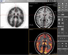
Positron emission tomography (PET) is a functional imaging technique that uses radioactive substances known as radiotracers to visualize and measure changes in metabolic processes, and in other physiological activities including blood flow, regional chemical composition, and absorption. Different tracers are used for various imaging purposes, depending on the target process within the body.

<span class="mw-page-title-main">Fluorescent tag</span>

In molecular biology and biotechnology, a fluorescent tag, also known as a fluorescent label or fluorescent probe, is a molecule that is attached chemically to aid in the detection of a biomolecule such as a protein, antibody, or amino acid. Generally, fluorescent tagging, or labeling, uses a reactive derivative of a fluorescent molecule known as a fluorophore. The fluorophore selectively binds to a specific region or functional group on the target molecule and can be attached chemically or biologically. Various labeling techniques such as enzymatic labeling, protein labeling, and genetic labeling are widely utilized. Ethidium bromide, fluorescein and green fluorescent protein are common tags. The most commonly labelled molecules are antibodies, proteins, amino acids and peptides which are then used as specific probes for detection of a particular target.

<span class="mw-page-title-main">Fluorophore</span> Agents that emit light after excitation by light

A fluorophore is a fluorescent chemical compound that can re-emit light upon light excitation. Fluorophores typically contain several combined aromatic groups, or planar or cyclic molecules with several π bonds.

<span class="mw-page-title-main">Near-infrared spectroscopy</span> Analytical method

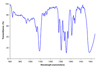
Near-infrared spectroscopy (NIRS) is a spectroscopic method that uses the near-infrared region of the electromagnetic spectrum. Typical applications include medical and physiological diagnostics and research including blood sugar, pulse oximetry, functional neuroimaging, sports medicine, elite sports training, ergonomics, rehabilitation, neonatal research, brain computer interface, urology, and neurology. There are also applications in other areas as well such as pharmaceutical, food and agrochemical quality control, atmospheric chemistry, combustion research and knowledge.

<span class="mw-page-title-main">Two-photon excitation microscopy</span> Fluorescence imaging technique

Two-photon excitation microscopy is a fluorescence imaging technique that is particularly well-suited to image scattering living tissue of up to about one millimeter in thickness. Unlike traditional fluorescence microscopy, where the excitation wavelength is shorter than the emission wavelength, two-photon excitation requires simultaneous excitation by two photons with longer wavelength than the emitted light. The laser is focused onto a specific location in the tissue and scanned across the sample to sequentially produce the image. Due to the non-linearity of two-photon excitation, mainly fluorophores in the micrometer-sized focus of the laser beam are excited, which results in the spatial resolution of the image. This contrasts with confocal microscopy, where the spatial resolution is produced by the interaction of excitation focus and the confined detection with a pinhole.

Cyanines, also referred to as tetramethylindo(di)-carbocyanines are a synthetic dye family belonging to the polymethine group. Although the name derives etymologically from terms for shades of blue, the cyanine family covers the electromagnetic spectrum from near IR to UV.

<span class="mw-page-title-main">Neuroimaging</span> Set of techniques to measure and visualize aspects of the nervous system

Neuroimaging is the use of quantitative (computational) techniques to study the structure and function of the central nervous system, developed as an objective way of scientifically studying the healthy human brain in a non-invasive manner. Increasingly it is also being used for quantitative research studies of brain disease and psychiatric illness. Neuroimaging is highly multidisciplinary involving neuroscience, computer science, psychology and statistics, and is not a medical specialty. Neuroimaging is sometimes confused with neuroradiology.

In medicine, a biomarker is a measurable indicator of the severity or presence of some disease state. It may be defined as a "cellular, biochemical or molecular alteration in cells, tissues or fluids that can be measured and evaluated to indicate normal biological processes, pathogenic processes, or pharmacological responses to a therapeutic intervention." More generally a biomarker is anything that can be used as an indicator of a particular disease state or some other physiological state of an organism. According to the WHO, the indicator may be chemical, physical, or biological in nature - and the measurement may be functional, physiological, biochemical, cellular, or molecular.

Functional imaging is a medical imaging technique of detecting or measuring changes in metabolism, blood flow, regional chemical composition, and absorption.

<span class="mw-page-title-main">Fluorescence in the life sciences</span> Scientific investigative technique

Fluorescence is used in the life sciences generally as a non-destructive way of tracking or analysing biological molecules. Some proteins or small molecules in cells are naturally fluorescent, which is called intrinsic fluorescence or autofluorescence. Alternatively, specific or general proteins, nucleic acids, lipids or small molecules can be "labelled" with an extrinsic fluorophore, a fluorescent dye which can be a small molecule, protein or quantum dot. Several techniques exist to exploit additional properties of fluorophores, such as fluorescence resonance energy transfer, where the energy is passed non-radiatively to a particular neighbouring dye, allowing proximity or protein activation to be detected; another is the change in properties, such as intensity, of certain dyes depending on their environment allowing their use in structural studies.

Preclinical imaging is the visualization of living animals for research purposes, such as drug development. Imaging modalities have long been crucial to the researcher in observing changes, either at the organ, tissue, cell, or molecular level, in animals responding to physiological or environmental changes. Imaging modalities that are non-invasive and in vivo have become especially important to study animal models longitudinally. Broadly speaking, these imaging systems can be categorized into primarily morphological/anatomical and primarily molecular imaging techniques. Techniques such as high-frequency micro-ultrasound, magnetic resonance imaging (MRI) and computed tomography (CT) are usually used for anatomical imaging, while optical imaging, positron emission tomography (PET), and single photon emission computed tomography (SPECT) are usually used for molecular visualizations.

<span class="mw-page-title-main">Brain positron emission tomography</span> Form of positron emission tomography

Brain positron emission tomography is a form of positron emission tomography (PET) that is used to measure brain metabolism and the distribution of exogenous radiolabeled chemical agents throughout the brain. PET measures emissions from radioactively labeled metabolically active chemicals that have been injected into the bloodstream. The emission data from brain PET are computer-processed to produce multi-dimensional images of the distribution of the chemicals throughout the brain.

Multi-spectral optoacoustic tomography (MSOT), also known as functional photoacoustic tomography (fPAT), is an imaging technology that generates high-resolution optical images in scattering media, including biological tissues. MSOT illuminates tissue with light of transient energy, typically light pulses lasting 1-100 nanoseconds. The tissue absorbs the light pulses, and as a result undergoes thermo-elastic expansion, a phenomenon known as the optoacoustic or photoacoustic effect. This expansion gives rise to ultrasound waves (photoechoes) that are detected and formed into an image. Image formation can be done by means of hardware or computed tomography. Unlike other types of optoacoustic imaging, MSOT involves illuminating the sample with multiple wavelengths, allowing it to detect ultrasound waves emitted by different photoabsorbing molecules in the tissue, whether endogenous or exogenous. Computational techniques such as spectral unmixing deconvolute the ultrasound waves emitted by these different absorbers, allowing each emitter to be visualized separately in the target tissue. In this way, MSOT can allow visualization of hemoglobin concentration and tissue oxygenation or hypoxia. Unlike other optical imaging methods, MSOT is unaffected by photon scattering and thus can provide high-resolution optical images deep inside biological tissues.

Fluorescence guided surgery (FGS), also called fluorescence image-guided surgery, or in the specific case of tumor resection, fluorescence guided resection, is a medical imaging technique used to detect fluorescently labelled structures during surgery. Similarly to standard image-guided surgery, FGS has the purpose of guiding the surgical procedure and providing the surgeon of real time visualization of the operating field. When compared to other medical imaging modalities, FGS is cheaper and superior in terms of resolution and number of molecules detectable. As a drawback, penetration depth is usually very poor in the visible wavelengths, but it can reach up to 1–2 cm when excitation wavelengths in the near infrared are used.

<span class="mw-page-title-main">FMISO</span> Chemical compound

18F-FMISO or fluoromisonidazole is a radiopharmaceutical used for PET imaging of hypoxia. It consists of a 2-nitroimidazole molecule labelled with the positron-emitter fluorine-18.

Preclinical or small-animal Single Photon Emission Computed Tomography (SPECT) is a radionuclide based molecular imaging modality for small laboratory animals. Although SPECT is a well-established imaging technique that is already for decades in use for clinical application, the limited resolution of clinical SPECT (~10 mm) stimulated the development of dedicated small animal SPECT systems with sub-mm resolution. Unlike in clinics, preclinical SPECT outperforms preclinical coincidence PET in terms of resolution and, at the same time, allows to perform fast dynamic imaging of animals.

<span class="mw-page-title-main">Fluorescence imaging</span> Type of non-invasive imaging technique

Fluorescence imaging is a type of non-invasive imaging technique that can help visualize biological processes taking place in a living organism. Images can be produced from a variety of methods including: microscopy, imaging probes, and spectroscopy.

Ultrasound-switchable fluorescence (USF) imaging is a deep optics imaging technique. In last few decades, fluorescence microscopy has been highly developed to image biological samples and live tissues. However, due to light scattering, fluorescence microscopy is limited to shallow tissues. Since fluorescence is characterized by high contrast, high sensitivity, and low cost which is crucial to investigate deep tissue information, developing fluorescence imaging technique with high depth-to-resolution ratio would be promising.. Recently, ultrasound-switchable fluorescence imaging has been developed to achieve high signal-to-noise ratio (SNR) and high spatial resolution imaging without sacrificing image depth.

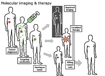
Theranostics, also known as theragnostics, is an emerging field in precision medicine that combines diagnostic and therapeutic approaches to provide the potential for personalized treatment and real-time monitoring of the effectiveness of treatments. Improvements in imaging techniques and targeted therapies are the basis of the field of theranostics. When medical imaging is coupled with the development of novel radiotracers and contrast agents, theranostics may provide opportunities for precise diagnosis and targeted therapy.

References

  1. Gallagher, F.A. (2010). "An introduction to functional and molecular imaging with MRI". Clinical Radiology. 65 (7): 557–566. doi:10.1016/j.crad.2010.04.006. ISSN   0009-9260. PMID   20541655.
  2. Shenghui, Xue; Jingjuan Qiao; Fan Pu; Mathew Cameron; Jenny J. Yang (17 Jan 2013). "Design of a novel class of protein-based magnetic resonance imaging contrast agents for the molecular imaging of cancer biomarkers". Wiley Interdiscip Rev Nanomed Nanobiotechnol. 5 (2): 163–79. doi:10.1002/wnan.1205. PMC   4011496 . PMID   23335551.
  3. Gauberti M, Montagne A, Quenault A, Vivien D (2014). "Molecular magnetic resonance imaging of brain-immune interactions". Front Cell Neurosci. 8: 389. doi: 10.3389/fncel.2014.00389 . PMC   4245913 . PMID   25505871.
  4. Weissleder R, Mahmood U (May 2001). "Molecular imaging". Radiology. 219 (2): 316–33. doi:10.1148/radiology.219.2.r01ma19316. PMID   11323453.
  5. Kovar JL, Simpson MA, Schutz-Geschwender A, Olive DM (August 2007). "A systematic approach to the development of fluorescent contrast agents for optical imaging of mouse cancer models". Anal. Biochem. 367 (1): 1–12. doi:10.1016/j.ab.2007.04.011. PMID   17521598. as PDF Archived February 11, 2009, at the Wayback Machine
  6. Acharya, B; Wang, K; Kim, IS; Kang, W; Moon, C; Lee, BH (2013). "In vivo imaging of myocardial cell death using a peptide probe and assessment of long-term heart function". Journal of Controlled Release. 172 (1): 367–73. doi:10.1016/j.jconrel.2013.08.294. PMID   24021357.
  7. Houston JP, Ke S, Wang W, Li C, Sevick-Muraca EM (2005). "Quality analysis of in vivo near-infrared fluorescence and conventional gamma images acquired using a dual-labeled tumor-targeting probe". J Biomed Opt. 10 (5): 054010. Bibcode:2005JBO....10e4010H. doi: 10.1117/1.2114748 . PMID   16292970.
  8. Chen K, Xie J, Chen X (2009). "RGD-human serum albumin conjugates as efficient tumor targeting probes". Mol Imaging. 8 (2): 65–73. doi:10.2310/7290.2009.00011. PMC   6366843 . PMID   19397852. Archived from the original on 2014-03-26.
  9. Kovar JL, Johnson MA, Volcheck WM, Chen J, Simpson MA (October 2006). "Hyaluronidase expression induces prostate tumor metastasis in an orthotopic mouse model". Am. J. Pathol. 169 (4): 1415–26. doi:10.2353/ajpath.2006.060324. PMC   1698854 . PMID   17003496.
  10. Adams KE, Ke S, Kwon S, et al. (2007). "Comparison of visible and near-infrared wavelength-excitable fluorescent dyes for molecular imaging of cancer". J Biomed Opt. 12 (2): 024017. Bibcode:2007JBO....12b4017A. doi: 10.1117/1.2717137 . PMID   17477732. S2CID   39806507.
  11. Zaheer A, Lenkinski RE, Mahmood A, Jones AG, Cantley LC, Frangioni JV (December 2001). "In vivo near-infrared fluorescence imaging of osteoblastic activity". Nat. Biotechnol. 19 (12): 1148–54. doi:10.1038/nbt1201-1148. PMID   11731784. S2CID   485155.
  12. Humblet V, Lapidus R, Williams LR, et al. (2005). "High-affinity near-infrared fluorescent small-molecule contrast agents for in vivo imaging of prostate-specific membrane antigen". Mol Imaging. 4 (4): 448–62. doi: 10.2310/7290.2005.05163 . PMID   16285907.
  13. Ohnishi S, Lomnes SJ, Laurence RG, Gogbashian A, Mariani G, Frangioni JV (2005). "Organic alternatives to quantum dots for intraoperative near-infrared fluorescent sentinel lymph node mapping". Mol Imaging. 4 (3): 172–81. doi: 10.1162/15353500200505127 . PMID   16194449.
  14. Kovar JL, Volcheck W, Sevick-Muraca E, Simpson MA, Olive DM (January 2009). "Characterization and performance of a near-infrared 2-deoxyglucose optical imaging agent for mouse cancer models". Anal. Biochem. 384 (2): 254–62. doi:10.1016/j.ab.2008.09.050. PMC   2720560 . PMID   18938129. as PDF Archived July 13, 2011, at the Wayback Machine