Related Research Articles

Tuberculosis (TB) is an infectious disease usually caused by Mycobacterium tuberculosis (MTB) bacteria. Tuberculosis generally affects the lungs, but it can also affect other parts of the body. Most infections show no symptoms, in which case it is known as latent tuberculosis. Around 10% of latent infections progress to active disease which, if left untreated, kill about half of those affected. Typical symptoms of active TB are chronic cough with blood-containing mucus, fever, night sweats, and weight loss. It was historically referred to as consumption due to the weight loss associated with the disease. Infection of other organs can cause a wide range of symptoms.

Silicosis is a form of occupational lung disease caused by inhalation of crystalline silica dust. It is marked by inflammation and scarring in the form of nodular lesions in the upper lobes of the lungs. It is a type of pneumoconiosis. Silicosis is characterized by shortness of breath, cough, fever, and cyanosis. It may often be misdiagnosed as pulmonary edema, pneumonia, or tuberculosis. Using workplace controls, silicosis is almost always a preventable disease.
Nontuberculous mycobacteria (NTM), also known as environmental mycobacteria, atypical mycobacteria and mycobacteria other than tuberculosis (MOTT), are mycobacteria which do not cause tuberculosis or leprosy. NTM do cause pulmonary diseases that resemble tuberculosis. Mycobacteriosis is any of these illnesses, usually meant to exclude tuberculosis. They occur in many animals, including humans and are commonly found in soil and water.
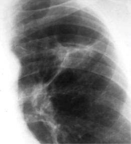
Radiology (X-rays) is used in the diagnosis of tuberculosis. Abnormalities on chest radiographs may be suggestive of, but are never diagnostic of TB, but can be used to rule out pulmonary TB.
Tuberculosis is diagnosed by finding Mycobacterium tuberculosis bacteria in a clinical specimen taken from the patient. While other investigations may strongly suggest tuberculosis as the diagnosis, they cannot confirm it.

Tuberculosis management describes the techniques and procedures utilized for treating tuberculosis (TB).

A chest radiograph, called a chest X-ray (CXR), or chest film, is a projection radiograph of the chest used to diagnose conditions affecting the chest, its contents, and nearby structures. Chest radiographs are the most common film taken in medicine.

World Tuberculosis Day, observed on 24 March each year, is designed to build public awareness about the global epidemic of tuberculosis (TB) and efforts to eliminate the disease. In 2018, 10 million people fell ill with TB, and 1.5 million died from the disease, mostly in low and middle-income countries. This also makes it the leading cause of death from an infectious disease.

Miliary tuberculosis is a form of tuberculosis that is characterized by a wide dissemination into the human body and by the tiny size of the lesions (1–5 mm). Its name comes from a distinctive pattern seen on a chest radiograph of many tiny spots distributed throughout the lung fields with the appearance similar to millet seeds—thus the term "miliary" tuberculosis. Miliary TB may infect any number of organs, including the lungs, liver, and spleen. Miliary tuberculosis is present in about 2% of all reported cases of tuberculosis and accounts for up to 20% of all extra-pulmonary tuberculosis cases.
Ghon's complex is a lesion seen in the lung that is caused by tuberculosis. The lesions consist of a Ghon focus along with pulmonary lymphadenopathy within a nearby pulmonary lymph node. A Ghon's complex retains viable bacteria, making them sources of long-term infection, which may reactivate and trigger secondary tuberculosis later in life.

Tuberculous meningitis, also known as TB meningitis or tubercular meningitis, is a specific type of bacterial meningitis caused by the Mycobacterium tuberculosis infection of the meninges—the system of membranes which envelop the central nervous system.
Tuberculosis (TB) vaccines are vaccinations intended for the prevention of tuberculosis. Immunotherapy as a defence against TB was first proposed in 1890 by Robert Koch. Today, the only effective tuberculosis vaccine in common use is the Bacillus Calmette-Guérin (BCG) vaccine, first used on humans in 1921. About three out of every 10,000 people who get the vaccine experience side effects, which are usually minor except in severely immuno-depressed individuals. While BCG immunization provides fairly effective protection for infants and young children, its efficacy in adults is variable, ranging from 0% to 80%. Several variables have been considered as responsible for the varying outcomes. Demand for TB immunotherapy advancement exists because the disease has become increasingly drug-resistant.
Caplan's syndrome is a combination of rheumatoid arthritis (RA) and pneumoconiosis that manifests as intrapulmonary nodules, which appear homogeneous and well-defined on chest X-ray.

High-resolution computed tomography (HRCT) is a type of computed tomography (CT) with specific techniques to enhance image resolution. It is used in the diagnosis of various health problems, though most commonly for lung disease, by assessing the lung parenchyma. On the other hand, HRCT of the temporal bone is used to diagnose various middle ear diseases such as otitis media, cholesteatoma, and evaluations after ear operations.

A lung nodule or pulmonary nodule is a relatively small focal density in the lung. A solitary pulmonary nodule (SPN) or coin lesion, is a mass in the lung smaller than three centimeters in diameter. A pulmonary micronodule has a diameter of less than three millimetres. There may also be multiple nodules.

Chronic pulmonary aspergillosis is a long-term fungal infection caused by members of the genus Aspergillus—most commonly Aspergillusfumigatus. The term describes several disease presentations with considerable overlap, ranging from an aspergilloma—a clump of Aspergillus mold in the lungs—through to a subacute, invasive form known as chronic necrotizing pulmonary aspergillosis which affects people whose immune system is weakened. Many people affected by chronic pulmonary aspergillosis have an underlying lung disease, most commonly tuberculosis, allergic bronchopulmonary aspergillosis, asthma, or lung cancer.

Ground-glass opacity (GGO) is a finding seen on chest x-ray (radiograph) or computed tomography (CT) imaging of the lungs. It is typically defined as an area of hazy opacification (x-ray) or increased attenuation (CT) due to air displacement by fluid, airway collapse, fibrosis, or a neoplastic process. When a substance other than air fills an area of the lung it increases that area's density. On both x-ray and CT, this appears more grey or hazy as opposed to the normally dark-appearing lungs. Although it can sometimes be seen in normal lungs, common pathologic causes include infections, interstitial lung disease, and pulmonary edema.

Tuberculosis in India is a major health problem, causing about 220,000 deaths every year. In 2020, the Indian government made statements to eliminate tuberculosis from the country by 2025 through its National TB Elimination Program. Interventions in this program include major investment in health care, providing supplemental nutrition credit through the Nikshay Poshan Yojana, organizing a national epidemiological survey for tuberculosis, and organizing a national campaign to tie together the Indian government and private health infrastructure for the goal of eliminating the disease.

Prisons in Russia consist of four types of facilities: pre-trial institutions; educative or juvenile colonies; corrective colonies; and prisons.

A focal lung pneumatosis, is an enclosed pocket of air or gas in the lung and includes blebs, bullae, pulmonary cysts, and lung cavities. Blebs and bullae can be classified by their wall thickness.
References
- 1 2 Leung, AN (February 1999). "Pulmonary tuberculosis: the essentials". Radiology. 210 (2): 307–22. doi:10.1148/radiology.210.2.r99ja34307. PMID 10207408.
- ↑ Robbins Basic Pathology (8th ed.). Elsevier. 2007. ISBN 978-1-4160-2973-1.
- ↑ "Assmann focus". Medicine.academic.ru. Retrieved 4 March 2015.
| | This infectious disease article is a stub. You can help Wikipedia by expanding it. |