Solar urticaria

Last updated
Solar urticaria
EMminor2010.JPG
Formation of wheals on the arm
Specialty Dermatology   OOjs UI icon edit-ltr-progressive.svg

Solar urticaria (SU) is a rare condition in which exposure to ultraviolet or UV radiation, or sometimes even visible light, induces a case of urticaria or hives that can appear in both covered and uncovered areas of the skin. [1] [2] It is classified as a type of physical urticaria. [3] The classification of disease types is somewhat controversial. One classification system distinguished various types of SU based on the wavelength of the radiation that causes the breakout; another classification system is based on the type of allergen that initiates a breakout. [4] [5]

Contents

The agent in the human body responsible for the reaction to radiation, known as the photoallergen, has not yet been identified. [6] The disease itself can be difficult to diagnose properly because it is so similar to other dermatological disorders, such as polymorphic light eruption or PMLE. [7] The most helpful test is a diagnostic phototest, a specialized test which confirms the presence of an abnormal sunburn reaction. Once recognized, treatment of the disease commonly involves the administration of antihistamines, and desensitization treatments such as phototherapy. [1] In more extreme cases, the use of immunosuppressive drugs and even plasmapheresis may be considered. [8]

The initial discovery of the disease is credited to P. Merklen in 1904, but it did not have a name until the suggestion of "solar urticaria" was given by William Waddell Duke in 1923. [9] [10] However, their research contributed to the study of this uncommon disease. More than one hundred cases have been reported in the past century. [11]

Signs and symptoms

Generally, the areas affected are exposed skin not usually protected by clothing; however it can also occur in areas covered by clothing. [1] [2] [12] Areas constantly subjected to the sun's rays may only be slightly affected if at all. People with extreme cases will also have reactions to artificial light sources that emit a UV wavelength. Parts of the body only thinly covered can also potentially be subjected to an outbreak.

Life with SU can be difficult. Patients are subject to constant itching and pain, as within minutes of the initial exposure to UV radiation a rash will appear. The urticarial reaction begins in the form of pruritus, later progressing to erythema and edema in the exposed areas of the skin. If vast areas of the body are affected, the loss of fluid into the skin could lead to light-headedness, headache, nausea, and vomiting. [1] [13] Extremely rarely, patients have been reported to experience an increase in heart rate that can cause a stroke or heart attack due to the body cavity swelling. Other rare side effects can be bronchospasm and glucose instability issues. Also, if a large area of the body is suddenly exposed the person may be subject to an anaphylactic reaction. Once free of exposure, the rash will usually fade away within several hours; rare and extreme cases can take a day or two to normalize depending on severity of the reaction. [14]

Causes

Solar urticaria is an immunoglobulin E-mediated hypersensitivity that can be introduced through primary or secondary factors, or induced by exogenous photosensitization. [15] [16] Primary SU is believed to be a type I hypersensitivity (a mild to severe reaction to an antigen including anaphylaxis) in which an antigen, or substance provoking an immune response, is "induced by UV or visible radiation." [15] Secondary SU can occur when a person comes into contact with chemicals such as tar, pitch, and dyes. People who use drugs such as benoxaprofen or patients with erythropoietic protoporphyria may also contract this secondary form. [15] These items that cause this photosensitivity are exogenous photosensitizers because they are outside of the body and cause it to have a greater sensitivity to light. [17]

Also, there have been a few unorthodox (unusual) causes of solar urticaria. For those susceptible to visible light, white T-shirts may increase the chances of experiencing an outbreak. In one case, doctors found that the white T-shirt absorbed UVA radiation from the sun and transformed it into visible light which caused the reaction. [18] Another patient was being treated with the antibiotic tetracycline for a separate dermatological disorder and broke out in hives when exposed to the sun, the first case to implicate tetracycline as a solar urticaria inducing agent. [19]

It is not yet known what specific agent in the body brings about the allergic reaction to the radiation. When patients with SU were injected with an irradiated autologous serum, many developed urticaria within the area of injection. When people who did not have SU were injected, they did not demonstrate similar symptoms. This indicates that the reaction is only a characteristic of the patients with solar urticaria and that it is not phototoxic. [6] It is possible that this photoallergen is located on the binding sites of IgE that are found on the surface of mast cells. [20] The photoallergen is believed to begin its configuration through the absorption of radiation by a chromophore. The molecule, because of the radiation, is transformed resulting in the formation of a new photoallergen. [21]

Diagnosis

Patch test Epikutanni-test.jpg
Patch test

Solar urticaria can be difficult to diagnose, but its presence can be confirmed by the process of phototesting. [22] There are several forms of these tests including photopatch tests, phototests, photoprovocation tests, and laboratory tests. All of these are necessary to determine the exact condition of the patient. Photopatch tests are patch tests conducted when it is believed that a patient is experiencing certain symptoms due to an allergy that will only occur when in contact with sunlight. After the procedure, the patient is given a low dosage of UVA radiation.

Another test known as a phototest is the most useful in identifying solar urticaria. In this test, one centimeter segments of skin are subject to varying amounts of UVA and UVB radiation in order to determine the specific dosage of the certain form of radiation that causes the urticaria to form. When testing for its less intense form (fixed solar urticaria), phototesting should be conducted only in the areas where the hives have appeared to avoid the possibility of getting false-negative results. [1] [23]

A third form of testing is the photoprovocation test which is used to identify disorders instigated by sun burns. The process of this test involves exposing one area of a patient's arm to certain dosage of UVB radiation and one area on the other arm to a certain dosage of UVA radiation. The amount of radiation that the patient is exposed to is equal to that "received in an hour of midday summer sun." If the procedure produces a rash, then the patient will undergo a biopsy. Finally, there are laboratory tests which generally involve procedures such as blood, urine, and fecal biochemical tests. In some situations, a skin biopsy may be performed. [1]

Classification

Solar urticaria, due to its particular features, is considered to be a type of physical urticaria or light sensitivity. Physical urticaria arises from physical factors in the environment, which in the case of solar urticaria is UV radiation or light. [24] [25] SU may be classified based on the wavelength of the radiative energy that causes the allergic reaction; known as Harber's classification, six types have been identified in this system. [11] Type I solar urticaria is caused by UVB (ultraviolet B) radiation, with wavelengths ranging from 290 to 320  nm. Type II is induced by UVA (ultraviolet A) radiation with wavelengths that can range from 320 to 400 nm. The wavelength range of type III and IV spans from 400 to 500 nm, while type V can be caused by UVB radiation to visible light (280–600 nm). Type VI has only been known to occur at 400 nm. [4]

Another classification distinguishes two types. The first is a hypersensitivity caused by a reaction to photoallergens located only in people with SU; while the second is caused by photoallergens that can be found in both people with SU and people without it. [5]

A subgroup of solar urticaria, fixed solar urticaria, has also been identified. It is a rare, less intense form of the disease with wheals (swollen areas of the skin) that affect certain, fixed areas of the body. Fixed solar urticaria is induced by a broad spectrum of radiative energy with wavelengths ranging from 300 to 700 nm. [23] [26]

Differential diagnosis

Polymorphous light eruption (PMLE) is the easiest disease to mistake for solar urticaria because the locations of the lesions are similar (the V of the neck and the arms). However, patients with SU are more likely to develop lesions on the face. Also, a reaction with PMLE will take a greater amount of time to appear than with solar urticaria. Lupus erythematosus has been mistaken for SU; however, lesions from lupus erythematosus will take a longer amount of time to go away. Furthermore, when being tested for the two diseases, patients with SU have a reaction immediately while patients with lupus erythematosus will have a delayed reaction. Patients who have experienced solar urticarial symptoms from a young age could mistakenly be thought to have erythropoietic protoporphyria. However, the main symptom for this disease is pain and patients with have been found to have abnormal levels of protoporphyrin in their blood while these levels are normal in SU patients. Finally, cholinergic urticaria, or urticaria induced by heat, can occasionally appear to be solar urticaria because the heat from the sun will cause a person with the disease to have a reaction. [7] [27]

Management

Antihistamines

Histamines are proteins associated with many allergic reactions. When the UV radiation or light comes in contact with a person with solar urticaria, histamine is released from mast cells. When this occurs, the permeability of vessels near the area of histamine release is increased. This allows blood fluid to enter the vessels and cause inflammation. Antihistamines suppress the activity of the histamine. [28]

Diphenhydramine, a first-generation H1 receptor antagonist or medicine that combats the H1 receptor that is associated with many allergic reactions, [29] has been found to be the most potent antihistamine for this particular disease. Patients prescribed 50 milligrams four times per day have been able to sustain normal exposure to the sun without developing a reaction.

Patients with less potent forms of solar urticaria such as fixed solar urticaria can be treated with the medication fexofenadine, which may also be used prophylactically to prevent recurrence. [23]

Desensitization

This form of treatment is meant to reduce the intensity or altogether eliminate the allergic reactions people have by gradually increasing exposure to the form of radiation that brings about the reaction. In the case of solar urticaria, phototherapy and photochemotherapy are the two major desensitization treatments. [30]

Phototherapy can be used for prevention. Exposure to a certain form of light or UV radiation enables the patient to build up a tolerance and outbreaks can be reduced. This type of treatment is generally conducted in the spring. [31] However, the benefits of this therapy only last for two to three days. [21]

Photochemotherapy, or PUVA, is considered superior to phototherapy because it produces a longer-lasting tolerance of the radiation that initiates the outbreak. When treatment first begins, the main goal is to build up the patient's tolerance to UVA radiation enough so that they can be outdoors without having an episode of solar urticaria. Therefore, treatments are regulated at three per week while constantly increasing the exposure to UVA radiation. Once the patient has reached an adequate level of desensitization, treatments are reduced to once or twice per week. [30]

Immunoglobulin E (IgE) Therapy

Some patients and researchers have successfully treated solar urticaria with Omalizumab (trade name Xolair) which is commonly used to treat Idiopathic Urticaria. Omalizumab is a recombinant humanized monoclonal antibody against IgE. It acts by binding free IgE at the same site that IgE would bind to its high-affinity receptor (FcεRI) on mast cells, thereby reducing free IgE in the serum [32]

Immunosuppression

Doctors will sometimes prescribe immunosuppressive drugs such as prednisolone and ciclosporin if the patient has an intense form of solar urticaria. However, the side effects of these medicines can be severe which is why they are reserved for the most extreme of cases. [8] [12]

Plasmapheresis

In more extreme cases, plasmapheresis can be considered. This technique is used to remove the blood plasma or fluid in the red blood cells and then return the cells to the body. It "removes a circulating factor from the blood that may be involved in causing the urticaria," but is still being tested and is not always effective. [8] When the treatment is a success, the patient's photosensitivity is decreased to the degree that they can undergo PUVA which can result in the relief of the urticarial outbreaks for an extended period of time. The major setback to this treatment is that the side effects can be severe and may include anaphylactoid reactions. [33]

Epidemiology

In the United States, only about 4% of patients with photosensitive disorders are reported to have been diagnosed with solar urticaria. Internationally, the number is slightly larger at 5.3%. [31] Solar urticaria may occur in all races but studies monitoring 135 African Americans and 110 Caucasians with photodermatoses found that 2.2% of the African Americans had SU and 8% of the Caucasians had the disease showing that Caucasians have a better chance of getting the disease. [34] The age ranges anywhere from 5–70 years old, but the average age is 35 and cases have been reported with children that are still in infancy. [31] [35] Solar urticaria accounts for less than one percent of the many documented urticaria cases. [13] To put that into a better perspective, since its first documented case in Japan in 1916, over one hundred other instances of the disease have been reported. [11]

History

Solar urticaria was first identified by P. Merklen in 1904. [10] Just a year later, in 1905, Ward became the first to induce urticaria through exposure to the sun in a controlled environment. The first documented case came in Japan in 1916. The name "solar urticaria" was proposed in 1923. In 1928, urticaria was induced for the first time. This was carried out by phototesting with increasing amounts of radiation of varying wavelengths. In 1942, the disease was passively transferred to normal volunteers using serum from patients with solar urticaria. [6] [11]

Related Research Articles

<span class="mw-page-title-main">Ultraviolet</span> Form of electromagnetic radiation

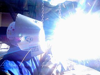
Ultraviolet (UV) is a form of electromagnetic radiation with wavelength shorter than that of visible light, but longer than X-rays. UV radiation is present in sunlight, and constitutes about 10% of the total electromagnetic radiation output from the Sun. It is also produced by electric arcs; Cherenkov radiation; and specialized lights; such as mercury-vapor lamps, tanning lamps, and black lights. Although long-wavelength ultraviolet is not considered an ionizing radiation because its photons lack the energy to ionize atoms, it can cause chemical reactions and causes many substances to glow or fluoresce. Many practical applications, including chemical and biological effects, derive from the way that UV radiation can interact with organic molecules. These interactions can involve absorption or adjusting energy states in molecules, but do not necessarily involve heating.

<span class="mw-page-title-main">Vitiligo</span> Skin condition where patches lose pigment

Vitiligo is a chronic autoimmune disorder that causes patches of skin to lose pigment or color. The cause of vitiligo is unknown, but it may be related to immune system changes, genetic factors, stress, or sun exposure. Treatment options include topical medications, light therapy, surgery and cosmetics.

<span class="mw-page-title-main">Sunscreen</span> Topical skin product that helps protect against sunburn

Sunscreen, also known as sunblock or sun cream, is a photoprotective topical product for the skin that helps protect against sunburn and most importantly prevent skin cancer. Sunscreens come as lotions, sprays, gels, foams, sticks, powders and other topical products. Sunscreens are common supplements to clothing, particularly sunglasses, sunhats and special sun protective clothing, and other forms of photoprotection.

<span class="mw-page-title-main">Blacklight</span> Light fixture that emits long-wave ultraviolet light and very little visible light

A blacklight, also called a UV-A light, Wood's lamp, or ultraviolet light, is a lamp that emits long-wave (UV-A) ultraviolet light and very little visible light. One type of lamp has a violet filter material, either on the bulb or in a separate glass filter in the lamp housing, which blocks most visible light and allows through UV, so the lamp has a dim violet glow when operating. Blacklight lamps which have this filter have a lighting industry designation that includes the letters "BLB". This stands for "blacklight blue". A second type of lamp produces ultraviolet but does not have the filter material, so it produces more visible light and has a blue color when operating. These tubes are made for use in "bug zapper" insect traps, and are identified by the industry designation "BL". This stands for "blacklight".

<span class="mw-page-title-main">Indoor tanning</span> Tanning using an artificial source of ultraviolet light

Indoor tanning involves using a device that emits ultraviolet radiation to produce a cosmetic tan. Typically found in tanning salons, gyms, spas, hotels, and sporting facilities, and less often in private residences, the most common device is a horizontal tanning bed, also known as a sunbed or solarium. Vertical devices are known as tanning booths or stand-up sunbeds.

<span class="mw-page-title-main">Contact dermatitis</span> Human disease

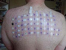
Contact dermatitis is a type of acute or chronic inflammation of the skin caused by exposure to chemical or physical agents. Symptoms of contact dermatitis can include itchy or dry skin, a red rash, bumps, blisters, or swelling. These rashes are not contagious or life-threatening, but can be very uncomfortable.

<span class="mw-page-title-main">Light therapy</span> Therapy involving intentional exposure to sunlight

Light therapy, also called phototherapy or bright light therapy is the exposure to direct sunlight or artificial light at controlled wavelengths in order to treat a variety of medical disorders, including seasonal affective disorder (SAD), circadian rhythm sleep-wake disorders, cancers, and skin wound infections. Treating skin conditions such as neurodermatitis, psoriasis, acne vulgaris, and eczema with ultraviolet light is called ultraviolet light therapy.

<span class="mw-page-title-main">Hives</span> Skin disease characterized by red, raised, and itchy bumps

Hives, also known as urticaria, is a kind of skin rash with red, raised, itchy bumps. Hives may burn or sting. The patches of rash may appear on different body parts, with variable duration from minutes to days, and does not leave any long-lasting skin change. Fewer than 5% of cases last for more than six weeks. The condition frequently recurs.

<span class="mw-page-title-main">Ultraviolet light therapy</span> Form of treatment for skin disorders

Ultraviolet light therapy or ultraviolet phototherapy is a treatment for psoriasis, atopic skin disorder, vitiligo and other skin diseases.

<span class="mw-page-title-main">Cold urticaria</span> Allergic reaction to low temperatures

Cold urticaria is a disorder in which large red welts called hives (urticaria) form on the skin after exposure to a cold stimulus. The hives are usually itchy and often the hands and feet will become itchy and swollen as well. Hives vary in size from about 7 mm in diameter to as big as about 27 mm or larger.

PUVA is an ultraviolet light therapy treatment for skin diseases: vitiligo, eczema, psoriasis, graft-versus-host disease, mycosis fungoides, large plaque parapsoriasis, and cutaneous T-cell lymphoma, using the sensitizing effects of the drug psoralen. The psoralen is applied or taken orally to sensitize the skin, then the skin is exposed to UVA.

Light sensitivity or photosensitivity refers to a notable or increased reactivity to light. Apart from vision, human beings have many physiological and psychological responses to light. In rare individuals an atypical response may result in serious discomfort, disease, or injury. Some drugs have a photosensitizing effect. Properties of natural or artificial light that may abnormally affect people include:

Photodermatoses is a skin condition resulting in abnormal skin reactions to ultraviolet (UV) radiation. These abnormal skin reactions may develop into rashes. While reactions to UV radiation is common, true photodermatoses is considered when an abnormal reaction is caused by UV-A rays or radiation between 320–400 nm.

<span class="mw-page-title-main">Polymorphous light eruption</span> Medical condition

Polymorphous light eruption (PLE) presents with itchy red small bumps on sun-exposed skin, particularly face, neck, forearms and legs. It generally appears 30 minutes to a few hours after sun exposure and may last between one and 14 days. The bumps may become small blisters or plaques and may appear bloody,often healing with minimal scarring.

Actinic prurigo is a rare sunlight-induced, pruritic, papular, or nodular skin eruption. Some medical experts use the term actinic prurigo to denote a rare photodermatosis that develops in childhood and is chronic and persistent; this rare photodermatosis, associated with the human leukocyte antigen HLA-DR4, is often called "Familial polymorphous light eruption of American Indians" or "Hereditary polymorphous light eruption of American Indians" but some experts consider it to be a variant of the syndrome known as polymorphous light eruption (PMLE). Some experts use the term actinic prurigo for Hutchinson's summer prurigo and several other photodermatoses that might, or might not, be distinct clinical entities.

<span class="mw-page-title-main">Sunburn</span> Burning of the skin by the suns radiation

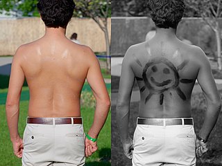
Sunburn is a form of radiation burn that affects living tissue, such as skin, that results from an overexposure to ultraviolet (UV) radiation, usually from the Sun. Common symptoms in humans and other animals include red or reddish skin that is hot to the touch or painful, general fatigue, and mild dizziness. Other symptoms include blistering, peeling skin, swelling, itching, and nausea. Excessive UV radiation is the leading cause of (primarily) non-malignant skin tumors, which in extreme cases can be life-threatening. Sunburn is an inflammatory response in the tissue triggered by direct DNA damage by UV radiation. When the cells' DNA is overly damaged by UV radiation, type I cell-death is triggered and the tissue is replaced.

Acne aestivalis also called as acne mallorca, is a special kind of polymorphous light eruption induced by ultra violet A radiation. This condition is said to be seasonal, usually affecting people in springtime and goes away in autumn when there is less sun light. This photo induced skin reaction leads to a mono-morphous eruption that consists of multiple, uniform, red, papular lesions. This skin reaction is classified as a delayed-type hypersensitivity because the onset is 24–72 hours after sun exposure. The condition equally affects men and women between the ages of 20–40 years old with no prior history of acne vulgaris. The eruption is unusual because it spares the face but it affects the lateral aspects of the upper arms, shoulder girdle, back, and chest. This condition's pathogenesis is not very well understood but scientists believe it an unfortunate side effect that results from a strong immune response to potentially cancer-causing cell damage.

<span class="mw-page-title-main">Photoaging</span> Ultraviolet light-induced biological development over time

Photoaging or photoageing is a term used for the characteristic changes to skin induced by chronic UVA and UVB exposure. Tretinoin is the best studied retinoid in the treatment of photoaging.

<span class="mw-page-title-main">Health effects of sunlight exposure</span>

Exposing skin to the ultraviolet radiation in sunlight has both positive and negative health effects. On the positive side, exposure allows for the synthesis of vitamin D3. Vitamin D has been suggested as having a wide range of positive health effects, which include strengthening bones and possibly inhibiting the growth of some cancers. A dietary supplement can also supply vitamin D, but there are also benefits to exposure not obtainable through Vitamin D supplementation. Long-term sun exposure is associated with reduced all-cause mortality and reduced mortality risk from cardiovascular disease (CVD), some forms of cancer, and non-CVD/noncancer related disease, with indications in these studies that Vitamin D is not the mediator. Supplementation offers limited bioavailability and no synthesis of subdermal nitric oxide. UV exposure also has positive effects for endorphin levels, and possibly for protection against multiple sclerosis. Abundant visible light to the eyes gives health benefits through its association with the timing of melatonin synthesis, maintenance of normal and robust circadian rhythms, and reduced risk of seasonal affective disorder.

Bernadette Eberlein is a dermatologist, allergologist, academic, and researcher. She is the Senior Physician of the private outpatient clinic, the Photodermatology unit, the unit for Soft X-Ray Therapy at Technical University of Munich.

References

  1. 1 2 3 4 5 6 Ngan, Vanessa (2008-03-10). "Solar urticaria". New Zealand Dermatological Society Incorporated. Retrieved 2008-10-06.
  2. 1 2 Lugović, Mihić L; Bulat, V; Situm, M; Cavka, V; Krolo, I (October 2008). "Allergic hypersensitivity skin reactions following sun exposure". Collegium Antropologicum. 32 (2): 153–7. PMID   19138019.
  3. Grammer, Leslie; Patterson, Roy; Greenberger, Paul A. (2002). Patterson's Allergic Diseases: Treatment and Prevention. Lippincott Williams & Wilkins. p. 244. ISBN   978-0-7817-2386-2.
  4. 1 2 Lim, Henry; Soter, Nicholas (1993). Clinical Photomedicine. CRC Press. p. 183. ISBN   978-0-8247-8862-9.
  5. 1 2 Leenutaphong, V; Hölzle, E; Plewig, G (August 1989). "Pathogenesis and classification of solar urticaria: a new concept". Journal of the American Academy of Dermatology. 21 (2 Pt 1): 237–40. doi:10.1016/S0190-9622(89)70167-5. PMID   2671065.
  6. 1 2 3 Lim, Henry; Honigsmann, Herbert; Hawk, John L. M. (2007). Photodermatology. CRC Press. pp. 5–190. ISBN   978-0-8493-7496-8.
  7. 1 2 Leung, Donald; Greaves, Malcolm W. (2000). Allergic Skin Disease: A Multidisciplinary Approach. Informa Health Care. p. 205. ISBN   978-0-8247-0287-8.
  8. 1 2 3 "Solar urticaria". urticaria.org.uk. 2006. Archived from the original on 2008-11-21. Retrieved 2008-10-25.
  9. Duke, William Waddell (1923-06-23). "Urticaria Caused by Light: Preliminary Report". Journal of the American Medical Association . 80 (25): 1835–1838. doi:10.1001/jama.1923.02640520017004. ISSN   0002-9955.
  10. 1 2 Hölzle, Erhard; Dawe, Robert (2019). "The Idiopathic Photodermatoses and Skin Testing". Harper's Textbook of Pediatric Dermatology (4th ed.). John Wiley & Sons. pp. 943–956. doi:10.1002/9781119142812.ch77. ISBN   9781119142812. S2CID   213560400.
  11. 1 2 3 4 Ozaki, Hirotake; Matsuyama, Takashi; Kawakubo, Yo; Miyahara, Motomi; Ozawa, Akira (2003-03-27). "A Case of Type IV Solar Urticaria Identified by Reversed in Vitro Serum Test" (PDF). Tokai Journal of Experimental and Clinical Medicine. 28 (2): 51–55. PMID   14714829. Archived from the original (PDF) on 2009-03-19. Retrieved 2009-01-30.
  12. 1 2 Gambichler, T; Al-Muhammadi, R; Boms, S (2009). "Immunologically mediated photodermatoses: diagnosis and treatment". American Journal of Clinical Dermatology. 10 (3): 169–80. doi:10.2165/00128071-200910030-00003. PMID   19354331. S2CID   38248032.
  13. 1 2 LeMura, Linda; Von Duvillard, Serge P. (2004). Clinical Exercise Physiology: Application and Physiological Principles. Lippincott Williams & Wilkins. p. 436. ISBN   978-0-7817-2680-1.
  14. Kane, Kay; Ryder, Jen Bissonette; Johnson, Richard Allen; Baden, Howard P.; Stratigos, Alexander (2001). Color Atlas & Synopsis of Pediatric Dermatology. McGraw-Hill Professional. p. 376. ISBN   978-0-07-006294-8.
  15. 1 2 3 Ostler, H. Bruce; Howard Maibach; Axel Hoke; Ivan Schwab (2003). Diseases of the Eye and Skin: A Color Atlas. Lippincott Williams & Wilkins. p. 21. ISBN   978-0-7817-4999-2.
  16. Botto, NC; Warshaw, EM (December 2008). "Solar urticaria". Journal of the American Academy of Dermatology. 59 (6): 909–20, quiz 921–2. doi:10.1016/j.jaad.2008.08.020. PMID   19022098.
  17. Ring, Johannes (2005). Allergy in Practice. Springer. p. 170. ISBN   978-3-540-00219-2.
  18. Gardeazabal, J; González-Pérez, R; Bilbao, I; Alvarez-Hernández, MI; Aguirre, A; Díaz-Pérez, JL (1998). "Solar urticaria enhanced through clothing". Photodermatology, Photoimmunology & Photomedicine. 14 (5–6): 164–6. doi:10.1111/j.1600-0781.1998.tb00036.x. PMID   9826887. S2CID   21203362.
  19. Yap, LM; Foley, PA; Crouch, RB; Baker, CS (August 2000). "Drug-induced solar urticaria due to tetracycline". The Australasian Journal of Dermatology. 41 (3): 181–4. doi:10.1046/j.1440-0960.2000.00435.x. PMID   10954992. S2CID   13162267.
  20. Zweiman, Burton; Schwartz, Lawrence B. (2001). Inflammatory Mechanisms in Allergic Diseases. Informa Health Care. p. 389. ISBN   978-0-8247-0540-4.
  21. 1 2 Braun-Falco, Otto; Plewig, G.; Wolff, H. H.; Burgdorf, W. H. (2000). Dermatology. Springer. p. 548. ISBN   978-3-540-59452-9.
  22. Roelandts, R (2003). "Diagnosis and treatment of solar urticaria". Dermatologic Therapy. 16 (1): 52–6. doi:10.1046/j.1529-8019.2003.01608.x. PMID   12919127. S2CID   22585366.
  23. 1 2 3 Schwarze, HP; Marguery, MC; Journé, F; Loche, E; Bazex, J (February 2001). "Fixed solar urticaria to visible light successfully treated with fexofenadine". Photodermatology, Photoimmunology & Photomedicine. 17 (1): 39–41. doi:10.1034/j.1600-0781.2001.017001039.x. PMID   11169175. S2CID   37465313.
  24. Schafer, C. M. (22 June 1995). "Physical urticaria". Immunology and Allergy Clinics of North America. 15 (4): 679–699. doi:10.1016/S0889-8561(22)00812-8. S2CID   251087252 . Retrieved 2009-05-08.
  25. Dice, JP (May 2004). "Physical urticaria". Immunology and Allergy Clinics of North America. 24 (2): 225–46, vi. doi:10.1016/j.iac.2004.01.005. PMID   15120149.
  26. Tuchinda, C; Leenutaphong, V; Sudtim, S; Lim, HW (April 2005). "Fixed solar urticaria induced by UVA and visible light: a report of a case". Photodermatology, Photoimmunology & Photomedicine. 21 (2): 97–9. doi:10.1111/j.1600-0781.2005.00148.x. PMID   15752128. S2CID   7791752.
  27. Ryckaert, S; Roelandts, R (January 1998). "Solar urticaria. A report of 25 cases and difficulties in phototesting". Archives of Dermatology. 134 (1): 71–4. doi:10.1001/archderm.134.1.71. PMID   9449912. S2CID   43137927.
  28. "Histamine". Davidson College. 2000. Retrieved 2008-11-01.
  29. Taranova, NP (1975). "[Effect of whole-body x-irradiation on the composition and metabolism of rat brain lipids]". Radiobiologiia (in Russian). 15 (6): 821–5. PMID   1219830.
  30. 1 2 Krutmann, Jean; Hönigsmann, Herbert; Elmets, Craig A.; Bergstresser, Paul R. (2001). Dermatological Phototherapy and Photodiagnostic Methods. Springer. pp. 117–118. ISBN   978-3-540-67789-5.
  31. 1 2 3 Baron, Elma; Taylor, Charles R. (2007-03-23). "Urticaria, Solar". emedicine. Retrieved 2008-10-06.
  32. Güzelbey, O.; Ardelean, E.; Magerl, M.; Zuberbier, T.; Maurer, M.; Metz, M. (2008-11-01). "Successful treatment of solar urticaria with anti-immunoglobulin E therapy". Allergy. 63 (11): 1563–1565. doi:10.1111/j.1398-9995.2008.01879.x. ISSN   1398-9995. PMID   18925897. S2CID   32623877.
  33. Katsambas, Andreas; Lotti, Torello M. (2003). European Handbook of Dermatological Treatments. Springer. pp. 499–500. ISBN   978-3-540-00878-1.
  34. Kerr, HA; Lim, HW (October 2007). "Photodermatoses in African Americans: a retrospective analysis of 135 patients over a 7-year period". Journal of the American Academy of Dermatology. 57 (4): 638–43. doi:10.1016/j.jaad.2007.05.043. PMID   17630044.
  35. Norris, PG; Hawk, JL (March 1990). "The acute idiopathic photodermatoses". Seminars in Dermatology. 9 (1): 32–8. PMID   2203441.