Sphingolipid

Last updated
General structures of sphingolipids Sphingolipids general structures.png
General structures of sphingolipids

Sphingolipids are a class of lipids containing a backbone of sphingoid bases, which are a set of aliphatic amino alcohols that includes sphingosine. They were discovered in brain extracts in the 1870s and were named after the mythological sphinx because of their enigmatic nature. [1] These compounds play important roles in signal transduction and cell recognition. Sphingolipidoses, or disorders of sphingolipid metabolism, have particular impact on neural tissue. A sphingolipid with a terminal hydroxyl group is a ceramide. Other common groups bonded to the terminal oxygen atom include phosphocholine, yielding a sphingomyelin, and various sugar monomers or dimers, yielding cerebrosides and globosides, respectively. Cerebrosides and globosides are collectively known as glycosphingolipids.

Contents

Structure

The long-chain bases, sometimes simply known as sphingoid bases, are the first non-transient products of de novo sphingolipid synthesis in both yeast and mammals. These compounds, specifically known as phytosphingosine and dihydrosphingosine (also known as sphinganine, [2] although this term is less common), are mainly C18 compounds, with somewhat lower levels of C20 bases. [3] Ceramides and glycosphingolipids are N-acyl derivatives of these compounds. [4]

The sphingosine backbone is O-linked to a (usually) charged head group such as ethanolamine, serine, or choline.[ citation needed ]

The backbone is also amide-linked to an acyl group, such as a fatty acid.[ citation needed ]

Types

Simple sphingolipids, which include the sphingoid bases and ceramides, make up the early products of the sphingolipid synthetic pathways.

Complex sphingolipids may be formed by addition of head groups to ceramide or phytoceramide:

Mammalian sphingolipid metabolism

De novo sphingolipid synthesis begins with formation of 3-keto-dihydrosphingosine by serine palmitoyltransferase. [7] The preferred substrates for this reaction are palmitoyl-CoA and serine. However, studies have demonstrated that serine palmitoyltransferase has some activity toward other species of fatty acyl-CoA [8] and alternative amino acids, [9] and the diversity of sphingoid bases has recently been reviewed. [10] Next, 3-keto-dihydrosphingosine is reduced to form dihydrosphingosine. Dihydrosphingosine is acylated by one of six (dihydro)-ceramide synthase, CerS - originally termed LASS - to form dihydroceramide. [11] The six CerS enzymes have different specificity for acyl-CoA substrates, resulting in the generation of dihydroceramides with differing chain lengths (ranging from C14-C26). Dihydroceramides are then desaturated to form ceramide. [12]

Metabolic pathways of various forms of sphingolipids. Sphingolipidoses are labeled at corresponding stages that are deficient. Sphingolipidoses.svg
Metabolic pathways of various forms of sphingolipids. Sphingolipidoses are labeled at corresponding stages that are deficient.

De novo generated ceramide is the central hub of the sphingolipid network and subsequently has several fates. It may be phosphorylated by ceramide kinase to form ceramide-1-phosphate. Alternatively, it may be glycosylated by glucosylceramide synthase or galactosylceramide synthase. Additionally, it can be converted to sphingomyelin by the addition of a phosphorylcholine headgroup by sphingomyelin synthase. Diacylglycerol is generated by this process. Finally, ceramide may be broken down by a ceramidase to form sphingosine. Sphingosine may be phosphorylated to form sphingosine-1-phosphate. This may be dephosphorylated to reform sphingosine. [13]

Breakdown pathways allow the reversion of these metabolites to ceramide. The complex glycosphingolipids are hydrolyzed to glucosylceramide and galactosylceramide. These lipids are then hydrolyzed by beta-glucosidases and beta-galactosidases to regenerate ceramide. Similarly, sphingomyelin may be broken down by sphingomyelinase to form ceramide.[ citation needed ]

The only route by which sphingolipids are converted to non-sphingolipids is through sphingosine-1-phosphate lyase. This forms ethanolamine phosphate and hexadecenal. [14]

Functions of mammalian sphingolipids

Sphingolipids are commonly believed to protect the cell surface against harmful environmental factors by forming a mechanically stable and chemically resistant outer leaflet of the plasma membrane lipid bilayer. Certain complex glycosphingolipids were found to be involved in specific functions, such as cell recognition and signaling. Cell recognition depends mainly on the physical properties of the sphingolipids, whereas signaling involves specific interactions of the glycan structures of glycosphingolipids with similar lipids present on neighboring cells or with proteins.[ citation needed ]

Recently, simple sphingolipid metabolites, such as ceramide and sphingosine-1-phosphate, have been shown to be important mediators in the signaling cascades involved in apoptosis, proliferation, stress responses, necrosis, inflammation, autophagy, senescence, and differentiation. [15] [16] [17] [18] [19] [20] Ceramide-based lipids self-aggregate in cell membranes and form separate phases less fluid than the bulk phospholipids. These sphingolipid-based microdomains, or "lipid rafts" were originally proposed to sort membrane proteins along the cellular pathways of membrane transport. At present, most research focuses on the organizing function during signal transduction. [21]

Sphingolipids are synthesized in a pathway that begins in the ER and is completed in the Golgi apparatus, but these lipids are enriched in the plasma membrane and in endosomes, where they perform many of their functions. [22] Transport occurs via vesicles and monomeric transport in the cytosol. Sphingolipids are virtually absent from mitochondria and the ER, but constitute a 20-35 molar fraction of plasma membrane lipids. [23]

In experimental animals, feeding sphingolipids inhibits colon carcinogenesis, reduces LDL cholesterol and elevates HDL cholesterol. [24]

Other sphingolipids

Sphingolipids are universal in eukaryotes but are rare in bacteria and archaea. Bacteria that do produce sphingolipids are found in family Sphingomonadaceae, some members of the FCB group, some members of the Bdellovibrionota, and some members of the Myxococcota. [25]

Yeast sphingolipids

Because of the incredible complexity of mammalian systems, yeast are often used as a model organism for working out new pathways. These single-celled organisms are often more genetically tractable than mammalian cells, and strain libraries are available to supply strains harboring almost any non-lethal open reading frame single deletion. The two most commonly used yeasts are Saccharomyces cerevisiae and Schizosaccharomyces pombe , although research is also done in the pathogenic yeast Candida albicans .[ citation needed ]

In addition to the important structural functions of complex sphingolipids (inositol phosphorylceramide and its mannosylated derivatives), the sphingoid bases phytosphingosine and dihydrosphingosine (sphinganine) play vital signaling roles in S. cerevisiae. These effects include regulation of endocytosis, ubiquitin-dependent proteolysis (and, thus, regulation of nutrient uptake [26] ), cytoskeletal dynamics, the cell cycle, translation, posttranslational protein modification, and the heat stress response. [27] Additionally, modulation of sphingolipid metabolism by phosphatidylinositol (4,5)-bisphosphate signaling via Slm1p and Slm2p and calcineurin has recently been described. [28] Additionally, a substrate-level interaction has been shown between complex sphingolipid synthesis and cycling of phosphatidylinositol 4-phosphate by the phosphatidylinositol kinase Stt4p and the lipid phosphatase Sac1p. [29]

Plant sphingolipids

Higher plants contain a wider variety of sphingolipids than animals and fungi.[ citation needed ]

Disorders

There are several disorders of sphingolipid metabolism, known as sphingolipidoses. The main members of this group are Niemann-Pick disease, Fabry disease, Krabbe disease, Gaucher disease, Tay–Sachs disease and Metachromatic leukodystrophy. They are generally inherited in an autosomal recessive fashion, but notably Fabry disease is X-linked. Taken together, sphingolipidoses have an incidence of approximately 1 in 10,000, but substantially more in certain populations such as Ashkenazi Jews. Enzyme replacement therapy is available to treat mainly Fabry disease and Gaucher disease, and people with these types of sphingolipidoses may live well into adulthood. The other types are generally fatal by age 1 to 5 years for infantile forms, but progression may be mild for juvenile- or adult-onset forms.[ citation needed ]

Sphingolipids have also been implicated with the frataxin protein (Fxn), the deficiency of which is associated with Friedreich's ataxia (FRDA). Loss of Fxn in the nervous system in mice also activates an iron/sphingolipid/PDK1/Mef2 pathway, indicating that the mechanism is evolutionarily conserved. Furthermore, sphingolipid levels and PDK1 activity are also increased in hearts of FRDA patients, suggesting that a similar pathway is affected in FRDA. [30] Other research has demonstrated that iron accumulation in the nervous systems of flies enhances the synthesis of sphingolipids, which in turn activates 3-phosphoinositide dependent protein kinase-1 (Pdk1) and myocyte enhancer factor-2 (Mef2) to trigger neurodegeneration of adult photoreceptors. [31]

Sphingolipids play a key role in neuronal survival in Parkinson's Disease (PD) and their catabolic pathway alteration in the brain is partly represented in cerebrospinal fluid and blood tissues (Table1) and have the diagnostic potential. [32]

Additional images

See also

Related Research Articles

<span class="mw-page-title-main">Lipid</span> Substance of biological origin that is soluble in nonpolar solvents

Lipids are a broad group of naturally-occurring molecules which includes fats, waxes, sterols, fat-soluble vitamins, monoglycerides, diglycerides, phospholipids, and others. The functions of lipids include storing energy, signaling, and acting as structural components of cell membranes. Lipids have applications in the cosmetic and food industries, and in nanotechnology.

<span class="mw-page-title-main">Sphingomyelin</span>

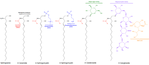
Sphingomyelin is a type of sphingolipid found in animal cell membranes, especially in the membranous myelin sheath that surrounds some nerve cell axons. It usually consists of phosphocholine and ceramide, or a phosphoethanolamine head group; therefore, sphingomyelins can also be classified as sphingophospholipids. In humans, SPH represents ~85% of all sphingolipids, and typically make up 10–20 mol % of plasma membrane lipids.

Sphingosine (2-amino-4-trans-octadecene-1,3-diol) is an 18-carbon amino alcohol with an unsaturated hydrocarbon chain, which forms a primary part of sphingolipids, a class of cell membrane lipids that include sphingomyelin, an important phospholipid.

<span class="mw-page-title-main">Fumonisin B1</span> Chemical compound

Fumonisin B1 is the most prevalent member of a family of toxins, known as fumonisins, produced by several species of Fusarium molds, such as Fusarium verticillioides, which occur mainly in maize (corn), wheat and other cereals. Fumonisin B1 contamination of maize has been reported worldwide at mg/kg levels. Human exposure occurs at levels of micrograms to milligrams per day and is greatest in regions where maize products are the dietary staple.

<span class="mw-page-title-main">Ceramide</span> Family of waxy lipid molecules

Ceramides are a family of waxy lipid molecules. A ceramide is composed of N-acetylsphingosine and a fatty acid. Ceramides are found in high concentrations within the cell membrane of eukaryotic cells, since they are component lipids that make up sphingomyelin, one of the major lipids in the lipid bilayer. Contrary to previous assumptions that ceramides and other sphingolipids found in cell membrane were purely supporting structural elements, ceramide can participate in a variety of cellular signaling: examples include regulating differentiation, proliferation, and programmed cell death (PCD) of cells.

<span class="mw-page-title-main">Sphingosine kinase</span> Class of enzymes

Sphingosine kinase (SphK) is a conserved lipid kinase that catalyzes formation sphingosine-1-phosphate (S1P) from the precursor sphingolipid sphingosine. Sphingolipid metabolites, such as ceramide, sphingosine and sphingosine-1-phosphate, are lipid second messengers involved in diverse cellular processes. There are two forms of SphK, SphK1 and SphK2. SphK1 is found in the cytosol of eukaryotic cells, and migrates to the plasma membrane upon activation. SphK2 is localized to the nucleus.

<span class="mw-page-title-main">Lipid signaling</span> Biological signaling using lipid molecules

Lipid signaling, broadly defined, refers to any biological signaling event involving a lipid messenger that binds a protein target, such as a receptor, kinase or phosphatase, which in turn mediate the effects of these lipids on specific cellular responses. Lipid signaling is thought to be qualitatively different from other classical signaling paradigms because lipids can freely diffuse through membranes. One consequence of this is that lipid messengers cannot be stored in vesicles prior to release and so are often biosynthesized "on demand" at their intended site of action. As such, many lipid signaling molecules cannot circulate freely in solution but, rather, exist bound to special carrier proteins in serum.

<span class="mw-page-title-main">Sphingomyelin phosphodiesterase</span> Class of enzymes

Sphingomyelin phosphodiesterase is a hydrolase enzyme that is involved in sphingolipid metabolism reactions. SMase is a member of the DNase I superfamily of enzymes and is responsible for breaking sphingomyelin (SM) down into phosphocholine and ceramide. The activation of SMase has been suggested as a major route for the production of ceramide in response to cellular stresses.

Sphingosine-1-phosphate (S1P) is a signaling sphingolipid, also known as lysosphingolipid. It is also referred to as a bioactive lipid mediator. Sphingolipids at large form a class of lipids characterized by a particular aliphatic aminoalcohol, which is sphingosine.

<span class="mw-page-title-main">Phosphatidate phosphatase</span>

The enzyme phosphatidate phosphatase (PAP, EC 3.1.3.4) is a key regulatory enzyme in lipid metabolism, catalyzing the conversion of phosphatidate to diacylglycerol:

<span class="mw-page-title-main">Serine C-palmitoyltransferase</span>

In enzymology, a serine C-palmitoyltransferase (EC 2.3.1.50) is an enzyme that catalyzes the chemical reaction:

In enzymology, sphingosine N-acyltransferases (ceramide synthases (CerS), EC 2.3.1.24) are enzymes that catalyze the chemical reaction of synthesis of ceramide:

In enzymology, a ceramide kinase, also abbreviated as CERK, is an enzyme that catalyzes the chemical reaction:

<span class="mw-page-title-main">COL4A3BP</span> Protein-coding gene in the species Homo sapiens

Collagen type IV alpha-3-binding protein, also known as ceramide transfer protein (CERT) or StAR-related lipid transfer protein 11 (STARD11) is a protein that in humans is encoded by the COL4A3BP gene. The protein contains a pleckstrin homology domain at its amino terminus and a START domain towards the end of the molecule. It is a member of the StarD2 subfamily of START domain proteins.

<span class="mw-page-title-main">SPTLC2</span> Protein-coding gene in the species Homo sapiens

Serine palmitoyltransferase, long chain base subunit 2, also known as SPTLC2, is a protein which in humans is encoded by the SPTLC2 gene. SPTLC2 belongs to the class-II pyridoxal-phosphate-dependent aminotransferase family.

Neutral ceramidase C also known as N-acylsphingosine amidohydrolase 2C or non-lysosomal ceramidase C or ASAH2C is a ceramidase enzyme which in humans is encoded by the ASAH2C gene.

<span class="mw-page-title-main">ACER1</span> Protein-coding gene in the species Homo sapiens

Alkaline ceramidase 1 also known as ACER1 is a ceramidase enzyme which in humans is encoded by the ACER1 gene.

<span class="mw-page-title-main">Ceramide synthase 3</span> Protein-coding gene in the species Homo sapiens

Ceramide synthase 3 (CersS3), also known as longevity assurance homologue 3, is an enzyme that is encoded in humans by the CERS3 gene.

Ceramide synthase 5 (CerS5) is the enzyme encoded in humans by the CERS5 gene.

The 1-deoxysphingolipids (1-deoxySLs) are an atypical and recently discovered class of sphingolipids (SLs). They are formed during the nove synthesis pathway and their essential C1-OH deficit causes the malfunctions of the following transformations to achieve complex sphingolipids. In general, sphingolipids are formed during a reaction that is catalyzed by the SPT enzyme (serine-palmitoyltransferase) where the condensation of serine and palmitoyl-CoA takes place. The origin of this rare sphingolipid, though, is due to a defect of the SPT which can also use alanine or glycine. This change is what forms the 1-deoxySL.

References

  1. Chun J, Hartung HP (2010). "Mechanism of action of oral fingolimod (FTY720) in multiple sclerosis". Clinical Neuropharmacology. 33 (2): 91–101. doi:10.1097/wnf.0b013e3181cbf825. PMC   2859693 . PMID   20061941.
  2. [http://www.sigmaaldrich.com/US/en/product/sigma/d3314 |SIGMA&N5=SEARCH_CONCAT_PNO|BRAND_KEY&F=SPEC Product page at Sigma Aldrich]
  3. Dickson RC (1998). "Sphingolipid functions in Saccharomyces cerevisiae: comparison to mammals". Annual Review of Biochemistry. 67: 27–48. doi: 10.1146/annurev.biochem.67.1.27 . PMID   9759481.
  4. A brief, very comprehensible review is given in Gunstone, F. (1996) Fatty Acid and Lipid Chemistry, pp 43-44. Blackie Academic and Professional. ISBN   0-7514-0253-2
  5. Dickson RC (May 2008). "Thematic review series: sphingolipids. New insights into sphingolipid metabolism and function in budding yeast". Journal of Lipid Research. 49 (5): 909–21. doi:10.1194/jlr.R800003-JLR200. PMC   2311445 . PMID   18296751.
  6. Bartke N, Hannun YA (April 2009). "Bioactive sphingolipids: metabolism and function". Journal of Lipid Research. 50 Suppl (Suppl): S91-6. doi:10.1194/jlr.R800080-JLR200. PMC   2674734 . PMID   19017611.
  7. Merrill AH (December 1983). "Characterization of serine palmitoyltransferase activity in Chinese hamster ovary cells". Biochimica et Biophysica Acta (BBA) - Lipids and Lipid Metabolism. 754 (3): 284–91. doi:10.1016/0005-2760(83)90144-3. PMID   6652105.
  8. Merrill AH, Williams RD (February 1984). "Utilization of different fatty acyl-CoA thioesters by serine palmitoyltransferase from rat brain". Journal of Lipid Research. 25 (2): 185–8. doi: 10.1016/S0022-2275(20)37838-X . PMID   6707526.
  9. Zitomer NC, Mitchell T, Voss KA, Bondy GS, Pruett ST, Garnier-Amblard EC, Liebeskind LS, Park H, Wang E, Sullards MC, Merrill AH, Riley RT (February 2009). "Ceramide synthase inhibition by fumonisin B1 causes accumulation of 1-deoxysphinganine: a novel category of bioactive 1-deoxysphingoid bases and 1-deoxydihydroceramides biosynthesized by mammalian cell lines and animals". The Journal of Biological Chemistry. 284 (8): 4786–95. doi: 10.1074/jbc.M808798200 . PMC   2643501 . PMID   19095642.
  10. Pruett ST, Bushnev A, Hagedorn K, Adiga M, Haynes CA, Sullards MC, Liotta DC, Merrill AH (August 2008). "Biodiversity of sphingoid bases ("sphingosines") and related amino alcohols". Journal of Lipid Research. 49 (8): 1621–39. doi:10.1194/jlr.R800012-JLR200. PMC   2444003 . PMID   18499644.
  11. Pewzner-Jung Y, Ben-Dor S, Futerman AH (September 2006). "When do Lasses (longevity assurance genes) become CerS (ceramide synthases)?: Insights into the regulation of ceramide synthesis". The Journal of Biological Chemistry. 281 (35): 25001–5. doi: 10.1074/jbc.R600010200 . PMID   16793762.
  12. Causeret C, Geeraert L, Van der Hoeven G, Mannaerts GP, Van Veldhoven PP (October 2000). "Further characterization of rat dihydroceramide desaturase: tissue distribution, subcellular localization, and substrate specificity". Lipids. 35 (10): 1117–25. doi:10.1007/s11745-000-0627-6. PMID   11104018. S2CID   3962533.
  13. Hannun YA, Obeid LM (February 2008). "Principles of bioactive lipid signalling: lessons from sphingolipids". Nature Reviews Molecular Cell Biology. 9 (2): 139–50. doi:10.1038/nrm2329. PMID   18216770. S2CID   8692993.
  14. Bandhuvula P, Saba JD (May 2007). "Sphingosine-1-phosphate lyase in immunity and cancer: silencing the siren". Trends in Molecular Medicine. 13 (5): 210–7. doi:10.1016/j.molmed.2007.03.005. PMID   17416206.
  15. Hannun YA, Obeid LM (July 2002). "The Ceramide-centric universe of lipid-mediated cell regulation: stress encounters of the lipid kind". The Journal of Biological Chemistry. 277 (29): 25847–50. doi: 10.1074/jbc.R200008200 . PMID   12011103.
  16. Spiegel S, Milstien S (July 2002). "Sphingosine 1-phosphate, a key cell signaling molecule". The Journal of Biological Chemistry. 277 (29): 25851–4. doi: 10.1074/jbc.R200007200 . PMID   12011102.
  17. Lavieu G, Scarlatti F, Sala G, Carpentier S, Levade T, Ghidoni R, Botti J, Codogno P (March 2006). "Regulation of autophagy by sphingosine kinase 1 and its role in cell survival during nutrient starvation". The Journal of Biological Chemistry. 281 (13): 8518–27. doi: 10.1074/jbc.M506182200 . PMID   16415355.
  18. Venable ME, Lee JY, Smyth MJ, Bielawska A, Obeid LM (December 1995). "Role of ceramide in cellular senescence". The Journal of Biological Chemistry. 270 (51): 30701–8. doi: 10.1074/jbc.270.51.30701 . PMID   8530509.
  19. Hetz CA, Hunn M, Rojas P, Torres V, Leyton L, Quest AF (December 2002). "Caspase-dependent initiation of apoptosis and necrosis by the Fas receptor in lymphoid cells: onset of necrosis is associated with delayed ceramide increase". Journal of Cell Science. 115 (Pt 23): 4671–83. doi: 10.1242/jcs.00153 . PMID   12415011.
  20. Snider AJ, Orr Gandy KA, Obeid LM (June 2010). "Sphingosine kinase: Role in regulation of bioactive sphingolipid mediators in inflammation". Biochimie. 92 (6): 707–15. doi:10.1016/j.biochi.2010.02.008. PMC   2878898 . PMID   20156522.
  21. Brown DA, London E (June 2000). "Structure and function of sphingolipid- and cholesterol-rich membrane rafts". The Journal of Biological Chemistry. 275 (23): 17221–4. doi: 10.1074/jbc.R000005200 . PMID   10770957.
  22. Futerman AH (December 2006). "Intracellular trafficking of sphingolipids: relationship to biosynthesis". Biochimica et Biophysica Acta (BBA) - Biomembranes. 1758 (12): 1885–92. doi: 10.1016/j.bbamem.2006.08.004 . PMID   16996025.
  23. van Meer G, Lisman Q (July 2002). "Sphingolipid transport: rafts and translocators". The Journal of Biological Chemistry. 277 (29): 25855–8. doi: 10.1074/jbc.R200010200 . PMID   12011105.
  24. Vesper H, Schmelz EM, Nikolova-Karakashian MN, Dillehay DL, Lynch DV, Merrill AH (July 1999). "Sphingolipids in food and the emerging importance of sphingolipids to nutrition". The Journal of Nutrition. 129 (7): 1239–50. doi: 10.1093/jn/129.7.1239 . PMID   10395583.
  25. Heaver, SL; Johnson, EL; Ley, RE (June 2018). "Sphingolipids in host-microbial interactions" (PDF). Current Opinion in Microbiology. 43: 92–99. doi:10.1016/j.mib.2017.12.011. PMID   29328957. S2CID   26181993.
  26. Chung N, Mao C, Heitman J, Hannun YA, Obeid LM (September 2001). "Phytosphingosine as a specific inhibitor of growth and nutrient import in Saccharomyces cerevisiae". The Journal of Biological Chemistry. 276 (38): 35614–21. doi: 10.1074/jbc.m105653200 . PMID   11468289.
  27. Cowart LA, Obeid LM (March 2007). "Yeast sphingolipids: recent developments in understanding biosynthesis, regulation, and function". Biochimica et Biophysica Acta (BBA) - Molecular and Cell Biology of Lipids. 1771 (3): 421–31. doi:10.1016/j.bbalip.2006.08.005. PMC   1868558 . PMID   16997623.
  28. Dickson RC (May 2008). "Thematic review series: sphingolipids. New insights into sphingolipid metabolism and function in budding yeast". Journal of Lipid Research. 49 (5): 909–21. doi:10.1194/jlr.R800003-JLR200. PMC   2311445 . PMID   18296751.
  29. Brice SE, Alford CW, Cowart LA (March 2009). "Modulation of sphingolipid metabolism by the phosphatidylinositol-4-phosphate phosphatase Sac1p through regulation of phosphatidylinositol in Saccharomyces cerevisiae". The Journal of Biological Chemistry. 284 (12): 7588–96. doi: 10.1074/jbc.M808325200 . PMC   2658053 . PMID   19139096.
  30. Chen K, Ho TS, Lin G, Tan KL, Rasband MN, Bellen HJ (November 2016). "Loss of Frataxin activates the iron/sphingolipid/PDK1/Mef2 pathway in mammals". eLife. 5. doi:10.7554/eLife.20732. PMC   5130293 . PMID   27901468.
  31. Chen K, Lin G, Haelterman NA, Ho TS, Li T, Li Z, Duraine L, Graham BH, Jaiswal M, Yamamoto S, Rasband MN, Bellen HJ (June 2016). "Loss of Frataxin induces iron toxicity, sphingolipid synthesis, and Pdk1/Mef2 activation, leading to neurodegeneration". eLife. 5. doi:10.7554/eLife.16043. PMC   4956409 . PMID   27343351.
  32. Esfandiary, Ali; Finkelstein, David Isaac; Voelcker, Nicolas Hans; Rudd, David (2022-04-15). "Clinical Sphingolipids Pathway in Parkinson's Disease: From GCase to Integrated-Biomarker Discovery". Cells. 11 (8): 1353. doi: 10.3390/cells11081353 . ISSN   2073-4409. PMC   9028315 . PMID   35456032.