Actin remodeling

Last updated

Actin remodeling is the biochemical process that allows for the dynamic alterations of cellular organization. The remodeling of actin filaments occurs in a cyclic pattern on cell surfaces and exists as a fundamental aspect to cellular life. During the remodeling process, actin monomers polymerize in response to signaling cascades that stem from environmental cues. [1] The cell's signaling pathways cause actin to affect intracellular organization of the cytoskeleton and often consequently, the cell membrane. Again triggered by environmental conditions, actin filaments break back down into monomers and the cycle is completed. Actin-binding proteins (ABPs) aid in the transformation of actin filaments throughout the actin remodeling process. [1] These proteins account for the diverse structure and changes in shape of Eukaryotic cells. Despite its complexity, actin remodeling may result in complete cytoskeletal reorganization in under a minute. [2]

Contents

Structural composition of actin

Thin filament formation depicting the polymerization mechanism for converting G-actin to F-actin; note the hydrolysis of the ATP. Thin filament formation.svg
Thin filament formation depicting the polymerization mechanism for converting G-actin to F-actin; note the hydrolysis of the ATP.

Actin remains one of the most abundant proteins in all of Eukarya and is an enzyme (ATPase) that gradually hydrolyzes ATP. It exists in two forms within eukaryotic cells: globular or G-actin and filament/filamentous or F-actin. Globular actin is the monomeric form of the protein while the filamentous actin is a linear polymer of globular subunits. The assembly of filamentous actin arises as a result of weak, noncovalent interactions between G-actin and appears in the arrangement of a two-stranded asymmetrical helical polymer. [2]

The asymmetrical nature of F-actin allows for distinct binding specificities at each terminus. The terminus that presents an actin subunit with an exposed ATP binding site is commonly labeled the "(-) end". Whereas, the opposite end of the polymer that presents a cleft and lacks a free ATP binding site is referred to as the "(+) end". [2] Additionally, the respective ends of the actin microfilament are often specified by their appearance under transmission electron microscopy during a technique known as "decoration", where the addition of myosin results in distinctive actin-myosin binding at each terminus. The terms "pointed end" and "barbed end" refer to the "(-) end" and "(+) end" respectively. [3]

Within the cell, the concentrations of G-actin and F-actin continuously fluctuate. The assembly and disassembly of F-actin is regularly known as "actin tread-milling". In this process, G-actin subunits primarily add to the "barbed end" of the filamentous polymer. This end proves to be both more thermodynamically favored for the addition of G-actin and kinetically dynamic as well. [4] Simultaneously, older G-actin monomers "fall off" of the pointed end of the microfilament. At the "pointed end" of the F-actin polymer, actin monomers are bound to ADP, which dissociates more readily and rapidly than ATP-bound actin, which is found at the "barbed end" of the polymer. Thus, in environments with high concentrations of free actin subunits, filamentous growth at the "barbed end" remains greater than that of the "pointed end". This "tread-milling", essentially exists as a simplified explanation of the actin remodeling process. [2]

Actin remodeling cycle

Cell surface (cortical) actin remodeling is a cyclic (9-step) process where each step is directly responsive to a cell signaling mechanism. Over the course of the cycle, actin begins as a monomer, elongates into a polymer with the help of attached actin-binding-proteins, and disassembles back into a monomer so the remodeling cycle may commence again. [1] [5] The dynamic function of actin remodeling is directly correlated to the immense variability of cell shape, structure, and behavior.

Cytoskeletal reorganization and cell motility in the form of actin remodeling to close a wound located on the human prostate.

Initiation

Consists of a number of different possible mechanisms that ultimately determine where and when actin filament elongation is to occur. In the mechanism that involves the uncapping of the barbed-end, diffusion-regulated actin polymerization of subunits bound to actin-monomer-sequestering proteins control initiation. Thymosin and Profilin both exist as actin-monomer-sequestering proteins that maintain the ability to limit spontaneous nucleation from occurring, thus halting the actin remodeling process and returning the cycle to its first step. [6] Additionally, the cell utilizes polyphosphoinositides to aid in the removal of all known "barbed end" capping proteins. [1]

Possible Mechanisms:

Elongation

Facilitated in vivo by polymerization promoters and barbed-end capping inhibitory proteins. The elongation phase begins when the concentration of short, F-actin polymers is significantly larger than at equilibrium. [7] At this point, both termini accept the addition of new monomers (although primarily at the "barbed end") and the actin microfilament lengthens. [4]

Termination

Involves the degradation of polyphosphoinositides and reactivation of "barbed end" capping proteins Hsp70 and CapZ, thereby reinitiating barbed-end capping and greatly diminishing elongation. Despite the presence of active capping proteins, certain inhibitors including profilin, formins, ENA and VASP promote elongation. [6] These inhibitors may function in a variety of different methods, however, most employ the inhibition of subunit depolymerization and actin-depolymerizing actin-binding-proteins. [1]

Branching amplification

Consists of the nucleation of new actin microfilaments from the existing sides of F-actin. The cell employs Arp2/3 complex to temporarily bind to existing polymers at a 70° angle. The Arp2/3 complex then elongates into a filamentous branch that proves essential for intracellular reorganization through cytoskeletal changes. [8] This change in infrastructure may alter cell shape and behavior and is often used to transport vesicles, pathogens, or other related structures. [1]

Actin filament crosslinking

Results in the overall stabilization of the actin filament network. The cell utilizes crosslinking proteins are various sizes to accomplish different means of stability within the binding network. Relatively small ABP's such as scruin, fimbrin, and espin function by solidifying actin filament bundles. [1] Larger ABPs that exhibit coil-like qualities such as filament function in the promotion of orthogonal organization. As a whole, actin crosslinking provides framework for which the cell may transport signaling intermediates needed for other steps within the actin remodeling cycle. [3]

Actin filament contraction and cargo motoring

Represents the ability for the actin filament network to react to environmental conditions and respond through various forms of vesicle and signal trafficking. Most commonly, the myosin protein exists as a "motor" that escorts cellular "cargo" throughout the cell. Myosin, primarily Myosin II, is also essential to the generation of contractile forces amongst the actin filaments. [1]

Membrane attachment to actin network

Attachment of the actin-orthogonal network to the cell's membrane proves essential to the locomotion, shape, and mechanical function of the cell. The dynamic nature of a cell remains directly related to the actin-filament network's ability to respond to the contractile forces that result from environmental and internal cues. [4]

Actin filament disassembly

The immobilization by interpenetration of actin filaments results from two distinct ABP families. The gelsolin protein family is believed to be the most efficient in the disruption of actin filaments and is considered a "strong severing protein". These proteins respond to an increase in Ca2+ and cap the "barbed end" of the recently severed F-actin. [8] The increased level of Ca2+ may also destabilize the actin-filament network by interfering with the binding of crosslinking proteins. [6] The ADF/Cofilin protein family also serves to severe actin-filament networks through the weak severing of actin networks. This form of weak severing does not tightly cap the "barbed ends" but does allow for the disassociation of actin monomers and thus the disassembly of F-actin. [3]

Monomer sequestration that prevents spontaneous nucleation

Exists as the turnover point in the actin remodeling cycle. The proteins thymosin and profilin prevent the spontaneous nucleation of new actin trimers. The absence or inhibition of these proteins results in the cell's ability to commence the actin remodeling cycle and produce elongated F-actin. [1]

See also

Related Research Articles

<span class="mw-page-title-main">Cytoskeleton</span> Network of filamentous proteins that forms the internal framework of cells

The cytoskeleton is a complex, dynamic network of interlinking protein filaments present in the cytoplasm of all cells, including those of bacteria and archaea. In eukaryotes, it extends from the cell nucleus to the cell membrane and is composed of similar proteins in the various organisms. It is composed of three main components, microfilaments, intermediate filaments and microtubules, and these are all capable of rapid growth or disassembly dependent on the cell's requirements.

<span class="mw-page-title-main">Microfilament</span> Filament in the cytoplasm of eukaryotic cells

Microfilaments, also called actin filaments, are protein filaments in the cytoplasm of eukaryotic cells that form part of the cytoskeleton. They are primarily composed of polymers of actin, but are modified by and interact with numerous other proteins in the cell. Microfilaments are usually about 7 nm in diameter and made up of two strands of actin. Microfilament functions include cytokinesis, amoeboid movement, cell motility, changes in cell shape, endocytosis and exocytosis, cell contractility, and mechanical stability. Microfilaments are flexible and relatively strong, resisting buckling by multi-piconewton compressive forces and filament fracture by nanonewton tensile forces. In inducing cell motility, one end of the actin filament elongates while the other end contracts, presumably by myosin II molecular motors. Additionally, they function as part of actomyosin-driven contractile molecular motors, wherein the thin filaments serve as tensile platforms for myosin's ATP-dependent pulling action in muscle contraction and pseudopod advancement. Microfilaments have a tough, flexible framework which helps the cell in movement.

<span class="mw-page-title-main">Myofibril</span> Contractile element of muscle

A myofibril is a basic rod-like organelle of a muscle cell. Skeletal muscles are composed of long, tubular cells known as muscle fibers, and these cells contain many chains of myofibrils. Each myofibril has a diameter of 1–2 micrometres. They are created during embryonic development in a process known as myogenesis.

<span class="mw-page-title-main">Actin</span> Family of proteins

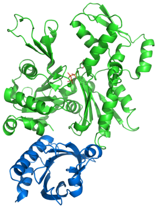
Actin is a family of globular multi-functional proteins that form microfilaments in the cytoskeleton, and the thin filaments in muscle fibrils. It is found in essentially all eukaryotic cells, where it may be present at a concentration of over 100 μM; its mass is roughly 42 kDa, with a diameter of 4 to 7 nm.

<span class="mw-page-title-main">Phalloidin</span> Chemical compound

Phalloidin belongs to a class of toxins called phallotoxins, which are found in the death cap mushroom (Amanita phalloides). It is a rigid bicyclic heptapeptide that is lethal after a few days when injected into the bloodstream. The major symptom of phalloidin poisoning is acute hunger due to the destruction of liver cells. It functions by binding and stabilizing filamentous actin (F-actin) and effectively prevents the depolymerization of actin fibers. Due to its tight and selective binding to F-actin, derivatives of phalloidin containing fluorescent tags are used widely in microscopy to visualize F-actin in biomedical research.

<span class="mw-page-title-main">Cytochalasin B</span> Chemical compound

Cytochalasin B, the name of which comes from the Greek cytos (cell) and chalasis (relaxation), is a cell-permeable mycotoxin. It was found that substoichiometric concentrations of cytochalasin B (CB) strongly inhibit network formation by actin filaments. Due to this, it is often used in cytological research. It inhibits cytoplasmic division by blocking the formation of contractile microfilaments. It inhibits cell movement and induces nuclear extrusion. Cytochalasin B shortens actin filaments by blocking monomer addition at the fast-growing end of polymers. Cytochalasin B inhibits glucose transport and platelet aggregation. It blocks adenosine-induced apoptotic body formation without affecting activation of endogenous ADP-ribosylation in leukemia HL-60 cells. It is also used in cloning through nuclear transfer. Here enucleated recipient cells are treated with cytochalasin B. Cytochalasin B makes the cytoplasm of the oocytes more fluid and makes it possible to aspirate the nuclear genome of the oocyte within a small vesicle of plasma membrane into a micro-needle. Thereby, the oocyte genome is removed from the oocyte, while preventing rupture of the plasma membrane.

<span class="mw-page-title-main">Profilin</span>

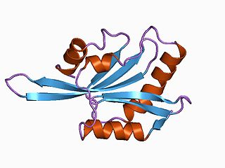
Profilin is an actin-binding protein involved in the dynamic turnover and reconstruction of the actin cytoskeleton. It is found in most eukaryotic organisms. Profilin is important for spatially and temporally controlled growth of actin microfilaments, which is an essential process in cellular locomotion and cell shape changes. This restructuring of the actin cytoskeleton is essential for processes such as organ development, wound healing, and the hunting down of infectious intruders by cells of the immune system.

The lamellipodium is a cytoskeletal protein actin projection on the leading edge of the cell. It contains a quasi-two-dimensional actin mesh; the whole structure propels the cell across a substrate. Within the lamellipodia are ribs of actin called microspikes, which, when they spread beyond the lamellipodium frontier, are called filopodia. The lamellipodium is born of actin nucleation in the plasma membrane of the cell and is the primary area of actin incorporation or microfilament formation of the cell.

<span class="mw-page-title-main">ADF/Cofilin family</span>

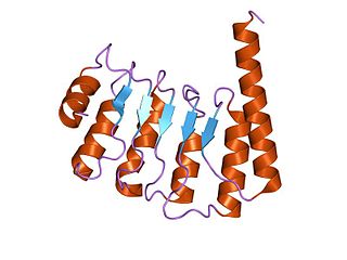
ADF/cofilin is a family of actin-binding proteins associated with the rapid depolymerization of actin microfilaments that give actin its characteristic dynamic instability. This dynamic instability is central to actin's role in muscle contraction, cell motility and transcription regulation.

Cytochalasins are fungal metabolites that have the ability to bind to actin filaments and block polymerization and the elongation of actin. As a result of the inhibition of actin polymerization, cytochalasins can change cellular morphology, inhibit cellular processes such as cell division, and even cause cells to undergo apoptosis. Cytochalasins have the ability to permeate cell membranes, prevent cellular translocation and cause cells to enucleate. Cytochalasins can also have an effect on other aspects of biological processes unrelated to actin polymerization. For example, cytochalasin A and cytochalasin B can also inhibit the transport of monosaccharides across the cell membrane, cytochalasin H has been found to regulate plant growth, cytochalasin D inhibits protein synthesis and cytochalasin E prevents angiogenesis.

<span class="mw-page-title-main">Treadmilling</span> Simultaneous growth and breakdown on opposite ends of a protein filament

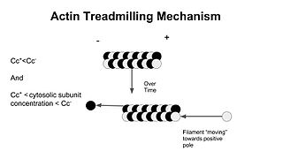
In molecular biology, treadmilling is a phenomenon observed within protein filaments of the cytoskeletons of many cells, especially in actin filaments and microtubules. It occurs when one end of a filament grows in length while the other end shrinks, resulting in a section of filament seemingly "moving" across a stratum or the cytosol. This is due to the constant removal of the protein subunits from these filaments at one end of the filament, while protein subunits are constantly added at the other end. Treadmilling was discovered by Wegner, who defined the thermodynamic and kinetic constraints. Wegner recognized that: “The equilibrium constant (K) for association of a monomer with a polymer is the same at both ends, since the addition of a monomer to each end leads to the same polymer.”; a simple reversible polymer can’t treadmill; ATP hydrolysis is required. GTP is hydrolyzed for microtubule treadmilling.

<span class="mw-page-title-main">Protein filament</span> Long chain of protein monomers

In biology, a protein filament is a long chain of protein monomers, such as those found in hair, muscle, or in flagella. Protein filaments form together to make the cytoskeleton of the cell. They are often bundled together to provide support, strength, and rigidity to the cell. When the filaments are packed up together, they are able to form three different cellular parts. The three major classes of protein filaments that make up the cytoskeleton include: actin filaments, microtubules and intermediate filaments.

CapZ, also known as CAPZ, CAZ1 and CAPPA1, is a capping protein that caps the barbed end of actin filaments in muscle cells.

<span class="mw-page-title-main">Cordon-bleu protein</span> Protein-coding gene in the species Homo sapiens

Protein cordon-bleu is a protein that in humans is encoded by the COBL gene.

<span class="mw-page-title-main">Actin assembly-inducing protein</span>

The Actin assembly-inducing protein (ActA) is a protein encoded and used by Listeria monocytogenes to propel itself through a mammalian host cell. ActA is a bacterial surface protein comprising a membrane-spanning region. In a mammalian cell the bacterial ActA interacts with the Arp2/3 complex and actin monomers to induce actin polymerization on the bacterial surface generating an actin comet tail. The gene encoding ActA is named actA or prtB.

Actin remodeling is a biochemical process in cells. In the actin remodeling of neurons, the protein actin is part of the process to change the shape and structure of dendritic spines. G-actin is the monomer form of actin, and is uniformly distributed throughout the axon and the dendrite. F-actin is the polymer form of actin, and its presence in dendritic spines is associated with their change in shape and structure. Actin plays a role in the formation of new spines as well as stabilizing spine volume increase. The changes that actin brings about lead to the formation of new synapses as well as increased cell communication.

<span class="mw-page-title-main">MDia1</span> Protein

mDia1 is a member of the protein family called the formins and is a Rho effector. It is the mouse version of the diaphanous homolog 1 of Drosophila. mDia1 localizes to cells' mitotic spindle and midbody, plays a role in stress fiber and filopodia formation, phagocytosis, activation of serum response factor, formation of adherens junctions, and it can act as a transcription factor. mDia1 accelerates actin nucleation and elongation by interacting with barbed ends of actin filaments. The gene encoding mDia1 is located on Chromosome 18 of Mus musculus and named Diap1.

<span class="mw-page-title-main">Arp2/3 complex</span> Macromolecular complex

Arp2/3 complex is a seven-subunit protein complex that plays a major role in the regulation of the actin cytoskeleton. It is a major component of the actin cytoskeleton and is found in most actin cytoskeleton-containing eukaryotic cells. Two of its subunits, the Actin-Related Proteins ARP2 and ARP3, closely resemble the structure of monomeric actin and serve as nucleation sites for new actin filaments. The complex binds to the sides of existing ("mother") filaments and initiates growth of a new ("daughter") filament at a distinctive 70 degree angle from the mother. Branched actin networks are created as a result of this nucleation of new filaments. The regulation of rearrangements of the actin cytoskeleton is important for processes like cell locomotion, phagocytosis, and intracellular motility of lipid vesicles.

<span class="mw-page-title-main">Cyclase-associated protein family</span>

In molecular biology, the cyclase-associated protein family (CAP) is a family of highly conserved actin-binding proteins present in a wide range of organisms including yeast, flies, plants, and mammals. CAPs are multifunctional proteins that contain several structural domains. CAP is involved in species-specific signalling pathways. In Drosophila, CAP functions in Hedgehog-mediated eye development and in establishing oocyte polarity. In Dictyostelium discoideum, CAP is involved in microfilament reorganisation near the plasma membrane in a PIP2-regulated manner and is required to perpetuate the cAMP relay signal to organise fruitbody formation. In plants, CAP is involved in plant signalling pathways required for co-ordinated organ expansion. In yeast, CAP is involved in adenylate cyclase activation, as well as in vesicle trafficking and endocytosis. In both yeast and mammals, CAPs appear to be involved in recycling G-actin monomers from ADF/cofilins for subsequent rounds of filament assembly. In mammals, there are two different CAPs that share 64% amino acid identity.

An actin nucleation core is a protein trimer with three actin monomers. It is called a nucleation core because it leads to the energetically favorable elongation reaction once a tetramer is formed from a trimer. Actin protein dimers and trimers are energetically unfavorable.Actin nucleators like the Arp2/3 complex of proteins from the formin family are most frequently involved in this process. Actin nucleation factors start the polymerization of actin within cells.

References

  1. 1 2 3 4 5 6 7 8 9 10 11 Thomas P. Stossel; Gabriel Fenteany; John H. Hartwig (2006). "Cell surface actin remodeling" (PDF). Journal of Cell Science. 119 (Pt 16): 3261–3264. doi: 10.1242/jcs.02994 . PMID   16899816. S2CID   30606964. Archived from the original (PDF) on 2010-06-18.
  2. 1 2 3 4 Amon; Berk; Bretscher; Kaiser; Krieger; Lodish; Ploegh; Scott (2013). Molecular Cell Biology (Seventh ed.). New York: W.H Freeman and Company. pp. 775–815. ISBN   978-1-4292-3413-9.
  3. 1 2 3 4 Begg, DA; Rodewald, R; Rebhun, LI (1 December 1978). "The visualization of actin filament polarity in thin sections. Evidence for the uniform polarity of membrane-associated filaments". The Journal of Cell Biology. 79 (3): 846–852. doi:10.1083/jcb.79.3.846. PMC   2110270 . PMID   569662.
  4. 1 2 3 Kuhn, JR; Pollard, TD (February 2005). "Real-Time Measurements of Actin Filament Polymerization by Total Internal Reflection Fluorescence Microscopy". Biophysical Journal. 88 (2): 1387–1402. Bibcode:2005BpJ....88.1387K. doi:10.1529/biophysj.104.047399. PMC   1305141 . PMID   15556992.
  5. Rottner, Klemens; Stradal, Theresia E.B. (2011). "Actin dynamics and turnover in cell motility". Current Opinion in Cell Biology. 23 (5): 569–578. doi:10.1016/j.ceb.2011.07.003. PMID   21807492.
  6. 1 2 3 Nicholson-Dykstra, S; Higgs, HN; Harris, ES (10 May 2005). "Actin Dynamics: Growth from Dendritic Branches". Current Biology. 15 (9): R346–R357. doi: 10.1016/j.cub.2005.04.029 . PMID   15886095. S2CID   16997184.
  7. Roselli, N; Castagnino, A; Pontrelli, G; Natalini, R; Barakat, A.I. (July 2022). "Modeling ATP-mediated endothelial cell elongation on line patterns". Biomechanics and Modeling in Mechanobiology . doi: 10.1007/s10237-022-01604-2 . PMID   35902488.
  8. 1 2 Kalwat, MA; Thurmond, DC (23 August 2013). "Signaling mechanisms of glucose-induced F-actin remodeling in pancreatic islet β cells". Experimental & Molecular Medicine . 45 (37): e37. doi:10.1038/emm.2013.73. PMC   3789261 . PMID   23969997.