Anal pore

Last updated
Diagram of a Ciliate with various cell structures (including anal pore) labelled Paramecium diagram.png
Diagram of a Ciliate with various cell structures (including anal pore) labelled

The anal pore or cytoproct is a structure in various single-celled eukaryotes where waste is ejected after the nutrients from food have been absorbed into the cytoplasm. [1]

Contents

In ciliates, the anal pore (cytopyge) and cytostome are the only regions of the pellicle that are not covered by ridges, cilia or rigid covering. They serve as analogues of, respectively, the anus and mouth of multicellular organisms. The cytopyge's thin membrane allows vacuoles to be merged into the cell wall and emptied.[ citation needed ]

Location

The anal pore is an exterior opening of microscopic organisms through which undigested food waste, water, or gas are expelled from the body. The anal pore is located on the ventral surface, usually in the posterior half of the cell. The anal pore itself is actually a structure made up of two components: piles of fibres, and microtubules.

This structure is found in different unicellular eukaryotes like paramecium organelles.

Function

Digested nutrients from the vacuole pass into the cytoplasm, making the vacuole shrink and moves to the anal pore, where it ruptures to release the waste content to the environment outside of the cell. The cytoproct is used for the excretion of indigestible debris contained in the food vacuoles.

Most microorganisms possess an anal pore for excretion, usually in the form of an opening on the pellicle to eject out indigestible debris. The opening and closing of the cytoproct resemble a reversible ring of tissue fusion occurring between the inner and outer layers located at the aboral end. An anal pore is not a permanently visible structure; it appears at the time of defecation and then disappears afterward.

In paramecium, the anal pore is a region of pellicle that is not covered by ridges and cilia, and the area has thin pellicles that allow the vacuoles to be merged into the cell surface to be emptied.

In ciliates, the anal cytostomes and cytopyge pore regions are not covered by either ridges or cilia or hard coatings like the other parts of the organism. As a food vacuole approaches the cytoproct region it actually starts to flatten out the surrounding cells, and a thin-membrane vacuole allows it to be combined in the cell wall. Once the vacuole attaches to the plasma membrane of the cell wall, the vacuole is emptied. The waste excreted by the cell can come as a membrane-bound packaged ball, or as a stream of debris behind the organism.

Directly after secretion of the waste products, deep invagination (deep, canyon-like structure that was the vacuole) is still present. About 10 to 30 seconds after secretion, the vacuole detaches, and a new thin plasma membrane is formed. After a minute has gone by the organisms cytoproct is closed up again and the process is ready to be repeated.

In marine animals

Anatomical diagram of a cydippid comb jelly. Ctenophore diagram - en.svg
Anatomical diagram of a cydippid comb jelly.

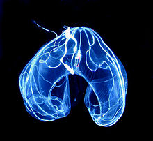
Ctnephores are marine animals which superficially resemble jellyfish, but have biradial symmetry and use eight bands of transverse ciliated plates to swim. All ctenophores possess a pair of small anal pores located adjacent to the apical sensory organ which is thought to control osmotic pressure. These animals are also with animal pore. Ctenophores have sometimes been interpreted as homologous with the anus of bilaterian animals (worms, humans, snails, fish, etc.). Furthermore, they possess a third tissue layer between the endoderm and ectoderm, another characteristic reminiscent of the Bilateria. Ctenophores possess a functional through-gut from which digested waste products and material distributed via the endodermal canals are expelled to the exterior environment through terminal anal pores, which are specialized to control outflow from the branched endodermal canal system. Ctenophores have no true anus; the central canal opens toward the aboral end by two small pores, through which a small amount of egestion can take place.[ citation needed ]

Bathocyroe fosteri with anal pore. Bathocyroe fosteri.jpg
Bathocyroe fosteri with anal pore.

Related Research Articles

<span class="mw-page-title-main">Cell (biology)</span> Basic unit of many life forms

The cell is the basic structural and functional unit of all forms of life. Every cell consists of cytoplasm enclosed within a membrane; many cells contain organelles, each with a specific function. The term comes from the Latin word cellula meaning 'small room'. Most cells are only visible under a microscope. Cells emerged on Earth about 4 billion years ago. All cells are capable of replication, protein synthesis, and motility.

<span class="mw-page-title-main">Ctenophora</span> Phylum of gelatinous marine animals

Ctenophora comprise a phylum of marine invertebrates, commonly known as comb jellies, that inhabit sea waters worldwide. They are notable for the groups of cilia they use for swimming, and they are the largest animals to swim with the help of cilia.

<i>Paramecium</i> Genus of unicellular ciliates, commonly studied as a representative of the ciliate group

Paramecium is a genus of eukaryotic, unicellular ciliates, commonly studied as a model organism of the ciliate group. Paramecium are widespread in freshwater, brackish, and marine environments and are often abundant in stagnant basins and ponds. Because some species are readily cultivated and easily induced to conjugate and divide, they have been widely used in classrooms and laboratories to study biological processes. The usefulness of Paramecium as a model organism has caused one ciliate researcher to characterize it as the "white rat" of the phylum Ciliophora.

<i>Stylonychia</i> Genus of single-celled organisms

Stylonychia is a genus of ciliates, in the subclass Hypotrichia. Species of Stylonychia are very common in fresh water and soil, and may be found on filamentous algae, surface films, and among particles of sediment. Like other Hypotrichs, Stylonychia has cilia grouped into membranelles alongside the mouth and cirri over the body. It is distinguished partly by long cirri at the posterior, usually a cluster of three. The largest can just be seen at a 25x magnification, and the smallest can just be seen at a 450x magnification.

The nephridium is an invertebrate organ, found in pairs and performing a function similar to the vertebrate kidneys. Nephridia remove metabolic wastes from an animal's body. Nephridia come in two basic categories: metanephridia and protonephridia. All nephridia- and kidney- having animals belong to the clade Nephrozoa.

<span class="mw-page-title-main">Contractile vacuole</span> Organelle used in regulating osmosis

A contractile vacuole (CV) is a sub-cellular structure (organelle) involved in osmoregulation. It is found predominantly in protists and in unicellular algae. It was previously known as pulsatile or pulsating vacuole.

<i>Mnemiopsis</i> Genus of comb jellies

Mnemiopsis leidyi, the warty comb jelly or sea walnut, is a species of tentaculate ctenophore. It is native to western Atlantic coastal waters, but has become established as an invasive species in European and western Asian regions. Three species have been named in the genus Mnemiopsis, but they are now believed to be different ecological forms of a single species M. leidyi by most zoologists.

<i>Vorticella</i> Genus of single-celled organisms

Vorticella is a genus of bell-shaped ciliates that have stalks to attach themselves to substrates. The stalks have contractile myonemes, allowing them to pull the cell body against substrates. The formation of the stalk happens after the free-swimming stage.

<i>Didinium</i> Genus of single-celled organisms

Didinium is a genus of unicellular ciliates with at least ten accepted species. All are free-living carnivores. Most are found in fresh and brackish water, but three marine species are known. Their diet consists largely of Paramecium, although they will also attack and consume other ciliates. Some species, such as D. gargantua, also feeds on non-ciliate protists, including dinoflagellates, cryptomonads, and green algae.

<i>Paramecium caudatum</i> Species of single-celled organism

Paramecium caudatum is a species of unicellular protist in the phylum Ciliophora. They can reach 0.33 mm in length and are covered with minute hair-like organelles called cilia. The cilia are used in locomotion and feeding. The species is very common, and widespread in marine, brackish and freshwater environments.

<i>Trichodina</i> Genus of single-celled organisms

Trichodina is a genus of ciliate alveolates that is ectocommensal or parasitic on aquatic animals, particularly fish. They are characterised by the presence of a ring of interlocking cytoskeletal denticles, which provide support for the cell and allow for adhesion to surfaces including fish tissue.

<span class="mw-page-title-main">Protozoan infection</span> Parasitic disease caused by a protozoan

Protozoan infections are parasitic diseases caused by organisms formerly classified in the kingdom Protozoa. These organisms are now classified in the supergroups Excavata, Amoebozoa, Harosa, and Archaeplastida. They are usually contracted by either an insect vector or by contact with an infected substance or surface.

<span class="mw-page-title-main">Protozoa</span> Single-celled eukaryotic organisms that feed on organic matter

Protozoa are a polyphyletic group of single-celled eukaryotes, either free-living or parasitic, that feed on organic matter such as other microorganisms or organic debris. Historically, protozoans were regarded as "one-celled animals".

<span class="mw-page-title-main">Ciliate</span> Taxon of protozoans with hair-like organelles called cilia

The ciliates are a group of alveolates characterized by the presence of hair-like organelles called cilia, which are identical in structure to eukaryotic flagella, but are in general shorter and present in much larger numbers, with a different undulating pattern than flagella. Cilia occur in all members of the group and are variously used in swimming, crawling, attachment, feeding, and sensation.

<i>Paramecium aurelia</i> Species of single-celled organism

Paramecium aurelia are unicellular organisms belonging to the genus Paramecium of the phylum Ciliophora. They are covered in cilia which help in movement and feeding.Paramecium can reproduce sexually, asexually, or by the process of endomixis. Paramecium aurelia demonstrate a strong "sex reaction" whereby groups of individuals will cluster together, and emerge in conjugant pairs. This pairing can last up to 12 hours, during which the micronucleus of each organism will be exchanged. In Paramecium aurelia, a cryptic species complex was discovered by observation. Since then, some have tried to decode this complex using genetic data.

<span class="mw-page-title-main">Intracellular digestion</span>

Every organism requires energy to be active. However, to obtain energy from its outside environment, cells must not only retrieve molecules from their surroundings but also break them down. This process is known as intracellular digestion. In its broadest sense, intracellular digestion is the breakdown of substances within the cytoplasm of a cell. In detail, a phagocyte's duty is obtaining food particles and digesting it in a vacuole. For example, following phagocytosis, the ingested particle fuses with a lysosome containing hydrolytic enzymes to form a phagolysosome; the pathogens or food particles within the phagosome are then digested by the lysosome's enzymes.

<span class="mw-page-title-main">Platyctenida</span> Order of benthic ctenophores

Platyctenida is an order of comb jellies in the class Tentaculata.

<span class="mw-page-title-main">Condylostoma</span> Genus of protists belonging to the ciliates phylum

Condylostoma is a genus of unicellular ciliate protists, belonging to the class Heterotrichea.

<span class="mw-page-title-main">Stentor roeselii</span> Species of single-celled organism

Stentor roeselii is a free-living ciliate species of the genus Stentor, in the class Heterotrichea. It is a common and widespread protozoan, found throughout the world in freshwater ponds, lakes, rivers and ditches.

<i>Euplokamis</i> Genus of ctenophores

Euplokamis is a genus of ctenophores, or comb jellies, belonging to the monotypic family Euplokamididae. It shares the common name sea gooseberry with species of the genus Pleurobrachia. Despite living for hundreds of millions of years in marine environments, there is minimal research regarding Euplokamis, because they are uncommon. Research on the evolution of the basic body structures of diploblastic metazoans revealed that there are four major phyla, including the Ctenophores. Although the morphology of Euplokamis often resembles the medusa stage of Cnidarians, their eight rows of combs are one distinguishing feature that led to the official classification of Ctenophores. After being originally described by Chun (1879), the family Euplokamididae was expanded by Mills (1987) due to the discovery of a new species, Euplokamis dunlapae. Further research indicated that Euplokamis should be identified from Mertensiidae due to the rows of combs and some compression. They may also be distinguished from the genus Pleurobrachia due to their more elongated shape. Additionally, various adaptations of Euplokamis have been observed such as the use of tentacles for movement/feeding, a complex nervous system, and bioluminescent capabilities. Other characteristics including a defined mesoderm, lack of stinging cells, developmental differences, and symmetry supported the reclassification of these organisms.

References

  1. Stuart Hogg (2005). "The Ciliates". Essential Microbiology (1 ed.). John Wiley & Sons. ISBN   978-0471497547 . Retrieved 16 January 2018.

Bibliography