The internal carotid artery is an artery in the neck which supplies the anterior and middle cerebral circulation.

The internal capsule is a white matter structure situated in the inferomedial part of each cerebral hemisphere of the brain. It carries information past the basal ganglia, separating the caudate nucleus and the thalamus from the putamen and the globus pallidus. The internal capsule contains both ascending and descending axons, going to and coming from the cerebral cortex. It also separates the caudate nucleus and the putamen in the dorsal striatum, a brain region involved in motor and reward pathways.

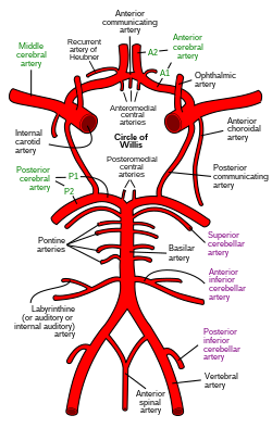
The anterior cerebral artery (ACA) is one of a pair of cerebral arteries that supplies oxygenated blood to most midline portions of the frontal lobes and superior medial parietal lobes of the brain. The two anterior cerebral arteries arise from the internal carotid artery and are part of the circle of Willis. The left and right anterior cerebral arteries are connected by the anterior communicating artery.

The subarachnoid cisterns are spaces formed by openings in the subarachnoid space, an anatomic space in the meninges of the brain. The space is situated between the two meninges, the arachnoid mater and the pia mater. These cisterns are filled with cerebrospinal fluid (CSF).
The middle cerebral artery (MCA) is one of the three major paired cerebral arteries that supply blood to the cerebrum. The MCA arises from the internal carotid artery and continues into the lateral sulcus where it then branches and projects to many parts of the lateral cerebral cortex. It also supplies blood to the anterior temporal lobes and the insular cortices.
The posterior cerebral artery (PCA) is one of a pair of cerebral arteries that supply oxygenated blood to the occipital lobe, part of the back of the human brain. The two arteries originate from the distal end of the basilar artery, where it bifurcates into the left and right posterior cerebral arteries. These anastomose with the middle cerebral arteries and internal carotid arteries via the posterior communicating arteries.

In human anatomy, the left and right posterior communicating arteries are small arteries at the base of the brain that form part of the circle of Willis.
In human anatomy, the anterior communicating artery is a blood vessel of the brain that connects the left and right anterior cerebral arteries.
The cerebral arteries describe three main pairs of arteries and their branches, which perfuse the cerebrum of the brain. The three main arteries are the:
The tuber cinereum is the portion of hypothalamus forming the floor of the third ventricle situated between the optic chiasm, and the mammillary bodies. The tuberal region is one of the three regions of the hypothalamus, the other two being the chiasmatic region and the mamillary region.
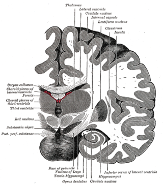
The posterior perforated substance is a layer of gray matter which is pierced by small apertures for the transmission of blood vessels. Its inferior part lies on the ventral aspect of the medial portions of the tegmenta and contains the interpeduncular nucleus; its superior part forms part of the floor of the third ventricle.

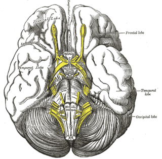
The anterior perforated substance is a part of the brain. It is bilateral. It is irregular and quadrilateral. It lies in front of the optic tract and behind the olfactory trigone.

The basal vein is a vein in the brain. It is formed at the anterior perforated substance by the union of
In anatomy, arterial tree is used to refer to all arteries and/or the branching pattern of the arteries. This article regards the human arterial tree. Starting from the aorta:
The interpeduncular cistern is the subarachnoid cistern situated between the dorsum sellae (anteriorly) and the two cerebral peduncles of the mesencephalon (midbrain). Its roof is represented by the floor of the third ventricle. Its floor is formed by the arachnoid membrane extending between the temporal lobes of either side. Anteriorly, it extends to the optic chiasm.
The anterolateral central arteries or lenticulostriate arteries are a group of small arteries mostly arising from the middle cerebral artery that enter the brain through the anterior perforated substance to provide arterial supply to parts of the basal ganglia. They are end arteries.
The recurrent artery of Heubner, Heubner's artery or distal medial striate artery is It is a branch of the anterior cerebral artery. It supplies the head of the caudate nucleus and adjacent part of the anterior limb of internal capsule, olfactory regions, and parts of the putamen and septal nuclei.
Central arteries may refer to:
The thalamoperforating arteries are posteromedial central arteries which supply parts of the thalamus.