Dendrites, also dendrons, are branched protoplasmic extensions of a nerve cell that propagate the electrochemical stimulation received from other neural cells to the cell body, or soma, of the neuron from which the dendrites project. Electrical stimulation is transmitted onto dendrites by upstream neurons via synapses which are located at various points throughout the dendritic tree. Dendrites play a critical role in integrating these synaptic inputs and in determining the extent to which action potentials are produced by the neuron. Dendritic arborization, also known as dendritic branching, is a multi-step biological process by which neurons form new dendritic trees and branches to create new synapses. The morphology of dendrites such as branch density and grouping patterns are highly correlated to the function of the neuron. Malformation of dendrites is also tightly correlated to impaired nervous system function. Some disorders that are associated with the malformation of dendrites are autism, depression, schizophrenia, Down syndrome and anxiety.

A neuron, also known as a neurone or nerve cell, is an electrically excitable cell that communicates with other cells via specialized connections called synapses. It is the main component of nervous tissue. All animals except sponges and placozoans have neurons, but other multicellular organisms such as plants do not.
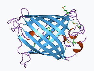
The green fluorescent protein (GFP) is a protein composed of 238 amino acid residues that exhibits bright green fluorescence when exposed to light in the blue to ultraviolet range. Although many other marine organisms have similar green fluorescent proteins, GFP traditionally refers to the protein first isolated from the jellyfish Aequorea victoria, avGFP. The GFP from A. victoria has a major excitation peak at a wavelength of 395 nm and a minor one at 475 nm. Its emission peak is at 509 nm, which is in the lower green portion of the visible spectrum. The fluorescence quantum yield (QY) of GFP is 0.79. The GFP from the sea pansy has a single major excitation peak at 498 nm. GFP makes for an excellent tool in many forms of biology due to its ability to form internal chromophore without requiring any accessory cofactors, gene products, or enzymes / substrates other than molecular oxygen.

In molecular biology and biotechnology, a fluorescent tag, also known as a fluorescent label or fluorescent probe, is a molecule that is attached chemically to aid in the detection of a biomolecule such as a protein, antibody, or amino acid. Generally, fluorescent tagging, or labeling, uses a reactive derivative of a fluorescent molecule known as a fluorophore. The fluorophore selectively binds to a specific region or functional group on the target molecule and can be attached chemically or biologically. Various labeling techniques such as enzymatic labeling, protein labeling, and genetic labeling are widely utilized. Ethidium bromide, fluorescein and green fluorescent protein are common tags. The most commonly labelled molecules are antibodies, proteins, amino acids and peptides which are then used as specific probes for detection of a particular target.
Behavioral neuroscience, also known as biological psychology, biopsychology, or psychobiology, is the application of the principles of biology to the study of physiological, genetic, and developmental mechanisms of behavior in humans and other animals.
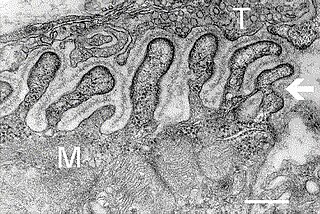
A neuromuscular junction is a chemical synapse formed by the contact between a motor neuron and a muscle fiber. It is at the neuromuscular junction that a motor neuron is able to transmit a signal to the muscle fiber, causing muscle contraction.

The axon hillock is a specialized part of the cell body of a neuron that connects to the axon. It can be identified using light microscopy from its appearance and location in a neuron and from its sparse distribution of Nissl substance.
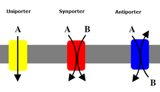
A uniporter is an integral membrane protein that transports a single type of substrate species across a cell membrane. It may use either facilitated diffusion and transport along a diffusion gradient or transport against one with an active transport process.. They can be either ion channels or carrier proteins.
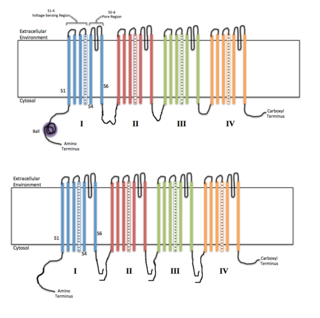
Voltage-gated ion channels are a class of transmembrane proteins that form ion channels that are activated by changes in the electrical membrane potential near the channel. The membrane potential alters the conformation of the channel proteins, regulating their opening and closing. Cell membranes are generally impermeable to ions, thus they must diffuse through the membrane through transmembrane protein channels. They have a crucial role in excitable cells such as neuronal and muscle tissues, allowing a rapid and co-ordinated depolarization in response to triggering voltage change. Found along the axon and at the synapse, voltage-gated ion channels directionally propagate electrical signals. Voltage-gated ion-channels are usually ion-specific, and channels specific to sodium (Na+), potassium (K+), calcium (Ca2+), and chloride (Cl−) ions have been identified. The opening and closing of the channels are triggered by changing ion concentration, and hence charge gradient, between the sides of the cell membrane.
The soma (somas), perikaryon, neurocyton, or cell body is the bulbous, non-process portion of a neuron or other brain cell type, containing the cell nucleus. The word 'soma' comes from the Greek 'σῶμα', meaning 'body'. Although it is often used to refer to neurons, it can also refer to other cell types as well, including astrocytes, oligodendrocytes, and microglia. There are many different specialized types of neurons, and their sizes vary from as small as about 5 micrometres to over 10 millimetre for some of the smallest and largest neurons of invertebrates, respectively.
Cameleon is an engineered protein based on variant of green fluorescent protein used to visualize calcium levels in living cells. It is a genetically encoded calcium sensor created by Roger Y. Tsien and coworkers. The name is a conflation of CaM and chameleon to indicate the fact that the sensor protein undergoes a conformation change and radiates at an altered wavelength upon calcium binding to the calmodulin element of the Cameleon. Cameleon was the first genetically encoded calcium sensor that could be used for ratiometric measurements and the first to be used in a transgenic animal to record activity in neurons and muscle cells. Cameleon and other genetically-encoded calcium indicators (GECIs) have found many applications in neuroscience and other fields of biology. It was created by fusing BFP, calmodulin, calmodulin-binding peptide M13 and EGFP.

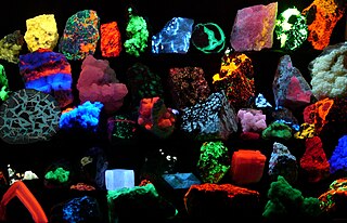
Microbial art, agar art, or germ art is artwork created by culturing microorganisms in certain patterns. The microbes used can be bacteria, yeast fungi, or less commonly, protists. The microbes can be chosen for their natural colours, or can be engineered to express fluorescent proteins and viewed under ultraviolet light to make them fluoresce in colour.

Brainbow is a process by which individual neurons in the brain can be distinguished from neighboring neurons using fluorescent proteins. By randomly expressing different ratios of red, green, and blue derivatives of green fluorescent protein in individual neurons, it is possible to flag each neuron with a distinctive color. This process has been a major contribution to the field of connectomics, traditionally known as hodology, which is the study of neural connections in the brain.
Optogenetics is a biological technique that involves the use of light to control cells in living tissue, typically neurons, that have been genetically modified to express light-sensitive ion channels. It is a neuromodulation method that uses a combination of techniques from optics and genetics to control and monitor the activities of individual neurons in living tissue—even within freely-moving animals—and to precisely measure these manipulation effects in real-time. The key reagents used in optogenetics are light-sensitive proteins. Neuronal control is achieved using optogenetic actuators like channelrhodopsin, halorhodopsin, and archaerhodopsin, while optical recording of neuronal activities can be made with the help of optogenetic sensors for calcium (GCaMP), vesicular release (synapto-pHluorin), neurotransmitter (GluSnFRs), or membrane voltage. Control of activity is restricted to genetically defined neurons and performed in a spatiotemporal-specific manner by light.

Sodium channel, voltage gated, type VIII, alpha subunit also known as SCN8A or Nav1.6 is a membrane protein encoded by the SCN8A gene. Nav1.6 is one sodium channel isoform and is the primary voltage-gated sodium channel at the nodes of Ranvier. The channels are highly concentrated in sensory and motor axons in the peripheral nervous system and cluster at the nodes in the central nervous system.

Potassium voltage-gated channel subfamily S member 3 (Kv9.3) is a protein that in humans is encoded by the KCNS3 gene. KCNS3 gene belongs to the S subfamily of the potassium channel family. It is highly expressed in pulmonary artery myocytes, placenta, and parvalbumin-containing GABA neurons in brain cortex. In humans, single-nucleotide polymorphisms of the KCNS3 gene are associated with airway hyperresponsiveness, whereas decreased KCNS3 mRNA expression is found in the prefrontal cortex of patients with schizophrenia.
Kaede is a photoactivatable fluorescent protein naturally originated from a stony coral, Trachyphyllia geoffroyi. Its name means "maple" in Japanese. With the irradiation of ultraviolet light (350–400 nm), Kaede undergoes irreversible photoconversion from green fluorescence to red fluorescence.
Fluorescent chloride sensors are used for chemical analysis. The discoveries of chloride (Cl−) participations in physiological processes stimulates the measurements of intracellular Cl− in live cells and the development of fluorescent tools referred below.
Calcium imaging is a scientific technique usually carried out in research which is designed to show the calcium (Ca2+) status of an isolated cell, tissue or medium. Calcium imaging techniques take advantage of so-called calcium indicators, fluorescent molecules that can respond to the binding of Ca2+ ions by changing their fluorescence properties. Two main classes of calcium indicators exist: chemical indicators and genetically encoded calcium indicators (GECI). Calcium imaging can be used to optically probe intracellular calcium in living animals. This technique has allowed studies of calcium signalling in a wide variety of cell types and neuronal activity in hundreds of neurons and glial cells within neuronal circuits.