Azurin

Last updated
Azurin
5azu.jpg
Azurin tetramer, Pseudomonas aeruginosa
Identifiers
SymbolCopper-bind
Pfam PF00127
SCOP2 5azu / SCOPe / SUPFAM
CDD cd13922
Available protein structures:
Pfam   structures / ECOD  
PDB RCSB PDB; PDBe; PDBj
PDBsum structure summary

Azurin is a small, periplasmic, bacterial blue copper protein found in Pseudomonas , Bordetella , or Alcaligenes bacteria. Azurin moderates single-electron transfer between enzymes associated with the cytochrome chain by undergoing oxidation-reduction between Cu(I) and Cu(II). Each monomer of an azurin tetramer has a molecular weight of approximately 14kDa, contains a single copper atom, is intensively blue, and has a fluorescence emission band centered at 308 nm.

Contents

Azurins and pseudoazurins participate in the denitrification processes in bacteria., [1] including the gram-negative bacteria Pseudomonas aeruginosa, by interacting with cytochrome c551. Azurin from P aeruginosa is a type I blue copper protein (cupredoxin), while cytochrome c551 (9 kDa) is a haem-containing cytochrome. Azurin possesses a relatively large hydrophobic patch close to the active site, and two residues in this hydrophobic patch, Met-44 and Met-64, are believed to be involved in its interaction with the redox partners cytochrome c551 and nitrite reductase. [2]

Although unrelated to its electron-transfer property, azurin has been found to have anticancer properties through its interaction with tumor-suppressor protein p53.

Protein function

In its oxidized form, azurin (Cu2+Az) receives an electron from its redox partner and is reduced according to the following reaction:

Cu2+Az + e → Cu+Az

The redox potential is 310 mV. [3]

The highly-interconnected beta-sheet structure of azurin is strongly coupled with its electron-transfer center (the copper-binding side). [4] Considerable experimental evidence exists to suggest that hydrogen bonds play a role in the long-distance electron transfer mechanism of azurin. Taken together, these observations suggest that electrons tunnel through the protein along its polypeptide and hydrogen bonds, making azurin a useful model system for studying long-range, intraprotein electron transfer (LRET). [4]

Protein structure

The copper binding domain of azurin rendered in PyMol, with four of the five copper-binding ligands labeled. The distance (in angstroms) from the copper atom to each individual ligand has been recorded. Azurin copper binding domain.png
The copper binding domain of azurin rendered in PyMol, with four of the five copper-binding ligands labeled. The distance (in angstroms) from the copper atom to each individual ligand has been recorded.

Azurin is a monomeric protein that weighs approximately 14 kDa and is composed of 128 amino acids forming eight beta-strands arranged in a beta-barrel formation. [5] The strands are connected by turns and a single alpha-helical insertion. [5] A single-atom copper binding site is located about 7 Å below each monomer’s surface towards its northern end; the copper atom that inhabits it is coordinated by five ligands surrounded by an extensive hydrophobic patch. [6]

The three equatorial copper ligands are composed of a thiolate (Cys112) and two imidazoles (His46, His117), and the carbonyl oxygen atoms of Gly45 and Met121 serve as the two weak axial ligands. [5] With the exception of Gly45, the copper-binding configuration above is common to the structures of all blue type 1 copper-binding proteins determined thus far. [7] Once coordinated, the ligand-metal complex assumes a distorted, trigonal bi-pyramidal geometry that stabilizes the cuprous (Cu(I)) reduced state of the protein relative to the cupric (Cu(II)) oxidized state. [5] Structurally imposed backbonding between the copper d orbitals and its ligand p orbitals may further stabilize the cuprous state. [8]

Existing structural information about azurin has largely been derived from X-ray crystallography studies of single-site mutated forms of the protein. Notable structural features elucidated by crystallography include the beta-sandwich motif formed from eight interlocking beta strands, [5] as well as an alpha-helical segment outside the barrel linking beta-sheets 4 and 5. [5]

Although the Cu(I)/Cu(II) redox potential is typically higher for azurin than most other copper complexes, structural studies in which Met121 (one of azurin’s equatorial copper-coordinating ligands) is replaced have demonstrated that the absence of a thiolate copper ligand does not preclude high reduction potentials, as large hydrophobic residues in position 121 also raise the redox potential of the copper atom. [8] Thus, the higher redox potentials have been attributed to the exclusion of water from the metal-binding site, a condition augmented by the presence of bulky hydrophobic residues. [8]

Conversely, negatively charged residues lower the redox potential, since they stabilize the more positively charged cupric form of the copper ion. [8]

Biological function

When expressed in nitrogen-fixing organisms, azurin serves as the electron donor to nitrite reductase, an enzyme in the denitrification pathway of the nitrogen cycle. [9]

Azurins support oxidative deamination of primary amines by passing electrons from aromatic amine dehydrogenase to cytochrome oxidase, as well as from some c-type cytochromes to nitrite reductases. [10]

Disease relevance

Azurin has garnered significant attention as a potential therapeutic for various diseases, including cancer. [11] In vivo, it has been demonstrated to induce regression of human melanoma and breast cancer tissue with minimal toxic effects to the organism. [11]

Azurin enters preferentially into cancer cells via the p28 domain of the enzyme, which roughly corresponds to the extended alpha-helical region of the enzyme. [11] In cancer cells, azurin complexes with p53, stabilizing it and preventing association with E3 ubiquitin ligases, which would otherwise bind and mark the protein for destruction. [12] Four azurin molecules bind each p53 monomer with high affinity. [12] The p53/azurin complex travels to the nucleus, where p53 upregulates the transcription of proapoptotic genes Bax and Noxa. [12] P53 also activates the expression of cell-cycle inhibitors, preventing tumor cells from progressing beyond the G1 or S phase. [12] Although this pathway plays a significant role in azurin’s anticancer activity, the details of the interaction between azurin and p53 are not well understood.

A phase I clinical trial in the United States demonstrated both partial and complete tumor regression effects in fifteen stage IV cancer patients treated with the p28 amino-acid fragment of azurin. [13] Another phase I trial with the p28 fragment demonstrated azurin’s therapeutic effects against pediatric patients with brain tumors; subsequently, the USFDA approved the designation of p28 as an orphan drug for glioma. [14]

Azurin’s other domains may also exhibit strong anticancer activity by binding to cell surface receptor tyrosine kinases such as EphB2 receptors, which induce angiogenesis in cancer cells. [14] This is another mechanism by which azurin has been proposed to exhibit its therapeutic effects.

See also

Related Research Articles

<span class="mw-page-title-main">Oxidative phosphorylation</span> Metabolic pathway

Oxidative phosphorylation or electron transport-linked phosphorylation or terminal oxidation is the metabolic pathway in which cells use enzymes to oxidize nutrients, thereby releasing chemical energy in order to produce adenosine triphosphate (ATP). In eukaryotes, this takes place inside mitochondria. Almost all aerobic organisms carry out oxidative phosphorylation. This pathway is so pervasive because it releases more energy than alternative fermentation processes such as anaerobic glycolysis.

<span class="mw-page-title-main">Electron transport chain</span> Energy-producing metabolic pathway

An electron transport chain (ETC) is a series of protein complexes and other molecules that transfer electrons from electron donors to electron acceptors via redox reactions (both reduction and oxidation occurring simultaneously) and couples this electron transfer with the transfer of protons (H+ ions) across a membrane. The electrons that are transferred from NADH and FADH2 to the ETC involves four multi-subunit large enzymes complexes and two mobile electron carriers. Many of the enzymes in the electron transport chain are membrane-bound.

<span class="mw-page-title-main">Cytochrome c oxidase</span> Complex enzyme found in bacteria, archaea, and mitochondria of eukaryotes

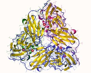
The enzyme cytochrome c oxidase or Complex IV, is a large transmembrane protein complex found in bacteria, archaea, and mitochondria of eukaryotes.

<span class="mw-page-title-main">Metalloprotein</span> Protein that contains a metal ion cofactor

Metalloprotein is a generic term for a protein that contains a metal ion cofactor. A large proportion of all proteins are part of this category. For instance, at least 1000 human proteins contain zinc-binding protein domains although there may be up to 3000 human zinc metalloproteins.

<span class="mw-page-title-main">Plastocyanin</span>

Plastocyanin is a copper-containing protein that mediates electron-transfer. It is found in a variety of plants, where it participates in photosynthesis. The protein is a prototype of the blue copper proteins, a family of intensely blue-colored metalloproteins. Specifically, it falls into the group of small type I blue copper proteins called "cupredoxins".

<span class="mw-page-title-main">Succinate dehydrogenase</span> Enzyme

Succinate dehydrogenase (SDH) or succinate-coenzyme Q reductase (SQR) or respiratory complex II is an enzyme complex, found in many bacterial cells and in the inner mitochondrial membrane of eukaryotes. It is the only enzyme that participates in both the citric acid cycle and the electron transport chain. Histochemical analysis showing high succinate dehydrogenase in muscle demonstrates high mitochondrial content and high oxidative potential.

Ferredoxins are iron–sulfur proteins that mediate electron transfer in a range of metabolic reactions. The term "ferredoxin" was coined by D.C. Wharton of the DuPont Co. and applied to the "iron protein" first purified in 1962 by Mortenson, Valentine, and Carnahan from the anaerobic bacterium Clostridium pasteurianum.

<span class="mw-page-title-main">Flavin adenine dinucleotide</span> Redox-active coenzyme

In biochemistry, flavin adenine dinucleotide (FAD) is a redox-active coenzyme associated with various proteins, which is involved with several enzymatic reactions in metabolism. A flavoprotein is a protein that contains a flavin group, which may be in the form of FAD or flavin mononucleotide (FMN). Many flavoproteins are known: components of the succinate dehydrogenase complex, α-ketoglutarate dehydrogenase, and a component of the pyruvate dehydrogenase complex.

<span class="mw-page-title-main">Protein contact map</span>

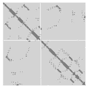
A protein contact map represents the distance between all possible amino acid residue pairs of a three-dimensional protein structure using a binary two-dimensional matrix. For two residues and , the element of the matrix is 1 if the two residues are closer than a predetermined threshold, and 0 otherwise. Various contact definitions have been proposed: The distance between the Cα-Cα atom with threshold 6-12 Å; distance between Cβ-Cβ atoms with threshold 6-12 Å ; and distance between the side-chain centers of mass.

Copper proteins are proteins that contain one or more copper ions as prosthetic groups. Copper proteins are found in all forms of air-breathing life. These proteins are usually associated with electron-transfer with or without the involvement of oxygen (O2). Some organisms even use copper proteins to carry oxygen instead of iron proteins. A prominent copper proteins in humans is in cytochrome c oxidase (cco). The enzyme cco mediates the controlled combustion that produces ATP.

Nitrite reductase refers to any of several classes of enzymes that catalyze the reduction of nitrite. There are two classes of NIR's. A multi haem enzyme reduces NO2 to a variety of products. Copper containing enzymes carry out a single electron transfer to produce nitric oxide.

<span class="mw-page-title-main">Nitrate reductase</span> Class of enzymes

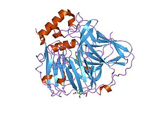
Nitrate reductases are molybdoenzymes that reduce nitrate to nitrite. This reaction is critical for the production of protein in most crop plants, as nitrate is the predominant source of nitrogen in fertilized soils.

<span class="mw-page-title-main">Nitrite reductase (NO-forming)</span> Class of enzymes

In enzymology, a nitrite reductase (NO-forming) (EC 1.7.2.1) is an enzyme that catalyzes the chemical reaction

<span class="mw-page-title-main">Nitrous-oxide reductase</span> Class of enzymes

In enzymology, a nitrous oxide reductase also known as nitrogen:acceptor oxidoreductase (N2O-forming) is an enzyme that catalyzes the final step in bacterial denitrification, the reduction of nitrous oxide to dinitrogen.

<span class="mw-page-title-main">Plastocyanin family of copper-binding proteins</span>

Plastocyanin/azurin family of copper-binding proteins is a family of small proteins that bind a single copper atom and that are characterised by an intense electronic absorption band near 600 nm. The most well-known members of this class of proteins are the plant chloroplastic plastocyanins, which exchange electrons with cytochrome c6, and the distantly related bacterial azurins, which exchange electrons with cytochrome c551. This family of proteins also includes amicyanin from bacteria such as Methylobacterium extorquens or Paracoccus versutus that can grow on methylamine; auracyanins A and B from Chloroflexus aurantiacus; blue copper protein from Alcaligenes faecalis; cupredoxin (CPC) from Cucumis sativus (Cucumber) peelings; cusacyanin from cucumber; halocyanin from Natronomonas pharaonis, a membrane-associated copper-binding protein; pseudoazurin from Pseudomonas; rusticyanin from Thiobacillus ferrooxidans; stellacyanin from Rhus vernicifera ; umecyanin from the roots of Armoracia rusticana (Horseradish); and allergen Ra3 from ragweed. This pollen protein has evolutary relation to the above proteins, but seems to have lost the ability to bind copper. Although there is an appreciable amount of divergence in the sequences of all these proteins, the copper ligand sites are conserved.

<span class="mw-page-title-main">Cytochrome c family</span> Protein family

Cytochromes c cytochromes, or heme-containing proteins, that have heme C covalently attached to the peptide backbone via one or two thioether bonds. These bonds are in most cases part of a specific Cys-X-X-Cys-His (CXXCH) binding motif, where X denotes a miscellaneous amino acid. Two thioether bonds of cysteine residues bind to the vinyl sidechains of heme, and the histidine residue coordinates one axial binding site of the heme iron. Less common binding motifs can include a single thioether linkage, a lysine or a methionine instead of the axial histidine or a CXnCH binding motif with n>2. The second axial site of the iron can be coordinated by amino acids of the protein, substrate molecules or water. Cytochromes c possess a wide range of properties and function as electron transfer proteins or catalyse chemical reactions involving redox processes. A prominent member of this family is mitochondrial cytochrome c.

<span class="mw-page-title-main">Multicopper oxidase</span> Class of enzymes

In molecular biology, multicopper oxidases are enzymes which oxidise their substrate by accepting electrons at a mononuclear copper centre and transferring them to a trinuclear copper centre; dioxygen binds to the trinuclear centre and, following the transfer of four electrons, is reduced to two molecules of water. There are three spectroscopically different copper centres found in multicopper oxidases: type 1, type 2 and type 3. Multicopper oxidases consist of 2, 3 or 6 of these homologous domains, which also share homology with the cupredoxins azurin and plastocyanin. Structurally, these domains consist of a cupredoxin-like fold, a beta-sandwich consisting of 7 strands in 2 beta-sheets, arranged in a Greek-key beta-barrel. Multicopper oxidases include:

<span class="mw-page-title-main">Galactose oxidase</span>

Galactose oxidase is an enzyme that catalyzes the oxidation of D-galactose in some species of fungi.

<span class="mw-page-title-main">Biliverdin reductase B</span> Protein-coding gene in the species Homo sapiens

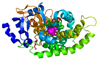
Biliverdin reductase B is a protein that in humans is encoded by the BLVRB gene.

<span class="mw-page-title-main">Cytochrome P450 aromatic O-demethylase</span>

Cytochrome P450 aromatic O-demethylase is a bacterial enzyme that catalyzes the demethylation of lignin and various lignols. The net reaction follows the following stoichiometry, illustrated with a generic methoxy arene:

References

  1. De Rienzo F, Gabdoulline RR, Menziani MC, Wade RC (August 2000). "Blue copper proteins: a comparative analysis of their molecular interaction properties". Protein Science. 9 (8): 1439–54. doi:10.1110/ps.9.8.1439. PMC   2144732 . PMID   10975566.
  2. Yamada T, Goto M, Punj V, Zaborina O, Kimbara K, Das Gupta TK, Chakrabarty AM (December 2002). "The bacterial redox protein azurin induces apoptosis in J774 macrophages through complex formation and stabilization of the tumor suppressor protein p53". Infection and Immunity. 70 (12): 7054–62. doi:10.1128/IAI.70.12.7054-7062.2002. PMC   133031 . PMID   12438386.
  3. van den Bosch M, Swart M, Snijders JG, Berendsen HJ, Mark AE, Oostenbrink C, van Gunsteren WF, Canters GW (April 2005). "Calculation of the redox potential of the protein azurin and some mutants". ChemBioChem. 6 (4): 738–46. doi:10.1002/cbic.200400244. PMID   15747387. S2CID   7672576.
  4. 1 2 Arcangeli C, Bizzarri AR, Cannistraro S (April 1999). "Long-term molecular dynamics simulation of copper azurin: structure, dynamics and functionality". Biophysical Chemistry. 78 (3): 247–57. doi:10.1016/S0301-4622(99)00029-0. PMID   17030312.
  5. 1 2 3 4 5 6 Inoue T, Nishio N, Suzuki S, Kataoka K, Kohzuma T, Kai Y (June 1999). "Crystal structure determinations of oxidized and reduced pseudoazurins from Achromobacter cycloclastes. Concerted movement of copper site in redox forms with the rearrangement of hydrogen bond at a remote histidine". The Journal of Biological Chemistry. 274 (25): 17845–52. doi: 10.1074/jbc.274.25.17845 . PMID   10364229.
  6. van de Kamp M, Silvestrini MC, Brunori M, Van Beeumen J, Hali FC, Canters GW (November 1990). "Involvement of the hydrophobic patch of azurin in the electron-transfer reactions with cytochrome C551 and nitrite reductase". European Journal of Biochemistry. 194 (1): 109–18. doi: 10.1111/j.1432-1033.1990.tb19434.x . PMID   2174771.
  7. Opella SJ, DeSilva TM, Veglia G (April 2002). "Structural biology of metal-binding sequences". Current Opinion in Chemical Biology. 6 (2): 217–23. doi:10.1016/S1367-5931(02)00314-9. PMID   12039007.
  8. 1 2 3 4 McLaughlin MP, Retegan M, Bill E, Payne TM, Shafaat HS, Peña S, Sudhamsu J, Ensign AA, Crane BR, Neese F, Holland PL (December 2012). "Azurin as a protein scaffold for a low-coordinate nonheme iron site with a small-molecule binding pocket". Journal of the American Chemical Society. 134 (48): 19746–57. doi:10.1021/ja308346b. PMC   3515693 . PMID   23167247.
  9. Vijgenboom E, Busch JE, Canters GW (September 1997). "In vivo studies disprove an obligatory role of azurin in denitrification in Pseudomonas aeruginosa and show that azu expression is under control of rpoS and ANR". Microbiology. 143 (9): 2853–63. doi: 10.1099/00221287-143-9-2853 . hdl: 1887/3239420 . PMID   9308169.
  10. Dodd FE, Hasnain SS, Hunter WN, Abraham ZH, Debenham M, Kanzler H, Eldridge M, Eady RR, Ambler RP, Smith BE (August 1995). "Evidence for two distinct azurins in Alcaligenes xylosoxidans (NCIMB 11015): potential electron donors to nitrite reductase". Biochemistry. 34 (32): 10180–6. doi:10.1021/bi00032a011. PMID   7640272.
  11. 1 2 3 Bernardes N, Ribeiro AS, Seruca R, Paredes J, Fialho AM (March 2011). "Bacterial protein azurin as a new candidate drug to treat untreatable breast cancers". 1st Portuguese Biomedical Engineering Meeting. pp. 1–4. doi:10.1109/ENBENG.2011.6026047. ISBN   978-1-4577-0522-9. S2CID   23396316.
  12. 1 2 3 4 Gao M, Zhou J, Su Z, Huang Y (December 2017). "Bacterial cupredoxin azurin hijacks cellular signaling networks: Protein-protein interactions and cancer therapy". Protein Science. 26 (12): 2334–2341. doi:10.1002/pro.3310. PMC   5699490 . PMID   28960574.
  13. Warso MA, Richards JM, Mehta D, Christov K, Schaeffer C, Rae Bressler L, Yamada T, Majumdar D, Kennedy SA, Beattie CW, Das Gupta TK (March 2013). "A first-in-class, first-in-human, phase I trial of p28, a non-HDM2-mediated peptide inhibitor of p53 ubiquitination in patients with advanced solid tumours". British Journal of Cancer. 108 (5): 1061–70. doi:10.1038/bjc.2013.74. PMC   3619084 . PMID   23449360.
  14. 1 2 Chaudhari A, Mahfouz M, Fialho AM, Yamada T, Granja AT, Zhu Y, Hashimoto W, Schlarb-Ridley B, Cho W, Das Gupta TK, Chakrabarty AM (February 2007). "Cupredoxin-cancer interrelationship: azurin binding with EphB2, interference in EphB2 tyrosine phosphorylation, and inhibition of cancer growth". Biochemistry. 46 (7): 1799–810. doi:10.1021/bi061661x. PMID   17249693.