Related Research Articles
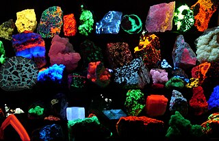
Fluorescence is one of two kinds of emission of light by a substance that has absorbed light or other electromagnetic radiation. When exposed to ultraviolet radiation, many substances will glow (fluoresce) with colored visible light. The color of the light emitted depends on the chemical composition of the substance. Fluorescent materials generally cease to glow nearly immediately when the radiation source stops. This distinguishes them from the other type of light emission, phosphorescence. Phosphorescent materials continue to emit light for some time after the radiation stops.

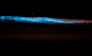
Bioluminescence is the production and emission of light by living organisms. It is a form of chemiluminescence. Bioluminescence occurs widely in marine vertebrates and invertebrates, as well as in some fungi, microorganisms including some bioluminescent bacteria, and terrestrial arthropods such as fireflies. In some animals, the light is bacteriogenic, produced by symbiotic bacteria such as those from the genus Vibrio; in others, it is autogenic, produced by the animals themselves.

Chemiluminescence is the emission of light (luminescence) as the result of a chemical reaction, i.e. a chemical reaction results in a flash or glow of light. A standard example of chemiluminescence in the laboratory setting is the luminol test. Here, blood is indicated by luminescence upon contact with iron in hemoglobin. When chemiluminescence takes place in living organisms, the phenomenon is called bioluminescence. A light stick emits light by chemiluminescence.

Luciferase is a generic term for the class of oxidative enzymes that produce bioluminescence, and is usually distinguished from a photoprotein. The name was first used by Raphaël Dubois who invented the words luciferin and luciferase, for the substrate and enzyme, respectively. Both words are derived from the Latin word lucifer, meaning "lightbearer", which in turn is derived from the Latin words for "light" (lux) and "to bring or carry" (ferre).
The term biophotonics denotes a combination of biology and photonics, with photonics being the science and technology of generation, manipulation, and detection of photons, quantum units of light. Photonics is related to electronics and photons. Photons play a central role in information technologies, such as fiber optics, the way electrons do in electronics.

Oxidative stress reflects an imbalance between the systemic manifestation of reactive oxygen species and a biological system's ability to readily detoxify the reactive intermediates or to repair the resulting damage. Disturbances in the normal redox state of cells can cause toxic effects through the production of peroxides and free radicals that damage all components of the cell, including proteins, lipids, and DNA. Oxidative stress from oxidative metabolism causes base damage, as well as strand breaks in DNA. Base damage is mostly indirect and caused by the reactive oxygen species generated, e.g., O−
2, OH and H2O2. Further, some reactive oxidative species act as cellular messengers in redox signaling. Thus, oxidative stress can cause disruptions in normal mechanisms of cellular signaling.

Alexander Gavrilovich Gurwitsch, sometimes Gurvich or Gurvitch was a Russian and later Soviet biologist and medical scientist who originated the morphogenetic field theory and discovered the biophoton.
Fritz-Albert Popp was a German researcher in biophysics, particularly in the study of biophotons.
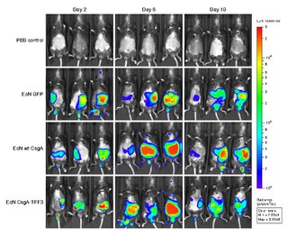
Bioluminescence imaging (BLI) is a technology developed over the past decades (1990's and onward). that allows for the noninvasive study of ongoing biological processes Recently, bioluminescence tomography (BLT) has become possible and several systems have become commercially available. In 2011, PerkinElmer acquired one of the most popular lines of optical imaging systems with bioluminescence from Caliper Life Sciences.

Biological pigments, also known simply as pigments or biochromes, are substances produced by living organisms that have a color resulting from selective color absorption. Biological pigments include plant pigments and flower pigments. Many biological structures, such as skin, eyes, feathers, fur and hair contain pigments such as melanin in specialized cells called chromatophores. In some species, pigments accrue over very long periods during an individual's lifespan.

Apoptosis signal-regulating kinase 1 (ASK1) also known as mitogen-activated protein kinase 5 (MAP3K5) is a member of MAP kinase family and as such a part of mitogen-activated protein kinase pathway. It activates c-Jun N-terminal kinase (JNK) and p38 mitogen-activated protein kinases in a Raf-independent fashion in response to an array of stresses such as oxidative stress, endoplasmic reticulum stress and calcium influx. ASK1 has been found to be involved in cancer, diabetes, rheumatoid arthritis, cardiovascular and neurodegenerative diseases.

Second-harmonic imaging microscopy (SHIM) is based on a nonlinear optical effect known as second-harmonic generation (SHG). SHIM has been established as a viable microscope imaging contrast mechanism for visualization of cell and tissue structure and function. A second-harmonic microscope obtains contrasts from variations in a specimen's ability to generate second-harmonic light from the incident light while a conventional optical microscope obtains its contrast by detecting variations in optical density, path length, or refractive index of the specimen. SHG requires intense laser light passing through a material with a noncentrosymmetric molecular structure, either inherent or induced externally, for example by an electric field.

Fluorescence is used in the life sciences generally as a non-destructive way of tracking or analysing biological molecules. Some proteins or small molecules in cells are naturally fluorescent, which is called intrinsic fluorescence or autofluorescence. The intrinsic DNA fluorescence is very weak.Alternatively, specific or general proteins, nucleic acids, lipids or small molecules can be "labelled" with an extrinsic fluorophore, a fluorescent dye which can be a small molecule, protein or quantum dot. Several techniques exist to exploit additional properties of fluorophores, such as fluorescence resonance energy transfer, where the energy is passed non-radiatively to a particular neighbouring dye, allowing proximity or protein activation to be detected; another is the change in properties, such as intensity, of certain dyes depending on their environment allowing their use in structural studies.
Bioelectrodynamics is a branch of medical physics and bioelectromagnetism which deals with rapidly changing electric and magnetic fields in biological systems, i.e. high frequency endogenous electromagnetic phenomena in living cells. Unlike the events studied by the electrophysiology, the generating mechanism of bioelectrodynamic phenomenon is not connected with currents of ions and its frequency is typically much higher. Examples include vibrations of electrically polar intracellular structures and non-thermal emission of photons as a result of metabolic activity.

Bioluminescent bacteria are light-producing bacteria that are predominantly present in sea water, marine sediments, the surface of decomposing fish and in the gut of marine animals. While not as common, bacterial bioluminescence is also found in terrestrial and freshwater bacteria. These bacteria may be free living or in symbiosis with animals such as the Hawaiian Bobtail squid or terrestrial nematodes. The host organisms provide these bacteria a safe home and sufficient nutrition. In exchange, the hosts use the light produced by the bacteria for camouflage, prey and/or mate attraction. Bioluminescent bacteria have evolved symbiotic relationships with other organisms in which both participants benefit each other equally. Bacteria also use luminescence reaction for quorum sensing, an ability to regulate gene expression in response to bacterial cell density.
Calcium imaging is a microscopy technique to optically measure the calcium (Ca2+) status of an isolated cell, tissue or medium. Calcium imaging takes advantage of calcium indicators, fluorescent molecules that respond to the binding of Ca2+ ions by fluorescence properties. Two main classes of calcium indicators exist: chemical indicators and genetically encoded calcium indicators (GECI). This technique has allowed studies of calcium signalling in a wide variety of cell types. In neurons, action potential generation is always accompanied by rapid influx of Ca2+ ions. Thus, calcium imaging can be used to monitor the electrical activity in hundreds of neurons in cell culture or in living animals, which has made it possible to observe the activity of neuronal circuits during ongoing behavior.
Scintillons are small structures in cytoplasm that produce light. Among bioluminescent organisms, only dinoflagellates have scintillons.
Howard Harold Seliger was a physicist, biochemist, and biology professor, known for his research on bioluminescence.
Chemiluminescence is the emission of light through a chemical reaction. It contrasts with fluorescence, which is excited by a light source. During chemiluminescence, the vibrationally excited product of an exoergic chemical reaction relaxes to its ground state with the emission of photons. Since the process does not require excitation light, problems in its application caused by light scattering or source instability are absent, and there is no concern about autofluorescence in the background, which can lead to highly sensitive deep tissue imaging.
Bioluminescence tomography (BLT) is a non-invasive imaging technique used to reconstruct the three-dimensional distribution of bioluminescent sources based on optical signals measured on the surface of a small animal. It is primarily applied in preclinical research to study molecular and cellular processes in living organisms. BLT was developed as an advancement over bioluminescence imaging (BLI), which only provides two-dimensional representations of bioluminescent sources.
References
- 1 2 Popp FA (May 2003). "Properties of biophotons and their theoretical implications". Indian Journal of Experimental Biology. 41 (5): 391–402. PMID 15244259.
- 1 2 Cifra M, Brouder C, Nerudová M, Kučera O (2015). "Biophotons, coherence and photocount statistics: A critical review". Journal of Luminescence. 164: 38–51. arXiv: 1502.07316 . Bibcode:2015JLum..164...38C. doi:10.1016/j.jlumin.2015.03.020. S2CID 97425113.
- 1 2 Takeda M, Kobayashi M, Takayama M, Suzuki S, Ishida T, Ohnuki K, et al. (August 2004). "Biophoton detection as a novel technique for cancer imaging". Cancer Science. 95 (8): 656–61. doi: 10.1111/j.1349-7006.2004.tb03325.x . PMC 11160017 . PMID 15298728. S2CID 21875229.
- ↑ Rastogi A, Pospísil P (August 2010). "Ultra-weak photon emission as a non-invasive tool for monitoring of oxidative processes in the epidermal cells of human skin: comparative study on the dorsal and the palm side of the hand". Skin Research and Technology. 16 (3): 365–70. doi:10.1111/j.1600-0846.2010.00442.x. PMID 20637006. S2CID 24243914.
- ↑ Niggli HJ (May 1993). "Artificial sunlight irradiation induces ultraweak photon emission in human skin fibroblasts". Journal of Photochemistry and Photobiology B: Biology. 18 (2–3): 281–5. doi:10.1016/1011-1344(93)80076-L. PMID 8350193.
- ↑ Bajpai R (2009). "Biophotons: a clue to unravel the mystery of "life"". In Meyer-Rochow VB (ed.). Bioluminescence in Focus - a collection of illuminating essays. Vol. 1. Kerala, India: Research Signpost. pp. 357–385. ISBN 9788130803579. OCLC 497860307.
- ↑ Zarkeshian P, Kumar S, Tuszynski J, Barclay P, Simon C (March 2018). "Are there optical communication channels in the brain?". Frontiers in Bioscience (Landmark Edition). 23 (8): 1407–1421. arXiv: 1708.08887 . doi:10.2741/4652. PMID 29293442. S2CID 29847303.
- ↑ Beloussov LV, Opitz JM, Gilbert SF (December 1997). "Life of Alexander G. Gurwitsch and his relevant contribution to the theory of morphogenetic fields". The International Journal of Developmental Biology. 41 (6): 771–7, comment 778–9. PMID 9449452.
- 1 2 Bennett M, Mehta M, Grant M (February 2005). "Biophoton imaging: a nondestructive method for assaying R gene responses". Molecular Plant-Microbe Interactions. 18 (2): 95–102. doi:10.1094/MPMI-18-0095. PMID 15720077.
- ↑ Yirka B (May 2012). "Research suggests cells communicate via biophotons" . Retrieved 26 January 2016.
- ↑ Kobayashi M, Kikuchi D, Okamura H (July 2009). "Imaging of ultraweak spontaneous photon emission from human body displaying diurnal rhythm". PLOS ONE. 4 (7): e6256. Bibcode:2009PLoSO...4.6256K. doi: 10.1371/journal.pone.0006256 . PMC 2707605 . PMID 19606225.
- ↑ Dotta BT, Saroka KS, Persinger MA (April 2012). "Increased photon emission from the head while imagining light in the dark is correlated with changes in electroencephalographic power: support for Bókkon's biophoton hypothesis". Neuroscience Letters. 513 (2): 151–4. doi:10.1016/j.neulet.2012.02.021. PMID 22343311. S2CID 207135123.
- ↑ Joines WT, Baumann SB, Kruth JG (2012). "Electromagnetic emission from humans during focused intent". Journal of Parapsychology. 76 (2): 275–294.
- ↑ Khaoua I, Graciani G, Kim A, Amblard F (February 2021). "Detectivity optimization to detect of ultraweak light fluxes with an EM-CCD as binary photon counter array". Scientific Reports. 11 (1): 3530. Bibcode:2021NatSR..11.3530K. doi:10.1038/s41598-021-82611-8. PMC 7878522 . PMID 33574351.
- ↑ Khaoua I, Graciani G, Kim A, Amblard F (May 2021). "Stochastic light concentration from 3D to 2D reveals ultraweak chemi- and bioluminescence". Scientific Reports. 11 (1): 10050. Bibcode:2021NatSR..1110050K. doi:10.1038/s41598-021-88091-0. PMC 8113247 . PMID 33976267.
- ↑ Cilento G, Adam W (July 1995). "From free radicals to electronically excited species". Free Radical Biology & Medicine. 19 (1): 103–14. doi:10.1016/0891-5849(95)00002-F. PMID 7635351.
- ↑ Ursini F, Barsacchi R, Pelosi G, Benassi A (July 1989). "Oxidative stress in the rat heart, studies on low-level chemiluminescence". Journal of Bioluminescence and Chemiluminescence. 4 (1): 241–4. doi:10.1002/bio.1170040134. PMID 2801215.
- ↑ Kataoka Y, Cui Y, Yamagata A, Niigaki M, Hirohata T, Oishi N, Watanabe Y (July 2001). "Activity-dependent neural tissue oxidation emits intrinsic ultraweak photons". Biochemical and Biophysical Research Communications. 285 (4): 1007–11. doi:10.1006/bbrc.2001.5285. PMID 11467852.
- ↑ Boveris A, Cadenas E, Reiter R, Filipkowski M, Nakase Y, Chance B (January 1980). "Organ chemiluminescence: noninvasive assay for oxidative radical reactions". Proceedings of the National Academy of Sciences of the United States of America. 77 (1): 347–51. Bibcode:1980PNAS...77..347B. doi: 10.1073/pnas.77.1.347 . PMC 348267 . PMID 6928628.
- ↑ Iniguez AL, Dong Y, Carter HD, Ahmer BM, Stone JM, Triplett EW (February 2005). "Regulation of enteric endophytic bacterial colonization by plant defenses". Molecular Plant-Microbe Interactions. 18 (2): 169–78. doi: 10.1094/MPMI-18-0169 . PMID 15720086.
- ↑ Kobayashi M, Sasaki K, Enomoto M, Ehara Y (2006). "Highly sensitive determination of transient generation of biophotons during hypersensitive response to cucumber mosaic virus in cowpea". Journal of Experimental Botany. 58 (3): 465–72. doi: 10.1093/jxb/erl215 . PMID 17158510.
- ↑ Kobayashi K, Okabe H, Kawano S, Hidaka Y, Hara K (2014). "Biophoton emission induced by heat shock". PLOS ONE. 9 (8): e105700. Bibcode:2014PLoSO...9j5700K. doi: 10.1371/journal.pone.0105700 . PMC 4143285 . PMID 25153902.
- ↑ Gurwitsch AA (July 1988). "A historical review of the problem of mitogenetic radiation". Experientia. 44 (7): 545–50. doi:10.1007/bf01953301. PMID 3294029. S2CID 10930945.
- ↑ Wijk RV, Wijk EP (April 2005). "An Introduction to Human Biophoton Emission". Forschende Komplementärmedizin und Klassische Naturheilkunde. 12 (2): 77–83. doi:10.1159/000083763. PMID 15947465. S2CID 25794113.
- ↑ Cifra M, Fields JZ, Farhadi A (May 2011). "Electromagnetic cellular interactions". Progress in Biophysics and Molecular Biology. 105 (3): 223–46. doi:10.1016/j.pbiomolbio.2010.07.003. PMID 20674588.
- ↑ Kučera O, Cifra M (November 2013). "Cell-to-cell signaling through light: just a ghost of chance?". Cell Communication and Signaling. 11 (87): 87. doi: 10.1186/1478-811X-11-87 . PMC 3832222 . PMID 24219796.