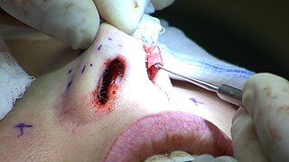
Rhinoplasty, commonly called nose job, medically called nasal reconstruction is a plastic surgery procedure for altering and reconstructing the nose. There are two types of plastic surgery used – reconstructive surgery that restores the form and functions of the nose and cosmetic surgery that changes the appearance of the nose. Reconstructive surgery seeks to resolve nasal injuries caused by various traumas including blunt, and penetrating trauma and trauma caused by blast injury. Reconstructive surgery can also treat birth defects, breathing problems, and failed primary rhinoplasties. Rhinoplasty may remove a bump, narrow nostril width, change the angle between the nose and the mouth, or address injuries, birth defects, or other problems that affect breathing, such as a deviated nasal septum or a sinus condition. Surgery only on the septum is called a septoplasty.
Facial feminization surgery (FFS) is a set of reconstructive surgical procedures that alter typically male facial features to bring them closer in shape and size to typical female facial features. FFS can include various bony and soft tissue procedures such as brow lift, rhinoplasty, cheek implantation, and lip augmentation.

A facelift, technically known as a rhytidectomy, is a type of cosmetic surgery procedure used to give a more youthful facial appearance. There are multiple surgical techniques and exercise routines. Surgery usually involves the removal of excess facial skin, with or without the tightening of underlying tissues, and the redraping of the skin on the patient's face and neck. Exercise routines tone underlying facial muscles without surgery. Surgical facelifts are effectively combined with eyelid surgery (blepharoplasty) and other facial procedures and are typically performed under general anesthesia or deep twilight sleep.
Chin augmentation using surgical implants can alter the underlying structure of the face, providing better balance to the facial features. The specific medical terms mentoplasty and genioplasty are used to refer to the reduction and addition of material to a patient's chin. This can take the form of chin height reduction or chin rounding by osteotomy, or chin augmentation using implants. Improving the facial balance is commonly performed by enhancing the chin using an implant inserted through the mouth. The goal is to provide a suitable projection of the chin as well as the correct height of the chin which is in balance with the other facial features.

Orthognathic surgery, also known as corrective jaw surgery or simply, jaw surgery, is surgery designed to correct conditions of the jaw and lower face related to structure, growth, airway issues including sleep apnea, TMJ disorders, malocclusion problems primarily arising from skeletal disharmonies, and other orthodontic dental bite problems that cannot be treated easily with braces, as well as the broad range of facial imbalances, disharmonies, asymmetries, and malproportions where correction may be considered to improve facial aesthetics and self esteem.

Microtia is a congenital deformity where the auricle is underdeveloped. A completely undeveloped pinna is referred to as anotia. Because microtia and anotia have the same origin, it can be referred to as microtia-anotia. Microtia can be unilateral or bilateral. Microtia occurs in 1 out of about 8,000–10,000 births. In unilateral microtia, the right ear is most commonly affected. It may occur as a complication of taking Accutane (isotretinoin) during pregnancy.

Hypertelorism is an abnormally increased distance between two organs or bodily parts, usually referring to an increased distance between the orbits (eyes), or orbital hypertelorism. In this condition the distance between the inner eye corners as well as the distance between the pupils is greater than normal. Hypertelorism should not be confused with telecanthus, in which the distance between the inner eye corners is increased but the distances between the outer eye corners and the pupils remain unchanged.

Cheek augmentation is a cosmetic surgical procedure that is intended to emphasize the cheeks on a person's face. To augment the cheeks, a plastic surgeon may place a solid implant over the cheekbone. Injections with the patients' own fat or a soft tissue filler, like Restylane, are also popular. Rarely, various cuts to the zygomatic bone (cheekbone) may be performed. Cheek augmentation is commonly combined with other procedures, such as a face lift or chin augmentation.
Velopharyngeal insufficiency is a disorder of structure that causes a failure of the velum to close against the posterior pharyngeal wall during speech in order to close off the nose during oral speech production. This is important because speech requires sound and airflow to be directed into the oral cavity (mouth) for the production of all speech sound with the exception of nasal sounds. If complete closure does not occur during speech, this can cause hypernasality and/or audible nasal emission during speech. In addition, there may be inadequate airflow to produce most consonants, making them sound weak or omitted.
Coccygectomy is a surgical procedure in which the coccyx or tailbone is removed. It is considered a required treatment for sacrococcygeal teratoma and other germ cell tumors arising from the coccyx. Coccygectomy is the treatment of last resort for coccydynia which has failed to respond to nonsurgical treatment. Non surgical treatments include use of seat cushions, external or internal manipulation and massage of the coccyx and the attached muscles, medications given by local injections under fluoroscopic guidance, and medications by mouth.
Jaw reduction or mandible angle reduction is a type of surgery to narrow the lower one-third of the face—particularly the contribution from the mandible and its muscular attachments. There are several techniques for treatment—including surgical and non-surgical methods. A square lower jaw can be considered a masculine trait, especially in Asian countries. As a result, whereas square lower jaws are often considered a positive trait in men, a wide mandible can be perceived as discordant or masculine on women, or sometimes in certain men, particularly when there is asymmetry.

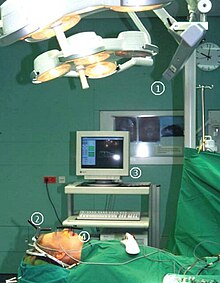
The surgical segment navigator (SSN) is a computer-based system for use in surgical navigation. It is integrated into a common platform, together with the surgical tool navigator (STN), the surgical microscope navigator (SMN) and the 6DOF manipulator (MKM), developed by Carl Zeiss.
Patient registration is used to correlate the reference position of a virtual 3D dataset gathered by computer medical imaging with the reference position of the patient. This procedure is crucial in computer assisted surgery, in order to insure the reproducitibility of the preoperative registration and the clinical situation during surgery. The use of the term "patient registration" out of this context can lead to a confusion with the procedure of registering a patient into the files of a medical institution.
Computer-assisted surgery (CAS) represents a surgical concept and set of methods, that use computer technology for surgical planning, and for guiding or performing surgical interventions. CAS is also known as computer-aided surgery, computer-assisted intervention, image-guided surgery, digital surgery and surgical navigation, but these are terms that are more or less synonymous with CAS. CAS has been a leading factor in the development of robotic surgery.

Surgical planning is the preoperative method of pre-visualising a surgical intervention, in order to predefine the surgical steps and furthermore the bone segment navigation in the context of computer-assisted surgery. The surgical planning is most important in neurosurgery and oral and maxillofacial surgery. The transfer of the surgical planning to the patient is generally made using a medical navigation system.

Laboratory Unit for Computer Assisted Surgery is a system used for virtual surgical planning. Starting with 1998, LUCAS was developed at the University of Regensburg, Germany, with the support of the Carl Zeiss Company. The resulting surgical planning is then reproduced onto the patient by using a navigation system. In fact, LUCAS is integrated into the same platform together with the Surgical Segment Navigator (SSN), the Surgical Tool Navigator (STN), the Surgical Microscope Navigator (SMN) and the 6DOF Manipulator, also from the Carl Zeiss Company.

Frontonasal dysplasia (FND) is a congenital malformation of the midface. For the diagnosis of FND, a patient should present at least two of the following characteristics: hypertelorism, a wide nasal root, vertical midline cleft of the nose and/or upper lip, cleft of the wings of the nose, malformed nasal tip, encephalocele or V-shaped hair pattern on the forehead. The cause of FND remains unknown. FND seems to be sporadic (random) and multiple environmental factors are suggested as possible causes for the syndrome. However, in some families multiple cases of FND were reported, which suggests a genetic cause of FND.
Zygoma reduction, also known as cheekbone reduction surgery, is a surgery used to reduce the facial width by excising part of the zygomatic bone and arch. Wide cheekbones are a characteristic facial trait of Asians, whose skull shapes tend to be more brachycephalic in comparison with Caucasian counterparts, whose skull shapes tend to be more dolichocephalic .This surgery is popular among Asians due to their inherent wide cheekbones. Due to the advanced surgical skills of Korean surgeons who perform facial contouring surgeries, the number of Asian people undergoing this surgery is increasing.
A facial cleft is an opening or gap in the face, or a malformation of a part of the face. Facial clefts is a collective term for all sorts of clefts. All structures like bone, soft tissue, skin etc. can be affected. Facial clefts are extremely rare congenital anomalies. There are many variations of a type of clefting and classifications are needed to describe and classify all types of clefting. Facial clefts hardly ever occur isolated; most of the time there is an overlap of adjacent facial clefts.

Trapeziometacarpal osteoarthritis (TMC OA) is, also known as osteoarthritis at the base of the thumb, thumb carpometacarpal osteoarthritis, basilar (or basal) joint arthritis, or as rhizarthrosis. This joint is formed by the trapezium bone of the wrist and the metacarpal bone of the thumb. This is one of the joints where most humans develop osteoarthritis with age. Osteoarthritis is age-related loss of the smooth surface of the bone where it moves against another bone (cartilage of the joint). In reaction to the loss of cartilage, the bones thicken at the joint surface, resulting in subchondral sclerosis. Also, bony outgrowths, called osteophytes (also known as “bone spurs”), are formed at the joint margins.


