Bristle sensilla

Last updated

Overview

Schematic cross-section of an insect bristle sensillum. Each bristle is composed of a hair with its base fixed to the dendrite of a sensory neuron. The hair acts as a lever that exerts force on the dendrite, inducing mechanotransduction channels to open and producing electrical currents. Bristle sensillum schematic.png
Schematic cross-section of an insect bristle sensillum. Each bristle is composed of a hair with its base fixed to the dendrite of a sensory neuron. The hair acts as a lever that exerts force on the dendrite, inducing mechanotransduction channels to open and producing electrical currents.
Bristle sensilla on the edge of a fruit fly wing. Green fluorescent labeling shows where sensory neurons innervate the bristles. In this image, some of the neurons are mechanosensory and some are gustatory. Flywingbristlewithneurons.jpg
Bristle sensilla on the edge of a fruit fly wing. Green fluorescent labeling shows where sensory neurons innervate the bristles. In this image, some of the neurons are mechanosensory and some are gustatory.

Bristle sensilla are a class of mechanoreceptors found in insects and other arthropods that respond to mechanical stimuli generated by the external world. [1] As a result, they are considered exteroceptors. Bristle sensilla can be divided into two main types, macrochaete and microchaete, based on their size and physiology. [2] [3] The larger macrochaete are thicker and stouter than the smaller microchaete. Macrochaete are also more consistent in their number and distribution across individuals of the same species. Between species, the organization of macrochaete is more conserved among closely related species, whereas the organization of microchaete is more variable and less correlated with phylogenetic relatedness. [4] [5]

Contents

Each bristle sensillum is composed of a hollow hair with its base fixed to the dendrite of a sensory neuron. The hair acts as a lever. When the hair is deflected, for example by dirt or parasites, force is exerted on the dendrite. This induces mechanotransduction channels to open, producing an electrical signal that is carried along an axon to the central nervous system. [1]

Bristles are directionally selective to mechanical deflection. Fly bristles are typically angled 45° relative to the cuticle, and most bristle neurons are most sensitive to forces that push the bristle toward the cuticle. [6] Movement of even a single bristle is sufficient to trigger an insect to groom. [7]

Bristle Neurons

Morphology

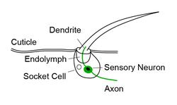
Bristle neurons generally refers to the mechanosensory neurons that can be found at the base of each bristle sensilla. The cell body is located at the base with a dendrite extending far enough into the hair that deflection of the hair will open mechanotransduction channels and produce electrical currents. While the cell body and dendrite are located peripherally, the axon of bristle neurons project to either the brain or the ventral nerve cord (VNC) of the animal - a correlate to the mammalian spinal cord. Axons from bristle sensilla on the head terminate in the subesophageal zone (SEZ) in the brain, whereas bristle neurons from the legs, thorax and abdomen project to and terminate in the ventral surface of the VNC. [8] It is thought that in both the SEZ and the VNC that the spatial organization of these axons represents a somatotopic map of different body segments. [8] [9]

Bristle neurons are also found at the base of chemosensory sensilla. These hairs tend to be longer with a distinct curve to their shape, they also contain a pore at the very tip that is used to sense various chemical signals. Instead of just a single mechanosensory neuron at the base of these hairs, they contain the mechanosensory neuron along with 2-4 gustatory receptor neurons. While gustatory and mechanosensory neurons project to different regions in the SEZ and VNC, it is still unclear which neuropil the mechanosensory neurons at the chemosensory sensilla project to.

Behavioral Responses

Stimulation of bristle neurons along the body elicit a range of behavioral responses across many invertebrate species. In flies, stimulation of bristle hairs along the body may result in targeted grooming, walking, kicking, jumping or flying away. [10] This variability in behavior is dependent on where on the body the bristles are, the number of bristles activated, how long the bristles are deflected along with many other spatial and temporal factors that are less understood.

If a single or few bristle neurons are activated on the leg by mechanical deflection or optogenetic activation, the fly will usually reach for that area of the leg and begin grooming its legs. [1] However, similarly targeted stimulation of bristles hairs on the wing was more likely to result in a kicking behavior. [10] It is thought that this spatial difference in behavior was due to wing stimulation mimicking the invading presence of mites.

Patches of bristle neuron activation on the leg through optogenetic activation result in a broad range of behaviors such as grooming, walking, or general leg movement. Which behavior was elicited was largely dependent on which segment and how many bristle neurons were activated.

Finally, if a fly is completely covered in dust (so all bristles are deflected), the fly will initiate a stereotyped grooming sequence to clean off the dust beginning at its head and ending with its legs. [11] Interestingly, as long as there is dust on a particular body segment, such as its head, the fly will not continue on to the next segment in the sequence. This stereotyped grooming behavior is also seen in response to broad optogenetic activation of bristle neurons all over the body.

Physiology

Upon mechanical stimulation, bristle neurons elicit strong spiking responses. This has been characterized previously with extracellular recordings of individual bristle neurons in the fly in response to mechanical and optogenetic stimulation. Besides direction selectivity, it is thought that bristle neurons can be categorized into two physiological types: rapidly adapting and slow adapting. While these two classes exist across insects, the density of distribution of these cells varies by species. For example in the Locust, slow adapting bristles are more numerous and present all along the leg whereas rapidly adapting bristles are limited to the tibia. [12] Furthermore, these categories exhibit different mechanical thresholds - slow adapting neurons respond to a lower threshold of about 10 degrees displacement, while rapidly adapting flies have a higher mechanical threshold at about 40 degrees. Similar findings have been observed in cockroaches and crickets. [13] [14] On the other hand, only slow adapting bristles have been observed in flies with an exceedingly low threshold of only 1 degree. It is still unclear whether they also possess rapidly adapting bristles.

Related Research Articles

<span class="mw-page-title-main">Dendrite</span> Small projection on a neuron that receives signals

A dendrite or dendron is a branched protoplasmic extension of a nerve cell that propagates the electrochemical stimulation received from other neural cells to the cell body, or soma, of the neuron from which the dendrites project. Electrical stimulation is transmitted onto dendrites by upstream neurons via synapses which are located at various points throughout the dendritic tree.

<span class="mw-page-title-main">Neuron</span> Electrically excitable cell found in the nervous system of animals

Within a nervous system, a neuron, neurone, or nerve cell is an electrically excitable cell that fires electric signals called action potentials across a neural network. Neurons communicate with other cells via synapses, which are specialized connections that commonly use minute amounts of chemical neurotransmitters to pass the electric signal from the presynaptic neuron to the target cell through the synaptic gap.

<span class="mw-page-title-main">Halteres</span> Pair of small club-shaped insect organs

Halteres are a pair of small club-shaped organs on the body of two orders of flying insects that provide information about body rotations during flight. Insects of the large order Diptera (flies) have halteres which evolved from a pair of ancestral hindwings, while males of the much smaller order Strepsiptera (stylops) have halteres which evolved from a pair of ancestral forewings.

<span class="mw-page-title-main">Motor neuron</span> Nerve cell sending impulse to muscle

A motor neuron is a neuron whose cell body is located in the motor cortex, brainstem or the spinal cord, and whose axon (fiber) projects to the spinal cord or outside of the spinal cord to directly or indirectly control effector organs, mainly muscles and glands. There are two types of motor neuron – upper motor neurons and lower motor neurons. Axons from upper motor neurons synapse onto interneurons in the spinal cord and occasionally directly onto lower motor neurons. The axons from the lower motor neurons are efferent nerve fibers that carry signals from the spinal cord to the effectors. Types of lower motor neurons are alpha motor neurons, beta motor neurons, and gamma motor neurons.

<span class="mw-page-title-main">Olfactory bulb</span> Neural structure

The olfactory bulb is a neural structure of the vertebrate forebrain involved in olfaction, the sense of smell. It sends olfactory information to be further processed in the amygdala, the orbitofrontal cortex (OFC) and the hippocampus where it plays a role in emotion, memory and learning. The bulb is divided into two distinct structures: the main olfactory bulb and the accessory olfactory bulb. The main olfactory bulb connects to the amygdala via the piriform cortex of the primary olfactory cortex and directly projects from the main olfactory bulb to specific amygdala areas. The accessory olfactory bulb resides on the dorsal-posterior region of the main olfactory bulb and forms a parallel pathway. Destruction of the olfactory bulb results in ipsilateral anosmia, while irritative lesions of the uncus can result in olfactory and gustatory hallucinations.

<span class="mw-page-title-main">Stimulus (physiology)</span> Detectable change in the internal or external surroundings

In physiology, a stimulus is a detectable change in the physical or chemical structure of an organism's internal or external environment. The ability of an organism or organ to detect external stimuli, so that an appropriate reaction can be made, is called sensitivity (excitability). Sensory receptors can receive information from outside the body, as in touch receptors found in the skin or light receptors in the eye, as well as from inside the body, as in chemoreceptors and mechanoreceptors. When a stimulus is detected by a sensory receptor, it can elicit a reflex via stimulus transduction. An internal stimulus is often the first component of a homeostatic control system. External stimuli are capable of producing systemic responses throughout the body, as in the fight-or-flight response. In order for a stimulus to be detected with high probability, its level of strength must exceed the absolute threshold; if a signal does reach threshold, the information is transmitted to the central nervous system (CNS), where it is integrated and a decision on how to react is made. Although stimuli commonly cause the body to respond, it is the CNS that finally determines whether a signal causes a reaction or not.

A mechanoreceptor, also called mechanoceptor, is a sensory receptor that responds to mechanical pressure or distortion. Mechanoreceptors are innervated by sensory neurons that convert mechanical pressure into electrical signals that, in animals, are sent to the central nervous system.

Neural adaptation or sensory adaptation is a gradual decrease over time in the responsiveness of the sensory system to a constant stimulus. It is usually experienced as a change in the stimulus. For example, if a hand is rested on a table, the table's surface is immediately felt against the skin. Subsequently, however, the sensation of the table surface against the skin gradually diminishes until it is virtually unnoticeable. The sensory neurons that initially respond are no longer stimulated to respond; this is an example of neural adaptation.

<span class="mw-page-title-main">Campaniform sensilla</span> Class of mechanoreceptors found in insects

Campaniform sensilla are a class of mechanoreceptors found in insects, which respond to local stress and strain within the animal's cuticle. Campaniform sensilla function as proprioceptors that detect mechanical load as resistance to muscle contraction, similar to mammalian Golgi tendon organs. Sensory feedback from campaniform sensilla is integrated in the control of posture and locomotion.

Chordotonal organs are stretch receptor organs found only in insects and crustaceans. They are located at most joints and are made up of clusters of scolopidia that either directly or indirectly connect two joints and sense their movements relative to one another. They can have both extero- and proprioceptive functions, for example sensing auditory stimuli or leg movement. The word was coined by Vitus Graber in 1882, though he interpreted them as being stretched between two points like a string, sensing vibrations through resonance.

Optogenetics is a biological technique to control the activity of neurons or other cell types with light. This is achieved by expression of light-sensitive ion channels, pumps or enzymes specifically in the target cells. On the level of individual cells, light-activated enzymes and transcription factors allow precise control of biochemical signaling pathways. In systems neuroscience, the ability to control the activity of a genetically defined set of neurons has been used to understand their contribution to decision making, learning, fear memory, mating, addiction, feeding, and locomotion. In a first medical application of optogenetic technology, vision was partially restored in a blind patient.

The Mauthner cells are a pair of big and easily identifiable neurons located in the rhombomere 4 of the hindbrain in fish and amphibians that are responsible for a very fast escape reflex. The cells are also notable for their unusual use of both chemical and electrical synapses.

<span class="mw-page-title-main">Proprioception</span> Sense of self-movement, force, and body position

Proprioception, also called kinaesthesia, is the sense of self-movement, force, and body position.

TRPN is a member of the transient receptor potential channel family of ion channels, which is a diverse group of proteins thought to be involved in mechanoreception. The TRPN gene was given the name no mechanoreceptor potential C (nompC) when it was first discovered in fruit flies, hence the N in TRPN. Since its discovery in fruit flies, TRPN homologs have been discovered and characterized in worms, frogs, and zebrafish.

<span class="mw-page-title-main">Insect olfaction</span> Function of chemical receptors

Insect olfaction refers to the function of chemical receptors that enable insects to detect and identify volatile compounds for foraging, predator avoidance, finding mating partners and locating oviposition habitats. Thus, it is the most important sensation for insects. Most important insect behaviors must be timed perfectly which is dependent on what they smell and when they smell it. For example, olfaction is essential for locating host plants and hunting prey in many species of insects, such as the moth Deilephila elpenor and the wasp Polybia sericea, respectively.

<span class="mw-page-title-main">Anion-conducting channelrhodopsin</span> Class of light-gated ion channels

Anion-conducting channelrhodopsins are light-gated ion channels that open in response to light and let negatively charged ions enter a cell. All channelrhodopsins use retinal as light-sensitive pigment, but they differ in their ion selectivity. Anion-conducting channelrhodopsins are used as tools to manipulate brain activity in mice, fruit flies and other model organisms (Optogenetics). Neurons expressing anion-conducting channelrhodopsins are silenced when illuminated with light, an effect that has been used to investigate information processing in the brain. For example, suppressing dendritic calcium spikes in specific neurons with light reduced the ability of mice to perceive a light touch to a whisker. Studying how the behavior of an animal changes when specific neurons are silenced allows scientists to determine the role of these neurons in the complex circuits controlling behavior.

Hair-plates are a type of proprioceptor found in the folds of insect joints. They consist of a cluster of hairs, in which each hair is innervated by a single mechanosensory neuron. Functionally, hair-plates operate as "limit-detectors" by signaling the extreme ranges of motion of a joint.

<span class="mw-page-title-main">Reinhard F. Stocker</span> Swiss biologist

Reinhard F. Stocker is a Swiss biologist. He pioneered the analysis of the sense of smell and taste in higher animals, using the fly Drosophila melanogaster as a study case. He provided a detailed account of the anatomy and development of the olfactory system, in particular across metamorphosis, for which he received the Théodore-Ott-Prize of the Swiss Academy of Medical Sciences in 2007, and pioneered the use of larval Drosophila for the brain and behavioural sciences.

<span class="mw-page-title-main">Femoral chordotonal organ</span> Sensory organ in insect legs

The femoral chordotonal organ is a group of mechanosensory neurons found in an insect leg that detects the movements and the position of the femur/tibia joint. It is thought to function as a proprioceptor that is critical for precise control of leg position by sending the information regarding the femur/tibia joint to the motor circuits in the ventral nerve cord and the brain

A descending neuron is a neuron that conveys signals from the brain to neural circuits in the spinal cord (vertebrates) or ventral nerve cord (invertebrates). As the sole conduits of information between the brain and the body, descending neurons play a key role in behavior. Their activity can initiate, maintain, modulate, and terminate behaviors such as locomotion. Because the number of descending neurons is several orders of magnitude smaller than the number of neurons in either the brain or spinal cord/ventral nerve cord, this class of cells represents a critical bottleneck in the flow of information from sensory systems to motor circuits.

References

  1. 1 2 3 Tuthill, John C.; Wilson, Rachel I. (2016-10-24). "Mechanosensation and Adaptive Motor Control in Insects". Current Biology. 26 (20): R1022–R1038. doi:10.1016/j.cub.2016.06.070. ISSN   0960-9822. PMC   5120761 . PMID   27780045.
  2. Keil, T. A. (1997-12-15). "Functional morphology of insect mechanoreceptors". Microscopy Research and Technique. 39 (6): 506–531. doi:10.1002/(SICI)1097-0029(19971215)39:6<506::AID-JEMT5>3.0.CO;2-B. ISSN   1059-910X. PMID   9438251. S2CID   5552615.
  3. Simpson, P.; Woehl, R.; Usui, K. (1999-04-01). "The development and evolution of bristle patterns in Diptera". Development. 126 (7): 1349–1364. doi:10.1242/dev.126.7.1349. ISSN   1477-9129. PMID   10068629.
  4. Sturtevant, A. H. (1970-01-01). "Studies on the bristle pattern of Drosophila". Developmental Biology. 21 (1): 48–61. doi:10.1016/0012-1606(70)90060-6. ISSN   0012-1606. PMID   5445760.
  5. Usui-Ishihara, Akiko; Simpson, Pat (2005-01-01). "Differences in sensory projections between macro- and microchaetes in Drosophilid flies". Developmental Biology. 277 (1): 170–183. doi: 10.1016/j.ydbio.2004.09.017 . ISSN   0012-1606. PMID   15572148.
  6. Walker, Richard G.; Willingham, Aarron T.; Zuker, Charles S. (2000-03-24). "A Drosophila Mechanosensory Transduction Channel". Science. 287 (5461): 2229–2234. Bibcode:2000Sci...287.2229W. doi:10.1126/science.287.5461.2229. ISSN   0036-8075. PMID   10744543.
  7. Corfas, G; Dudai, Y (1989-01-01). "Habituation and dishabituation of a cleaning reflex in normal and mutant Drosophila". The Journal of Neuroscience. 9 (1): 56–62. doi:10.1523/JNEUROSCI.09-01-00056.1989. ISSN   0270-6474. PMC   6569995 . PMID   2913213.
  8. 1 2 Newland, Philip L. (1991-10-22). "Morphology and somatotopic organisation of the central projections of afferents from tactile hairs on the hind leg of the locust". The Journal of Comparative Neurology. 312 (4): 493–508. doi:10.1002/cne.903120402. ISSN   0021-9967. PMID   1761738. S2CID   9246845.
  9. Eichler, Katharina; Hampel, Stefanie; Alejandro-García, Adrián; Calle-Schuler, Steven A.; Santana-Cruz, Alexis; Kmecova, Lucia; Blagburn, Jonathan M.; Hoopfer, Eric D.; Seeds, Andrew M. (2023-04-04). "Somatotopic organization among parallel sensory pathways that promote a grooming sequence in Drosophila". bioRxiv: The Preprint Server for Biology: 2023.02.11.528119. doi:10.1101/2023.02.11.528119. PMC   9934617 . PMID   36798384.
  10. 1 2 Vandervorst, Philippe; Ghysen, Alain (July 1980). "Genetic control of sensory connections in Drosophila". Nature. 286 (5768): 65–67. Bibcode:1980Natur.286...65V. doi:10.1038/286065a0. ISSN   1476-4687. PMID   7393324. S2CID   4307682.
  11. Zhang, Neil; Guo, Li; Simpson, Julie H. (2020-03-23). "Spatial Comparisons of Mechanosensory Information Govern the Grooming Sequence in Drosophila". Current Biology. 30 (6): 988–1001.e4. doi:10.1016/j.cub.2020.01.045. ISSN   0960-9822. PMC   7184881 . PMID   32142695.
  12. Newland, Philip L. (1991-01-01). "Physiological Properties of Afferents from Tactile Hairs on the Hindlegs of the Locust". Journal of Experimental Biology. 155 (1): 487–503. doi: 10.1242/jeb.155.1.487 . ISSN   0022-0949. PMID   2016576.
  13. Shimozawa, Tateo; Kanou, Masamichi (1984). "Varieties of filiform hairs: range fractionation by sensory afferents and cereal interneurons of a cricket". Journal of Comparative Physiology A. 155 (4): 485–493. doi:10.1007/bf00611913. ISSN   0340-7594. S2CID   42806812.
  14. Westin, Joanne (1979). "Responses to wind recorded from the cercal nerve of the cockroachPeriplaneta americana". Journal of Comparative Physiology ? A. 133 (2): 97–102. doi:10.1007/bf00657523. ISSN   0340-7594. S2CID   46107869.