Related Research Articles

The rib cage is an endoskeletal enclosure in the thorax of most vertebrate animals that comprises the ribs, vertebral column and sternum, which protects vital organs such as the heart, lungs and great vessels. The circumferential enclosure formed by left and right rib cages, together known as the thoracic cage, is a semi-rigid bony and cartilaginous structure which surrounds the thoracic cavity and supports the shoulder girdles to form the core part of the axial skeleton.

The tibia, also known as the shinbone or shankbone, is the larger, stronger, and anterior (frontal) of the two bones in the leg below the knee in vertebrates ; it connects the knee with the ankle. The tibia is found on the medial side of the leg next to the fibula and closer to the median plane. The tibia is connected to the fibula by the interosseous membrane of leg, forming a type of fibrous joint called a syndesmosis with very little movement. The tibia is named for the flute tibia. It is the second largest bone in the human body, after the femur. The leg bones are the strongest long bones as they support the rest of the body.

The pectoralis major is a thick, fan-shaped or triangular convergent muscle of the human chest. It makes up the bulk of the chest muscles and lies under the breast. Beneath the pectoralis major is the pectoralis minor muscle.

In human anatomy, the abdominal aorta is the largest artery in the abdominal cavity. As part of the aorta, it is a direct continuation of the descending aorta.

In tetrapods, cervical vertebrae are the vertebrae of the neck, immediately below the skull. Truncal vertebrae lie caudal of cervical vertebrae. In sauropsid species, the cervical vertebrae bear cervical ribs. In lizards and saurischian dinosaurs, the cervical ribs are large; in birds, they are small and completely fused to the vertebrae. The vertebral transverse processes of mammals are homologous to the cervical ribs of other amniotes. Most mammals have seven cervical vertebrae, with the only three known exceptions being the manatee with six, the two-toed sloth with five or six, and the three-toed sloth with nine.

The mediastinum is the central compartment of the thoracic cavity. Surrounded by loose connective tissue, it is an undelineated region that contains a group of structures within the thorax, namely the heart and its vessels, the esophagus, the trachea, the phrenic and cardiac nerves, the thoracic duct, the thymus and the lymph nodes of the central chest.
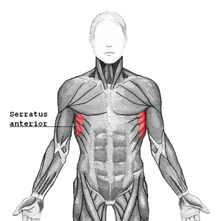
The serratus anterior is a muscle of the chest. It originates at the side of the chest from the upper 8 or 9 ribs; it inserts along the entire length of the anterior aspect of the medial border of the scapula. It is innervated by the long thoracic nerve from the brachial plexus. The serratus anterior acts to pull the scapula forward around the thorax.

The transverse abdominal muscle (TVA), also known as the transverse abdominis, transversalis muscle and transversus abdominis muscle, is a muscle layer of the anterior and lateral abdominal wall, deep to the internal oblique muscle. It is thought by most fitness instructors to be a significant component of the core.

The transversus thoracis muscle, also known as triangularis sterni, lies internal to the thoracic cage, anteriorly. It is usually a thin plane of muscular and tendinous fibers, however on athletic individuals it can be a thick 'slab of meat', situated upon the inner surface of the front wall of the chest. It is in the same layer as the subcostal muscles and the innermost intercostal muscles.

The abdominal internal oblique muscle, also internal oblique muscle or interior oblique, is an abdominal muscle in the abdominal wall that lies below the external oblique muscle and just above the transverse abdominal muscle.

The scalene muscles are a group of three muscles on each side of the neck, identified as the anterior, the middle, and the posterior. They are innervated by the third to the eighth cervical spinal nerves (C3-C8).

In the anatomy of humans and homologous primates, the ascending colon is the part of the colon located between the cecum and the transverse colon.
The abdominal external oblique muscle is the largest and outermost of the three flat abdominal muscles of the lateral anterior abdomen.

The quadratus lumborum muscle, informally called the QL, is a paired muscle of the left and right posterior abdominal wall. It is the deepest abdominal muscle, and commonly referred to as a back muscle. Each is irregular and quadrilateral in shape.

A respiratory examination, or lung examination, is performed as part of a physical examination, in response to respiratory symptoms such as shortness of breath, cough, or chest pain, and is often carried out with a cardiac examination.

The abdomen is the part of the body between the thorax (chest) and pelvis, in humans and in other vertebrates. The abdomen is the front part of the abdominal segment of the torso. The area occupied by the abdomen is called the abdominal cavity. In arthropods it is the posterior tagma of the body; it follows the thorax or cephalothorax.

The pelvis is the lower part of the trunk, between the abdomen and the thighs, together with its embedded skeleton.

The vertebral column, also known as the backbone or spine, is the core part of the axial skeleton in vertebrate animals. The vertebral column is the defining characteristic of vertebrate endoskeleton in which the notochord found in all chordates has been replaced by a segmented series of mineralized irregular bones called vertebrae, separated by fibrocartilaginous intervertebral discs. The dorsal portion of the vertebral column houses the spinal canal, a cavity formed by alignment of the neural arches that encloses and protects the spinal cord.

Each vertebra is an irregular bone with a complex structure composed of bone and some hyaline cartilage, that make up the vertebral column or spine, of vertebrates. The proportions of the vertebrae differ according to their spinal segment and the particular species.
Pump-handle is a movement of the ribs that results in a change in the anteroposterior diameter of the thorax.
References
- ↑ "28 : The Chest Wall and Pleura". www.oganatomy.org.
- ↑ Drake, Richard L.; Vogl, Wayne; Tibbitts, Adam W.M. Mitchell; illustrations by Richard; Richardson, Paul (2005). Gray's anatomy for students. Philadelphia: Elsevier/Churchill Livingstone. ISBN 978-0-8089-2306-0.
- 1 2 "Chapter 20: The thoracic wall and mediastinum". www.dartmouth.edu. Archived from the original on 2017-12-06. Retrieved 2015-06-29.
- ↑ "Figure 20-7". www.dartmouth.edu. Archived from the original on 2016-07-29. Retrieved 2015-06-29.