Cellular dewetting

Last updated
Figure 1. Snapshot images taken from a video showing the nucleation and growth of a TEM in an endothelial cell intoxicated with C3 exoenzyme from Clostridium botulinum for 24 hours, Bar = 10 mm. For the dynamics see Video. Cellular Dewetting.jpg
Figure 1. Snapshot images taken from a video showing the nucleation and growth of a TEM in an endothelial cell intoxicated with C3 exoenzyme from Clostridium botulinum for 24 hours, Bar = 10 μm. For the dynamics see Video.

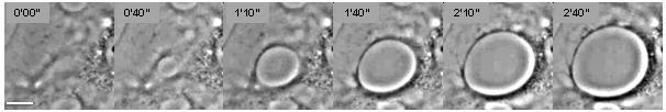
Cellular dewetting refers to the process of nucleation and enlargement of transendothelial cell macroaperture (TEM) tunnels in endothelial cells (Figure 1). [1] This phenomenon is analogous to the nucleation and growth of dry patches in viscous liquids spreading on a non-wettable substrate (Figure 2). [2] Cellular dewetting is triggered by several protein toxins from pathogenic bacteria, notably the EDIN-like factors from Staphylococcus aureus and from Clostridium botulinum, as well as edema toxin from Bacillus anthracis. [3] [4] TEMs form in response to the rupture of cytoskeleton physical connections through the cytoplasm due to inhibition of the RhoA/ROCK pathway or to induction of the flux of cyclic-AMP (cAMP) broad signaling molecule. [4] [5]

Contents

Physics behind cellular dewetting

Figure 2. Analogy between liquid dewetting and cellular dewetting. Liquid and cellular dewetting.png
Figure 2. Analogy between liquid dewetting and cellular dewetting.

The phenomenon of cellular dewetting can be interpreted by physical modeling (Figure 2). [6] The driving force responsible for the spontaneous formation of TEM tunnels and their opening is the membrane tension that results from the spreading of cells due to actomyosin relaxation. Opposite to liquid dewetting, TEMs reach a maximum diameter, at which the driving force is balanced by a resisting force that develops along TEM edges (Figure 2). This resisting force is referred to as line tension and is uncharacterized at the molecular level.

Physical parameters

Driving forces pulling on a tunnel of radius R, as depicted in Figure 2. Here, pulling is due to the tensioning of the cell membrane (σ) that is partly counteracted by a line tension around the tunnel (T). In these conditions, the net driving force (FD) consists of two contributions:

Dewetting proceeds if FD>0.

Membrane tension (σ) depends on the tunnel radius R. A tunnel increase in size relaxes the membrane, inducing a decrease in membrane tension, as described by Helfrich’s law.

Line tension (T) corresponds to the resisting force along the edge of the tunnel that opposes membrane tension and limits dewetting. This line tension can have physical and molecular components.

Related Research Articles

<i>Staphylococcus aureus</i> species of bacterium

Staphylococcus aureus is a Gram-positive, round-shaped bacterium that is a member of the Firmicutes, and it is a usual member of the microbiota of the body, frequently found in the upper respiratory tract and on the skin. It is often positive for catalase and nitrate reduction and is a facultative anaerobe that can grow without the need for oxygen. Although S. aureus usually acts as a commensal of the human microbiota it can also become an opportunistic pathogen, being a common cause of skin infections including abscesses, respiratory infections such as sinusitis, and food poisoning. Pathogenic strains often promote infections by producing virulence factors such as potent protein toxins, and the expression of a cell-surface protein that binds and inactivates antibodies. The emergence of antibiotic-resistant strains of S. aureus such as methicillin-resistant S. aureus (MRSA) is a worldwide problem in clinical medicine. Despite much research and development, no vaccine for S. aureus has been approved.

Cerebral edema is excess accumulation of fluid (edema) in the intracellular or extracellular spaces of the brain.

Exotoxin class of toxic proteins secreted by bacteria

An exotoxin is a toxin secreted by bacteria. An exotoxin can cause damage to the host by destroying cells or disrupting normal cellular metabolism. They are highly potent and can cause major damage to the host. Exotoxins may be secreted, or, similar to endotoxins, may be released during lysis of the cell. Gram negative pathogens may secrete outer membrane vesicles containing lipopolysaccharide endotoxin and some virulence proteins in the bounding membrane along with some other toxins as intra-vesicular contents, thus adding a previously unforeseen dimension to the well-known eukaryote process of membrane vesicle trafficking, which is quite active at the host-pathogen interface.

The Starling equation for fluid filtration is named for the British physiologist Ernest Starling, who is also recognised for the Frank–Starling law of the heart. The classic Starling equation has in recent years been revised. The Starling principle of fluid exchange is key to understanding how plasma fluid (solvent) within the bloodstream moves to the space outside the bloodstream. Starling can be credited with identifying that the "absorption of isotonic salt solutions by the blood vessels is determined by this osmotic pressure of the serum proteins." (1896)

Dewetting rupture of a thin liquid film on the substrate and the formation of droplets

In fluid mechanics, dewetting is one of the processes that can occur at a solid–liquid, solid-solid or liquid–liquid interface. Generally, dewetting describes the process of retraction of a fluid from a non-wettable surface it was forced to cover. The opposite process—spreading of a liquid on a substrate—is called wetting. The factor determining the spontaneous spreading and dewetting for a drop of liquid placed on a solid substrate with ambient gas, is the so-called spreading coefficient S:

Cell cortex The region of a cell that lies just beneath the plasma membrane and often, but not always, contains a network of actin filaments and associated proteins.

The cell cortex, also known as the actin cortex or actomyosin cortex, is a specialized layer of cytoplasmic proteins on the inner face of the cell membrane. It functions as a modulator of membrane behavior and cell surface properties. In most eukaryotic cells lacking a cell wall, the cortex is an actin-rich network consisting of F-actin filaments, myosin motors, and actin-binding proteins. The actomyosin cortex is attached to the cell membrane via membrane-anchoring proteins called ERM proteins and it plays a central role in cell shape control. The protein constituents of the cortex undergo rapid turnover, making the cortex both mechanically rigid and highly plastic, two properties essential to its function. In most cases, the cortex is in the range of 100 to 1000 nanometers thick.

Virulence factors are molecules produced by bacteria, viruses, fungi, and protozoa that add to their effectiveness and enable them to achieve the following:

Anthrax toxin tripartite protein complex secreted by virulent strains of Bacillus anthracis

Anthrax toxin is a three-protein exotoxin secreted by virulent strains of the bacterium, Bacillus anthracis—the causative agent of anthrax. The toxin was first discovered by Harry Smith in 1954. Anthrax toxin is composed of a cell-binding protein, known as protective antigen (PA), and two enzyme components, called edema factor (EF) and lethal factor (LF). These three protein components act together to impart their physiological effects. Assembled complexes containing the toxin components are endocytosed. In the endosome, the enzymatic components of the toxin translocate into the cytoplasm of a target cell. Once in the cytosol, the enzymatic components of the toxin disrupts various immune cell functions, namely cellular signaling and cell migration. The toxin may even induce cell lysis, as is observed for macrophage cells. Anthrax toxin allows the bacteria to evade the immune system, proliferate, and ultimately kill the host animal. Research on anthrax toxin also provides insight into the generation of macromolecular assemblies, and on protein translocation, pore formation, endocytosis, and other biochemical processes.

Panton–Valentine leukocidin cytotoxin

Panton–Valentine leukocidin (PVL) is a cytotoxin—one of the β-pore-forming toxins. The presence of PVL is associated with increased virulence of certain strains (isolates) of Staphylococcus aureus. It is present in the majority of community-associated Methicillin-resistant Staphylococcus aureus (CA-MRSA) isolates studied and is the cause of necrotic lesions involving the skin or mucosa, including necrotic hemorrhagic pneumonia. PVL creates pores in the membranes of infected cells. PVL is produced from the genetic material of a bacteriophage that infects Staphylococcus aureus, making it more virulent.

Pore-forming toxin class of proteins synthesized by one cell and secreted for insertion into the membrane of another cell where they form transmembrane pores

Pore-forming proteins are usually produced by bacteria, and include a number of protein exotoxins but may also be produced by other organisms such as earthworms, who produce lysenin. They are frequently cytotoxic, as they create unregulated pores in the membrane of targeted cells.

Hemolysin Molecule destroying the membrane of red blood cells

Hemolysins or haemolysins are lipids and proteins that cause lysis of red blood cells by destroying their cell membrane. Although the lytic activity of some microbe-derived hemolysins on red blood cells may be of great importance for nutrient acquisition, many hemolysins produced by pathogens do not cause significant destruction of red blood cells during infection. However, hemolysins are often capable of lysing red blood cells in vitro.

<i>Staphylococcus aureus</i> alpha toxin

Alpha-toxin, also known as alpha-hemolysin (Hla), is the major cytotoxic agent released by bacterium Staphylococcus aureus and the first identified member of the pore forming beta-barrel toxin family. This toxin consists mostly of beta-sheets (68%) with only about 10% alpha-helices. The hly gene on the S. aureus chromosome encodes the 293 residue protein monomer, which forms heptameric units on the cellular membrane to form a complete beta-barrel pore. This structure allows the toxin to perform its major function, development of pores in the cellular membrane, eventually causing cell death.

ANTXR1 protein-coding gene in the species Homo sapiens

Anthrax toxin receptor 1 is a protein that in humans is encoded by the ANTXR1 gene. Its molecular weight is predicted as about 63kDa.

Staphylococcus aureus beta toxin is a toxin produced by Staphylococcus aureus. It is a form of sphingomyelinase called sphingomyelinase C. This enzyme is toxic to a variety of cells, including erythrocytes, fibroblasts, leukocytes, and macrophages. Susceptible cells are subject to lysis of exposed sphingomyelin on their membrane surfaces.

'Staphylococcus aureus delta toxin is a toxin produced by Staphylococcus aureus. It has a wide spectrum of cytolytic activity.

Microbial toxins are toxins produced by micro-organisms, including bacteria and fungi. Microbial toxins promote infection and disease by directly damaging host tissues and by disabling the immune system. Some bacterial toxins, such as Botulinum neurotoxins, are the most potent natural toxins known. However, microbial toxins also have important uses in medical science and research. Potential applications of toxin research include combating microbial virulence, the development of novel anticancer drugs and other medicines, and the use of toxins as tools in neurobiology and cellular biology.

Rho-associated protein kinase

Rho-associated protein kinase (ROCK) is a kinase belonging to the AGC family of serine-threonine kinases. It is involved mainly in regulating the shape and movement of cells by acting on the cytoskeleton.

Aureolysin class of enzymes

Aureolysin is an extracellular metalloprotease expressed by Staphylococcus aureus. This protease is a major contributor to the bacterium's virulence, or ability to cause disease, by cleaving host factors of the innate immune system as well as regulating S. aureus secreted toxins and cell wall proteins. To catalyze its enzymatic activities, aureolysin requires zinc and calcium which it obtains from the extracellular environment within the host.

Actomyosin ring

The cell cycle is divided into two primary phases: DNA synthesis or S phase and Mitosis or M phase. During the S phase, duplication of chromosomes occurs whereas M phase is characterized by two processes known as nuclear (mitosis) and cytoplasmic (cytokinesis) divisions. The Actomyosin ring is a prominent structure during cytoplasmic division or cytokinesis. The ring forms perpendicular to the axis of the spindle apparatus. and occurs towards the later stage of mitosis, the telophase, in which sister chromatids are identically separated at the opposite sides of the spindle forming nuclei. The actomyosin ring follows an orderly sequence of identification of active division site, formation of the ring, constriction of the ring, and disassembly of the contractile ring. It is composed of actin and myosin II bundles, thus the term actomyosin, that operates in contractile motion although the mechanism on how or what triggers the constriction is still an evolving topic. Other cytoskeletal proteins are also involved in maintaining stability of the ring. Apart from cytokinesis in which the ring constricts as the cells divide, actomyosin ring constriction has also been found to activate during wound closure. During the process actin filaments are degraded, preserving the thickness of the ring. After cytokinesis is complete, one of the two daughter cells inherits a remnant, called the midbody ring.

References

  1. Lemichez, E. (2012). "Transcellular tunnel dynamics: Control of cellular dewetting by actomyosin contractility and I-BAR proteins". Biology of the Cell. 105 (3): 109–117. doi:10.1111/boc.201200063. PMID   23189935.
  2. De Gennes, P.-G. (2004). Capillarity and Wetting Phenomena. New York: Springer. ISBN   978-0387005928.
  3. Boyer, L. (2006). "Induction of transient macroapertures in endothelial cells through RhoA inhibition by Staphylococcus aureus factors". Journal of Cell Biology. 173 (5): 809–819. doi:10.1083/jcb.200509009. PMC   2063895 . PMID   16754962.
  4. 1 2 Maddugoda, M. P. (2011). "cAMP signaling by anthrax edema toxin induces transendothelial cell tunnels, which are resealed by MIM via Arp2/3-driven actin polymerization". Cell Host & Microbe. 10 (5): 464–474. doi:10.1016/j.chom.2011.09.014. PMID   22100162.
  5. Cai, Y. (2010). "Cytoskeletal coherence requires myosin-IIA contractility". Journal of Cell Science. 123 (3): 413–423. doi:10.1242/jcs.058297. PMC   2816186 . PMID   20067993.
  6. Gonzalez-Rodriguez, D. (2012). "Cellular dewetting: opening of macroapertures in endothelial cells". Physical Review Letters. 108 (21): 218105. doi:10.1103/PhysRevLett.108.218105. PMID   23003307.