Related Research Articles

A Geiger counter is an electronic instrument used for detecting and measuring ionizing radiation. It is widely used in applications such as radiation dosimetry, radiological protection, experimental physics and the nuclear industry.

X-ray fluorescence (XRF) is the emission of characteristic "secondary" X-rays from a material that has been excited by being bombarded with high-energy X-rays or gamma rays. The phenomenon is widely used for elemental analysis and chemical analysis, particularly in the investigation of metals, glass, ceramics and building materials, and for research in geochemistry, forensic science, archaeology and art objects such as paintings.
The Geiger–Müller tube or G–M tube is the sensing element of the Geiger counter instrument used for the detection of ionizing radiation. It is named after Hans Geiger, who invented the principle in 1908, and Walther Müller, who collaborated with Geiger in developing the technique further in 1928 to produce a practical tube that could detect a number of different radiation types.

A scintillation counter is an instrument for detecting and measuring ionizing radiation by using the excitation effect of incident radiation on a scintillating material, and detecting the resultant light pulses.
A semiconductor detector in ionizing radiation detection physics is a device that uses a semiconductor to measure the effect of incident charged particles or photons.

Photodetectors, also called photosensors, are sensors of light or other electromagnetic radiation. There are a wide variety of photodetectors which may be classified by mechanism of detection, such as photoelectric or photochemical effects, or by various performance metrics, such as spectral response. Semiconductor-based photodetectors typically use a p–n junction that converts photons into charge. The absorbed photons make electron–hole pairs in the depletion region. Photodiodes and photo transistors are a few examples of photo detectors. Solar cells convert some of the light energy absorbed into electrical energy.
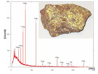
Gamma-ray spectroscopy is the qualitative study of the energy spectra of gamma-ray sources, such as in the nuclear industry, geochemical investigation, and astrophysics. Gamma-ray spectrometry, on the other hand, is the method used to acquire a quantitative spectrum measurement.

Gaseous ionization detectors are radiation detection instruments used in particle physics to detect the presence of ionizing particles, and in radiation protection applications to measure ionizing radiation.
Cadmium zinc telluride, (CdZnTe) or CZT, is a compound of cadmium, zinc and tellurium or, more strictly speaking, an alloy of cadmium telluride and zinc telluride. A direct bandgap semiconductor, it is used in a variety of applications, including semiconductor radiation detectors, photorefractive gratings, electro-optic modulators, solar cells, and terahertz generation and detection. The band gap varies from approximately 1.4 to 2.2 eV, depending on composition.

Neutron detection is the effective detection of neutrons entering a well-positioned detector. There are two key aspects to effective neutron detection: hardware and software. Detection hardware refers to the kind of neutron detector used and to the electronics used in the detection setup. Further, the hardware setup also defines key experimental parameters, such as source-detector distance, solid angle and detector shielding. Detection software consists of analysis tools that perform tasks such as graphical analysis to measure the number and energies of neutrons striking the detector.

Medipix is a family of photon counting and particle tracking pixel detectors developed by an international collaboration, hosted by CERN.
Optical heterodyne detection is a method of extracting information encoded as modulation of the phase, frequency or both of electromagnetic radiation in the wavelength band of visible or infrared light. The light signal is compared with standard or reference light from a "local oscillator" (LO) that would have a fixed offset in frequency and phase from the signal if the latter carried null information. "Heterodyne" signifies more than one frequency, in contrast to the single frequency employed in homodyne detection.

A transition-edge sensor (TES) is a type of cryogenic energy sensor or cryogenic particle detector that exploits the strongly temperature-dependent resistance of the superconducting phase transition.

Flat-panel detectors are a class of solid-state x-ray digital radiography devices similar in principle to the image sensors used in digital photography and video. They are used in both projectional radiography and as an alternative to x-ray image intensifiers (IIs) in fluoroscopy equipment.

X-ray detectors are devices used to measure the flux, spatial distribution, spectrum, and/or other properties of X-rays.

Photon counting is a technique in which individual photons are counted using a single-photon detector (SPD). A single-photon detector emits a pulse of signal for each detected photon. The counting efficiency is determined by the quantum efficiency and the system's electronic losses.
High energy X-ray imaging technology (HEXITEC) is a family of spectroscopic, single photon counting, pixel detectors developed for high energy X-ray and gamma ray spectroscopy applications.
Photon-counting computed tomography (PCCT) is a form of X-ray computed tomography (CT) in which X-rays are detected using a photon-counting detector (PCD) which registers the interactions of individual photons. By keeping track of the deposited energy in each interaction, the detector pixels of a PCD each record an approximate energy spectrum, making it a spectral or energy-resolved CT technique. In contrast, more conventional CT scanners use energy-integrating detectors (EIDs), where the total energy deposited in a pixel during a fixed period of time is registered. These EIDs thus register only photon intensity, comparable to black-and-white photography, whereas PCDs register also spectral information, similar to color photography.
Hybrid pixel detectors are a type of ionizing radiation detector consisting of an array of diodes based on semiconductor technology and their associated electronics. The term “hybrid” stems from the fact that the two main elements from which these devices are built, the semiconductor sensor and the readout chip, are manufactured independently and later electrically coupled by means of a bump-bonding process. Ionizing particles are detected as they produce electron-hole pairs through their interaction with the sensor element, usually made of doped silicon or cadmium telluride. The readout ASIC is segmented into pixels containing the necessary electronics to amplify and measure the electrical signals induced by the incoming particles in the sensor layer.
Photon-counting mammography was introduced commercially in 2003 and was the first widely available application of photon-counting detector technology in medical x-ray imaging. Photon-counting mammography improves dose efficiency compared to conventional technologies, and enables spectral imaging.
References
- ↑ Chmeissani, M.; Mikulec, B. (2001). "Performance limits of a single photon counting pixel system". Nuclear Instruments and Methods in Physics Research Section A: Accelerators, Spectrometers, Detectors and Associated Equipment. 460 (1): 81–90. Bibcode:2001NIMPA.460...81C. doi:10.1016/S0168-9002(00)01100-1.
- ↑ Myronakis, Marios E.; Darambara, Dimitra G. (2010-12-28). "Monte Carlo investigation of charge-transport effects on energy resolution and detection efficiency of pixelated CZT detectors for SPECT/PET applications: Investigation of charge-transport effects on pixelated CZT detectors". Medical Physics. 38 (1): 455–467. doi:10.1118/1.3532825. PMID 21361214.
- ↑ Bugby, S.L.; Koch-Mehrin, K.A.; Veale, M.C.; Wilson, M.D.; Lees, J.E. (2019). "Energy-loss correction in charge sharing events for improved performance of pixellated compound semiconductors" (PDF). Nuclear Instruments and Methods in Physics Research Section A: Accelerators, Spectrometers, Detectors and Associated Equipment. 940: 142–151. Bibcode:2019NIMPA.940..142B. doi: 10.1016/j.nima.2019.06.017 .
- ↑ Mohit Kumar Gupta (2006). EDA for IC implementation, circuit design, and process technology. CRC Press. ISBN 0-8493-7924-5.