Related Research Articles
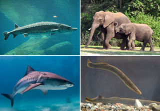
Vertebrates are deuterostomal animals with bony or cartilaginous axial endoskeleton — known as the vertebral column, spine or backbone — around and along the spinal cord, including all fish, amphibians, reptiles, birds and mammals. The vertebrates consist of all the taxa within the subphylum Vertebrata and represent the overwhelming majority of the phylum Chordata, with currently about 69,963 species described.

The mesoderm is the middle layer of the three germ layers that develops during gastrulation in the very early development of the embryo of most animals. The outer layer is the ectoderm, and the inner layer is the endoderm.

The skull is a bone protective cavity for the brain. The skull is composed of four types of bone i.e., cranial bones, facial bones, ear ossicles and hyoid bone, however two parts are more prominent: the cranium and the mandible. In humans, these two parts are the neurocranium (braincase) and the viscerocranium that includes the mandible as its largest bone. The skull forms the anterior-most portion of the skeleton and is a product of cephalisation—housing the brain, and several sensory structures such as the eyes, ears, nose, and mouth. In humans, these sensory structures are part of the facial skeleton.

In anatomy, the temporomandibular joints (TMJ) are the two joints connecting the jawbone to the skull. It is a bilateral synovial articulation between the temporal bone of the skull above and the mandible below; it is from these bones that its name is derived. This joint is unique in that it is a bilateral joint that functions as one unit. Since the TMJ is connected to the mandible, the right and left joints must function together and therefore are not independent of each other.

The maxilla in vertebrates is the upper fixed bone of the jaw formed from the fusion of two maxillary bones. In humans, the upper jaw includes the hard palate in the front of the mouth. The two maxillary bones are fused at the intermaxillary suture, forming the anterior nasal spine. This is similar to the mandible, which is also a fusion of two mandibular bones at the mandibular symphysis. The mandible is the movable part of the jaw.

The frontal bone or sincipital bone is a bone in the human skull. The bone consists of two portions. These are the vertically oriented squamous part, and the horizontally oriented orbital part, making up the bony part of the forehead, part of the bony orbital cavity holding the eye, and part of the bony part of the nose respectively. The name comes from the Latin word frons.

The temporal bones are situated at the sides and base of the skull, and lateral to the temporal lobes of the cerebral cortex.

Fish anatomy is the study of the form or morphology of fish. It can be contrasted with fish physiology, which is the study of how the component parts of fish function together in the living fish. In practice, fish anatomy and fish physiology complement each other, the former dealing with the structure of a fish, its organs or component parts and how they are put together, such as might be observed on the dissecting table or under the microscope, and the latter dealing with how those components function together in living fish.
A craniate is a member of the Craniata, a proposed clade of chordate animals with a skull of hard bone or cartilage. Living representatives are the Myxini (hagfishes), Hyperoartia, and the much more numerous Gnathostomata. Formerly distinct from vertebrates by excluding hagfish, molecular and anatomical research in the 21st century has led to the reinclusion of hagfish as vertebrates, making living craniates synonymous with living vertebrates.

In zoology and developmental anatomy, the notochord is an elastic, rod-like anatomical structure found in many deuterostomal animals. A notochord is one of five synapomorphies used to define a species as a chordate.
An endoskeleton is a structural frame (skeleton) on the inside of an animal, overlaid by soft tissues and usually composed of mineralized tissue. Endoskeletons serve as structural support against gravity and mechanical loads, and provide anchoring attachment sites for skeletal muscles to transmit force and allow movements and locomotion.

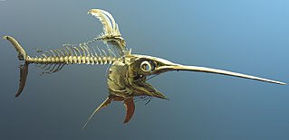
Cladoselache is an extinct genus of shark-like chondrichthyan from the Late Devonian (Famennian) of North America. It was similar in body shape to modern lamnid sharks, but was not closely related to lamnids or to any other modern (selachian) shark. As an early chondrichthyan, it had yet to evolve traits of modern sharks such as accelerated tooth replacement, a loose jaw suspension, enameloid teeth, and possibly claspers.
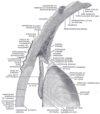
The lens capsule is a component of the globe of the eye. It is a clear elastic basement membrane composed of collagen IV laminin etc. a quality that keeps it under constant tension. As a result, the lens naturally tends towards a rounder or more globular configuration, a shape it must assume for the eye to focus at a near distance. The lens capsule is the thickest basement membrane in the body.

The pharyngeal arches, also known as visceral arches, are structures seen in the embryonic development of vertebrates that are recognisable precursors for many structures. In fish, the arches are known as the branchial arches, or gill arches.

The petrosquamous suture is a cranial suture between the petrous portion and the squama of the temporal bone. It forms the Koerner's septum. The petrous portion forms the medial component of the osseous margin, while the squama forms the lateral component. The anterolateral portion (squama) arises from the mesenchyme at 8 weeks of embryogenesis while the petromastoid portion develops later from a cartilaginous center at 6 months of fetal development.

In human anatomy, the neurocranium, also known as the braincase, brainpan, or brain-pan, is the upper and back part of the skull, which forms a protective case around the brain. In the human skull, the neurocranium includes the calvaria or skullcap. The remainder of the skull is the facial skeleton.

The endocranium in comparative anatomy is a part of the skull base in vertebrates and it represents the basal, inner part of the cranium. The term is also applied to the outer layer of the dura mater in human anatomy.

The vertebral column, also known as the backbone or spine, is the core part of the axial skeleton in vertebrate animals. The vertebral column is the defining characteristic of vertebrate endoskeleton in which the notochord found in all chordates has been replaced by a segmented series of mineralized irregular bones called vertebrae, separated by fibrocartilaginous intervertebral discs. The dorsal portion of the vertebral column houses the spinal canal, a cavity formed by alignment of the neural arches that encloses and protects the spinal cord.

Most bony fishes have two sets of jaws made mainly of bone. The primary oral jaws open and close the mouth, and a second set of pharyngeal jaws are positioned at the back of the throat. The oral jaws are used to capture and manipulate prey by biting and crushing. The pharyngeal jaws, so-called because they are positioned within the pharynx, are used to further process the food and move it from the mouth to the stomach.

Trabecular cartilages are paired, rod-shaped cartilages, which develop in the head of the vertebrate embryo. They are the primordia of the anterior part of the cranial base, and are derived from the cranial neural crest cells.
References
- 1 2 3 Salentijn, L. Biology of Mineralized Tissues: Prenatal Skull Development, Columbia University College of Dental Medicine post-graduate dental lecture series, 2007
- ↑ Kardong, Kenneth V. (2015). Vertebrates: Comparative Anatomy, Function, Evolution. New York: McGraw-Hill Education. p. 701.
- ↑ Kent, G.C & Miller, L. (1997): Comparative Anatomy of the Vertebrates. Wm. C. Brown Publishers. ISBN 0-697-24378-8.
- ↑ Romer, Alfred Sherwood; Parsons, Thomas S. (1977). The Vertebrate Body. Philadelphia, PA: Holt-Saunders International. pp. 216–247. ISBN 0-03-910284-X.
| | This human musculoskeletal system article is a stub. You can help Wikipedia by expanding it. |