Clinical neurochemistry

Last updated
Progression of Huntington's Diesase. A microscope image of Medium spiny neurons (yellow) with nuclear inclusions (orange), which occur as part of the disease process. Neuron with mHtt inclusion.jpg
Progression of Huntington's Diesase. A microscope image of Medium spiny neurons (yellow) with nuclear inclusions (orange), which occur as part of the disease process.

Clinical neurochemistry is the field of neurological biochemistry which relates biochemical phenomena to clinical symptomatic manifestations in humans. While neurochemistry is mostly associated with the effects of neurotransmitters and similarly functioning chemicals on neurons themselves, clinical neurochemistry relates these phenomena to system-wide symptoms. Clinical neurochemistry is related to neurogenesis, neuromodulation, neuroplasticity, neuroendocrinology, and neuroimmunology in the context of associating neurological findings at both lower and higher level organismal functions.

Contents

Neuropharmacology and drug action

Efficacy spectrum of receptor ligands. Efficacy spectrum.png
Efficacy spectrum of receptor ligands.

The integration of knowledge concerning the molecular and cellular actions of a drug within the brain circuitry leads to an overall understanding of a neurological drug's action mechanisms. This understanding of drug action in turn can be extrapolated to account for system-wide or clinical manifestations which are observed as symptoms. The clinical effects of a neural drug are due to both immediate changes in homeostasis and long-term neural adaptations characterized by the phenomena neural plasticity. [1]

The most basic and fundamental neurological phenomena in neuropharmacology is the binding of a drug or neurologically active substance to a cellular target. One assay to determine the extent at which a ligand binds to its receptor is the radioligand binding assay (RBA), in which specific binding of a radioactively-labeled ligand is denoted by the difference between saturated and non-saturated tissue samples. While the RBA assay assumes that the tissue prepared has just one molecular target per ligand, in actuality this may not be the case. For example, serotonin binds to many diverse serotonin receptors which makes the RIA assay quite difficult to interpret. Because many receptors are essentially enzymes, the field of pharmakinetics utilizes the Michaelis–Menten equation to describe drug affinity (dissociation constant Kd) and total binding (Bmax). Although Kd and Bmax can be determined pictorially in a normal or logarithmic plot of ligand binding vs drug concentration, Scatchard plots allow for mathematical representation of several ligand binding sites, each with its own Kd. [1]

Semi-log plots of two agonists with different Kd. DoseResponse000.jpg
Semi-log plots of two agonists with different Kd.

Drug potency is the measure of binding strength between a drug and a specific molecular target, whereas drug efficacy describes the biological effect exerted by the drug itself, at either a cellular or organismal level. Because drugs range widely in their potency and efficacy, drugs have been categorized on the spectrum of agonists and antagonists. Agonists bind to receptors and elicit the same effects as an endogenous neurotransmitter. For example, morphine is an agonist of the opioid receptor family. Conversely, antagonists bind to a receptor and elicit no cellular change. [2] Naloxone, an antagonist of the opioid receptors, exerts a biological effect only be interfering with endogenous neurotransmitter (morphine) binding. [2] Inverse agonists bind to receptors and elicit the opposite effect that an agonist would. The spectrum of drug continuum also includes partial agonists and partial inverse agonists, which comprise the wide majority of neurological clinical treatments. The ultimate clinical effect of a drug can be analyzed with a dose-response curve. [1]

Neurologically active substances

There are many biologically active chemicals which elicit an effect on the nervous system. Neurotransmitters and similarly functioning biochemical messengers elicit effects on postsynaptic neurons at neuronal synapses. Excitatory Amino Acids include Glutamate, whereas inhibitory Amino Acids include GABA and Glycine. Additionally, catecholamines, serotonin, acetylcholine, histamine, and orexins have widely-projecting effects and are often referred to as neuromodulators. Neuropeptides include bradykinin, cholecystokinin, corticotropin-releasing factor (CRF), galanin, MCH, MSH, Neuropeptide Y (NPY), Neurotensin, Opioids, orexin, oxytocin, somatostatin, tachykinins, TRH, CUP, and vasopressin. Purines, endogenous cannabinoids, gasses, neurotrophic factors, chemokines, and VEGF are all classified as atypical neurotransmitters. Major receptors of neurotransmitters include AMPA receptors, NMDA receptors, and Kainate Receptors. [1]

Clinical neurological disorders

Changes in the homeostatic levels of many neurologically active chemicals elicit clinical disorders and symptoms.

Pain and inflammation

Nociception is the form of somatic sensation that detects potentially tissue-damaging noxious stimuli. Peripheral nociceptors uniquely express transient receptor potentials which are sensitive to potentially-damaging mechanical, chemical, or thermal stimuli. Nociceptors also contain receptors for pain and inflammatory-related mediators or cytokines. Peripheral nociceptors transmit noxious stimuli to the dorsal root ganglia, the dorsal horn, and further to the trigeminal ganglia in the brain. Pain has both a localizing somatic sensory component and an aversive emotional and motivational component. Pain travels through a variety of pathways via first pain on Alpha Delta fibers and second pain on slowly conducting C-fibers. [3]

The dorsal horn of the spinal cord serves as a major integration center for both ascending nociceptive information and descending antinociceptive influences from the brain. Plasticity within the dorsal horn is mediated by NMDA glutamate receptors and key in the initiation of chronic pain by decreasing the excitability threshold in nociceptive pathways. Additionally, damage to neurons in nociceptive pathways leads to neuropathic pain. [4] Three families in northern Pakistan were congenitally unable to perceive pain due to their homozygous loss of function mutation in the SCN9A gene which codes for the voltage-gated sodium channel NaV1.7 . This key finding allowed pharmacologists to begin researching if the NaV1.7 is a substantial molecular target for analgesic (antipain) medications. [5]

Sensitization, in the clinical sense of the word, is a phenomenon in which nociceptors in an area beyond a tissue injury exhibit decreased thresholds for activation. Sensitization can be initiated by inflammatory prostaglandins or leukotrienes and are therefore the targets of nonsteroidal anti-inflammatory (NSAIDs) which block key enzymes in their synthesis. Additionally, opioid drugs suppress nociceptions by binding to endogenous opioid receptors. [1]

Sleep and arousal

Polysomnographic record of REM Sleep. EEG highlighted by red box with eye movement highlighted by red line. Sleep EEG REM.png
Polysomnographic record of REM Sleep. EEG highlighted by red box with eye movement highlighted by red line.

Sleep arousal are active brain processes medicated and associated with specific brain regions. Despite the fact that various stages of sleep are discrete and quantifiable, the exact function of sleep is unknown. Sleep is controlled both by circadian rhythms and the homeostatic drive produced by wakefulness. Circadian rhythms are produced in the suprachiasmatic nucleus by pacemaker cells which contain transcriptional regulation "clock genes" which have been highly conserved throughout evolution. [6]

Non-REM sleep is initiated by neurons in the preoptic and anterior hypothalamic area, whereas REM sleep is eventually elicited by the cells in the pontine tegmentum. Electroencephalography is used to analyze brain wave patterns during sleep and has the capability to differentiate between REM sleep from non-REM sleep. REM sleep cycles mimic conscious brain patterns to an extent. Night Terrors, for example, involve the partial arousal out of non-REM sleep. Similarly, REM Behavior Disorder occurs when patients have fits of violent behavior during REM sleep. Benzodiazepines are the most common treatments for sleep-related disorders.

Dyssomnia is a class of sleep disorders which includes Primary insomnia, primary hypersomnia, narcolepsy, breathing-related sleep disorders, circadian rhythm sleep disorder, and other conditions. Primary insomnia is a disorder in which a patient has difficulty initiating and maintaining sleep. Behavior modification and a reduction in neurologically active substances such as caffeine and alcohol seem to be among the most promising treatments. Although the mechanism is unknown, brain plasticity and behavior modification are utilized to train patients to only go to bed when tired, associating the bed itself with a sleepy state.

Narcolepsy is a condition characterized by abnormal transitions between REM and non-REM cycles during sleep and the awake cycle. Cataplexy, on the other hand, is an involuntary loss of muscle tone during wakefulness. The mechanism of narcolepsy is unknown, though recent findings suggest that orexin neurons in the lateral and posterior hypothalamus may play a critical role in reinforcing wakefulness. [7] Narcolepsy is often treated with psychostimulants or tricyclic antidepressants in order to suppress REM sleep patterns. Sleep apnea is a common breathing disorder during sleep and is related to a disability in the central respiratory drive mechanisms. Parasomnias are a class represented by nightmares, sleep terrors, night terrors, schizophrenia, certain mood disorders, and other conditions which arise during Stage 4 of sleep.

General anesthetics typically induce non-REM sleep characterized by amnesia, analgesia, immobility, and hypnosis by facilitating the inhibition of excitatory ion channels or the excitation of inhibitory ligand-gated channels.

Schizophrenia and other psychoses

Functional magnetic resonance imaging depicting differences in brain activity among people diagnosed with schizophrenia Functional magnetic resonance imaging.jpg
Functional magnetic resonance imaging depicting differences in brain activity among people diagnosed with schizophrenia

Schizophrenia is a psychological disorder in which a patient experiences symptoms including hallucinations, delusions, amotivation, social withdrawal, cognitive defects, and poor working memory. Heredity and gene inheritance is a highly important risk factor, especially for identical twins. [8] Schizophrenia is anatomically characterized by a deterioration and loss of gray matter in the temporal and frontal regions of the cerebral cortex, though the exact mechanism is unknown. [9] What is known is that the main two neurotransmitter systems implicated in schizophrenia are the dopamine and glutamate pathways.

Dopamine became a candidate for research when it was clinically noticed that antipsychotic drugs which are dopamine D2 receptor antagonists were noted to be quite successful in treating schizophrenia as well. [10] Increased levels of dopamine in people with schizophrenia tend to induce paranoid delusions, ideas of reference, and auditory hallucinations. The same dopaminergic pathway is also involved in psychosis.

Glutamate has become a candidate for treatment focus because glutamate blocks some NMDA receptors which, on their own, induce schizophrenic behavior. In animal models, NMDA antagonists increase glutamate release in the prefrontal cortex. It is postulated that this is a homeostatic response to NMDA receptor blockade, which in turn increases psychotic symptoms. A class of NMDA receptor antagonists have been denoted dissociative anesthetics because they produce a sense of depersonalization and dissociation of subjective experience from various forms of sensory input stimuli. As such, variants such as Ketamine (Angel Dust/Special K) and Phencyclidine (PCP) have become a commonly abused street-drug. These drugs are no longer used due to harmful behavioral and addictive effects. [11]

Neurodegeneration

Neurodegeneration is classified as a massive death of neurons, and encompasses diseases such as Alzheimer's, Parkinson's, and Huntington's. Although many cells die due to necrosis, many cells in neurodegenerative disorders are killed via the apoptotic pathway. Excitotoxicity, which involves overstimulation of a cell via its glutamate receptors, is one of the major processes which can initiate cell death. Other inducers include mitochondrial dysfunction, oxidative stress, and abnormal protein aggregation. Surprisingly, both necrotic and apoptotic processes utilize a similar intracellular signaling cascade which uses caspase proteins to induce cell death. Abnormal protein accumulation causes Alzheimer's, Parkinson's, and Huntington's diseases. [1]

Alzheimer's disease

Comparison of a normal aged brain (left) and an Alzheimer's patient's brain (right). Alzheimer's disease brain comparison.jpg
Comparison of a normal aged brain (left) and an Alzheimer's patient's brain (right).

Alzheimer's disease is the most common cause of severe memory impairment and is caused by senile plaques, neurofibrillary tangles, dystrophic neuritis, and neuronal loss. It is thought that Alzheimer's disease may be due to unnecessary protein accumulation of β Amyloid. [12] In fact, Senile plaques are dense, protein deposits composed of amyloid β peptide. The two types of senile plaques are diffuse plaques and neuritic plaques, and differ in morphology. In addition to the amyloid, the microtubule-associated Tau protein has also been in involved with Alzheimer's disease and a variety of other neurodegenerative diseases. [13] Inherited forms of Alzheimer's have been linked to mutation in the APP genes or presenilins which regulate APP processing. Because cholinergic neurons of the nucleus basalis are significantly altered during Alzheimer's progression, cholinergic agents such as choline and lecithin were hypothesized to augment the progression. However, these attempts were unsuccessful and the only clinically useful drugs used in the United States are cholinesterase inhibitors, which prolong the time before choline degradation. [12] Although aNMDA receptor antagonists and anti-inflammatory drugs were tested in a clinical environment, more promising clinical trials are underway to targeting the Aβ with the immune system.

Parkinson's disease

Parkinson's disease is caused by the loss of dopamine stimulation to the basal ganglia of the midbrain, resulting in tremor at rest and bradykinesia. In some rare forms, protein aggregation of alpha-synuclein and parkin can elicit symptoms of Parkinson's disease. [14] For a variety of drugs, restoring dopamine to the central nervous system remains the target therapy. Because dopamine cannot pass the blood brain barrier on its own, the dopamine precursor L-DOPA is used in its stead. However, synaptic plasticity renders this treatment decreasingly effective with time. Another treatment option is bromocriptine, which directly stimulates D2 Dopamine Receptors. [15] Bromocriptine is less effective than L-Dopa in reducing symptoms, but provides less dyskinesia. Often, the two drugs are used in concert with one another. New approaches to treatment include deep brain simulation and cell transplantation with stem cells. Deep brain stimulation offsets symptoms rather than cures, and stem cell studies have been extremely disappointing, despite relative success with animal models. Gene therapy may provide a novel route to introduce new dopamine production via viral-medicated gene transfection, but clinical trials are yet to be completed. [16]

Huntington's disease

Huntington's disease is a disease characterized by minor coordination problems, jerking eye movements, and uncontrollable movement of peripheral limbs. Symptoms generally occur at the age of 40, and are often accompanied by depression and psychosis. The disease is caused by a mutation in the Huntingtin gene, on chromosome 4, which causes abnormally large numbers of glutamate residues in the protein. Via an unknown mechanism, this accumulation leads to neurodegeneration in the caudate nucleus and putamen, selectively destroying GABAergic neurons which project to the globus pallidus. [17] There is also significant necrosis in the thalamus and cerebral cortex. Cholinergic interneurons and dopaminergic neurons in the midbrain are largely unaffected. Treatment for Huntington's disease is extremely limited due to the lack of knowledge concerning the pathogenesis of protein accumulation, though drugs used include dopamine receptor antagonists to minimize tremors and antidepressants to ameliorate symptoms of psychosis and depression.

See also

Related Research Articles

<span class="mw-page-title-main">Neurotransmitter</span> Chemical substance that enables neurotransmission

A neurotransmitter is a signaling molecule secreted by a neuron to affect another cell across a synapse. The cell receiving the signal, or target cell, may be another neuron, but could also be a gland or muscle cell.

<span class="mw-page-title-main">Psychopharmacology</span> Study of the effects of psychoactive drugs

Psychopharmacology is the scientific study of the effects drugs have on mood, sensation, thinking, behavior, judgment and evaluation, and memory. It is distinguished from neuropsychopharmacology, which emphasizes the correlation between drug-induced changes in the functioning of cells in the nervous system and changes in consciousness and behavior.

<span class="mw-page-title-main">NMDA receptor</span> Glutamate receptor and ion channel protein found in nerve cells

The N-methyl-D-aspartatereceptor (also known as the NMDA receptor or NMDAR), is a glutamate receptor and ion channel found in neurons. The NMDA receptor is one of three types of ionotropic glutamate receptors, the other two being AMPA and kainate receptors. Depending on its subunit composition, its ligands are glutamate and glycine (or D-serine). However, the binding of the ligands is typically not sufficient to open the channel as it may be blocked by Mg2+ ions which are only removed when the neuron is sufficiently depolarized. Thus, the channel acts as a “coincidence detector” and only once both of these conditions are met, the channel opens and it allows positively charged ions (cations) to flow through the cell membrane. The NMDA receptor is thought to be very important for controlling synaptic plasticity and mediating learning and memory functions.

<span class="mw-page-title-main">Excitatory synapse</span> Sort of synapse

An excitatory synapse is a synapse in which an action potential in a presynaptic neuron increases the probability of an action potential occurring in a postsynaptic cell. Neurons form networks through which nerve impulses travels, each neuron often making numerous connections with other cells of neurons. These electrical signals may be excitatory or inhibitory, and, if the total of excitatory influences exceeds that of the inhibitory influences, the neuron will generate a new action potential at its axon hillock, thus transmitting the information to yet another cell.

<span class="mw-page-title-main">Excitotoxicity</span> Process that kills nerve cells

In excitotoxicity, nerve cells suffer damage or death when the levels of otherwise necessary and safe neurotransmitters such as glutamate become pathologically high, resulting in excessive stimulation of receptors. For example, when glutamate receptors such as the NMDA receptor or AMPA receptor encounter excessive levels of the excitatory neurotransmitter, glutamate, significant neuronal damage might ensue. Excess glutamate allows high levels of calcium ions (Ca2+) to enter the cell. Ca2+ influx into cells activates a number of enzymes, including phospholipases, endonucleases, and proteases such as calpain. These enzymes go on to damage cell structures such as components of the cytoskeleton, membrane, and DNA. In evolved, complex adaptive systems such as biological life it must be understood that mechanisms are rarely, if ever, simplistically direct. For example, NMDA in subtoxic amounts induces neuronal survival of otherwise toxic levels of glutamate.

<span class="mw-page-title-main">Dizocilpine</span> Chemical compound

Dizocilpine (INN), also known as MK-801, is a pore blocker of the N-Methyl-D-aspartate (NMDA) receptor, a glutamate receptor, discovered by a team at Merck in 1982. Glutamate is the brain's primary excitatory neurotransmitter. The channel is normally blocked with a magnesium ion and requires depolarization of the neuron to remove the magnesium and allow the glutamate to open the channel, causing an influx of calcium, which then leads to subsequent depolarization. Dizocilpine binds inside the ion channel of the receptor at several of PCP's binding sites thus preventing the flow of ions, including calcium (Ca2+), through the channel. Dizocilpine blocks NMDA receptors in a use- and voltage-dependent manner, since the channel must open for the drug to bind inside it. The drug acts as a potent anti-convulsant and probably has dissociative anesthetic properties, but it is not used clinically for this purpose because of the discovery of brain lesions, called Olney's lesions (see below), in laboratory rats. Dizocilpine is also associated with a number of negative side effects, including cognitive disruption and psychotic-spectrum reactions. It inhibits the induction of long term potentiation and has been found to impair the acquisition of difficult, but not easy, learning tasks in rats and primates. Because of these effects of dizocilpine, the NMDA receptor pore blocker ketamine is used instead as a dissociative anesthetic in human medical procedures. While ketamine may also trigger temporary psychosis in certain individuals, its short half-life and lower potency make it a much safer clinical option. However, dizocilpine is the most frequently used uncompetitive NMDA receptor antagonist in animal models to mimic psychosis for experimental purposes.
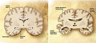
<span class="mw-page-title-main">Ligand-gated ion channel</span> Type of ion channel transmembrane protein

Ligand-gated ion channels (LICs, LGIC), also commonly referred to as ionotropic receptors, are a group of transmembrane ion-channel proteins which open to allow ions such as Na+, K+, Ca2+, and/or Cl to pass through the membrane in response to the binding of a chemical messenger (i.e. a ligand), such as a neurotransmitter.

Neuropharmacology is the study of how drugs affect function in the nervous system, and the neural mechanisms through which they influence behavior. There are two main branches of neuropharmacology: behavioral and molecular. Behavioral neuropharmacology focuses on the study of how drugs affect human behavior (neuropsychopharmacology), including the study of how drug dependence and addiction affect the human brain. Molecular neuropharmacology involves the study of neurons and their neurochemical interactions, with the overall goal of developing drugs that have beneficial effects on neurological function. Both of these fields are closely connected, since both are concerned with the interactions of neurotransmitters, neuropeptides, neurohormones, neuromodulators, enzymes, second messengers, co-transporters, ion channels, and receptor proteins in the central and peripheral nervous systems. Studying these interactions, researchers are developing drugs to treat many different neurological disorders, including pain, neurodegenerative diseases such as Parkinson's disease and Alzheimer's disease, psychological disorders, addiction, and many others.

Molecular neuroscience is a branch of neuroscience that observes concepts in molecular biology applied to the nervous systems of animals. The scope of this subject covers topics such as molecular neuroanatomy, mechanisms of molecular signaling in the nervous system, the effects of genetics and epigenetics on neuronal development, and the molecular basis for neuroplasticity and neurodegenerative diseases. As with molecular biology, molecular neuroscience is a relatively new field that is considerably dynamic.

<span class="mw-page-title-main">Kainate receptor</span> Class of ionotropic glutamate receptors

Kainate receptors, or kainic acid receptors (KARs), are ionotropic receptors that respond to the neurotransmitter glutamate. They were first identified as a distinct receptor type through their selective activation by the agonist kainate, a drug first isolated from the algae Digenea simplex. They have been traditionally classified as a non-NMDA-type receptor, along with the AMPA receptor. KARs are less understood than AMPA and NMDA receptors, the other ionotropic glutamate receptors. Postsynaptic kainate receptors are involved in excitatory neurotransmission. Presynaptic kainate receptors have been implicated in inhibitory neurotransmission by modulating release of the inhibitory neurotransmitter GABA through a presynaptic mechanism.

Neuroprotection refers to the relative preservation of neuronal structure and/or function. In the case of an ongoing insult the relative preservation of neuronal integrity implies a reduction in the rate of neuronal loss over time, which can be expressed as a differential equation. It is a widely explored treatment option for many central nervous system (CNS) disorders including neurodegenerative diseases, stroke, traumatic brain injury, spinal cord injury, and acute management of neurotoxin consumption. Neuroprotection aims to prevent or slow disease progression and secondary injuries by halting or at least slowing the loss of neurons. Despite differences in symptoms or injuries associated with CNS disorders, many of the mechanisms behind neurodegeneration are the same. Common mechanisms of neuronal injury include decreased delivery of oxygen and glucose to the brain, energy failure, increased levels in oxidative stress, mitochondrial dysfunction, excitotoxicity, inflammatory changes, iron accumulation, and protein aggregation. Of these mechanisms, neuroprotective treatments often target oxidative stress and excitotoxicity—both of which are highly associated with CNS disorders. Not only can oxidative stress and excitotoxicity trigger neuron cell death but when combined they have synergistic effects that cause even more degradation than on their own. Thus limiting excitotoxicity and oxidative stress is a very important aspect of neuroprotection. Common neuroprotective treatments are glutamate antagonists and antioxidants, which aim to limit excitotoxicity and oxidative stress respectively.

<span class="mw-page-title-main">Glutamate receptor</span> Cell-surface proteins that bind glutamate and trigger changes which influence the behavior of cells

Glutamate receptors are synaptic and non synaptic receptors located primarily on the membranes of neuronal and glial cells. Glutamate is abundant in the human body, but particularly in the nervous system and especially prominent in the human brain where it is the body's most prominent neurotransmitter, the brain's main excitatory neurotransmitter, and also the precursor for GABA, the brain's main inhibitory neurotransmitter. Glutamate receptors are responsible for the glutamate-mediated postsynaptic excitation of neural cells, and are important for neural communication, memory formation, learning, and regulation.

Neuropsychopharmacology, an interdisciplinary science related to psychopharmacology and fundamental neuroscience, is the study of the neural mechanisms that drugs act upon to influence behavior. It entails research of mechanisms of neuropathology, pharmacodynamics, psychiatric illness, and states of consciousness. These studies are instigated at the detailed level involving neurotransmission/receptor activity, bio-chemical processes, and neural circuitry. Neuropsychopharmacology supersedes psychopharmacology in the areas of "how" and "why", and additionally addresses other issues of brain function. Accordingly, the clinical aspect of the field includes psychiatric (psychoactive) as well as neurologic (non-psychoactive) pharmacology-based treatments. Developments in neuropsychopharmacology may directly impact the studies of anxiety disorders, affective disorders, psychotic disorders, degenerative disorders, eating behavior, and sleep behavior.

<span class="mw-page-title-main">Dopamine agonist</span> Compound that activates dopamine receptors

A dopamine agonist(DA) is a compound that activates dopamine receptors. There are two families of dopamine receptors, D2-like and D1-like, and they are all G protein-coupled receptors. D1- and D5-receptors belong to the D1-like family and the D2-like family includes D2, D3 and D4 receptors. Dopamine agonists are primarily used in the treatment of Parkinson's disease, and to a lesser extent, in hyperprolactinemia and restless legs syndrome. They are also used off-label in the treatment of clinical depression. The use of dopamine agonists is associated with impulse control disorders and dopamine agonist withdrawal syndrome (DAWS).

<span class="mw-page-title-main">NMDA receptor antagonist</span> Class of anesthetics

NMDA receptor antagonists are a class of drugs that work to antagonize, or inhibit the action of, the N-Methyl-D-aspartate receptor (NMDAR). They are commonly used as anesthetics for animals and humans; the state of anesthesia they induce is referred to as dissociative anesthesia.

<i>N</i>-Acetylaspartylglutamic acid Peptide neurotransmitter

N-Acetylaspartylglutamic acid is a peptide neurotransmitter and the third-most-prevalent neurotransmitter in the mammalian nervous system. NAAG consists of N-acetylaspartic acid (NAA) and glutamic acid coupled via a peptide bond.

The glutamate hypothesis of schizophrenia models the subset of pathologic mechanisms of schizophrenia linked to glutamatergic signaling. The hypothesis was initially based on a set of clinical, neuropathological, and, later, genetic findings pointing at a hypofunction of glutamatergic signaling via NMDA receptors. While thought to be more proximal to the root causes of schizophrenia, it does not negate the dopamine hypothesis, and the two may be ultimately brought together by circuit-based models. The development of the hypothesis allowed for the integration of the GABAergic and oscillatory abnormalities into the converging disease model and made it possible to discover the causes of some disruptions.

An H3 receptor antagonist is a classification of drugs used to block the action of histamine at the H3 receptor.

<span class="mw-page-title-main">Narcolepsy</span> Human sleep disorder that involves an excessive urge to sleep and other neurological features

Narcolepsy is a chronic neurological disorder that involves a decreased ability to regulate sleep–wake cycles. Symptoms often include periods of excessive daytime sleepiness and brief involuntary sleep episodes. About 70% of those affected also experience episodes of sudden loss of muscle strength, known as cataplexy. Narcolepsy paired with cataplexy is evidenced to be an autoimmune disorder. These experiences of cataplexy can be brought on by strong emotions. Less commonly, there may be vivid hallucinations or an inability to move while falling asleep or waking up. People with narcolepsy tend to sleep about the same number of hours per day as people without, but the quality of sleep tends to be lessened.

<span class="mw-page-title-main">Willardiine</span> Chemical compound

Willardiine (correctly spelled with two successive i's) or (S)-1-(2-amino-2-carboxyethyl)pyrimidine-2,4-dione is a chemical compound that occurs naturally in the seeds of Mariosousa willardiana and Acacia sensu lato. The seedlings of these plants contain enzymes capable of complex chemical substitutions that result in the formation of free amino acids (See: #Synthesis). Willardiine is frequently studied for its function in higher level plants. Additionally, many derivates of willardiine are researched for their potential in pharmaceutical development. Willardiine was first discovered in 1959 by R. Gmelin, when he isolated several free, non-protein amino acids from Acacia willardiana (another name for Mariosousa willardiana) when he was studying how these families of plants synthesize uracilyalanines. A related compound, Isowillardiine, was concurrently isolated by a different group, and it was discovered that the two compounds had different structural and functional properties. Subsequent research on willardiine has focused on the functional significance of different substitutions at the nitrogen group and the development of analogs of willardiine with different pharmacokinetic properties. In general, Willardiine is the one of the first compounds studied in which slight changes to molecular structure result in compounds with significantly different pharmacokinetic properties.

References

  1. 1 2 3 4 5 6 Nestler, Eric; Hyman, Steven; Malenka, Robert.Molecular Neurophamacology: A Foundation for Clinical Neuroscience. McGraw Hill Medical, 2009
  2. 1 2 Olofsen, Erik M.; van Dorp, Eveline; Teppema, Luc; Aarts, Leon; Smith, Terry; Dahan, Albert; Sarton, Elise.Naloxone Reversal of Morphine- and Morphine-6-Glucuronide-induced Respiratory Depression in Healthy Volunteers: A Mechanism-based Pharmacokinetic–Pharmacodynamic Modeling Study. Anesthesiology, 2010, pp. 1417–1427.
  3. Craig, ADLabeled Lines versus Convergence in Central Processing. Annu Rev Neurosci, 2003, pp. 1–30
  4. Coull, JA; Beggs, S; Boudreau D, et al.BDNF from microglia causes the shift in neuronal anion gradient underlying neuropathic pain. Nature, 2005, pp. 1017–1021
  5. Cox, JJ; Reimann, F; Nicholas, AK; et al.An SCN9A channelopathy causes congenital inability to experience pain. Nature, 2006, pp. 894–898
  6. King, D; Takahashi, JS.Molecular Genetics of Circadian Rhythms in Mammals. Annu Rev Neurosci, 2000, pp. 713–742
  7. Chemelli, RM; Willie, JT; Sinton, CM; et al.Narcolepsy in orexin knockout mice: molecular genetics of sleep regulation. Cell, 1999, pp. 437–452
  8. Cannon, TD; Thompson, PM; van Erp TG, et al.Cortex Mapping Reveals Regionally Specific Patterns of Genetic and Disease-Specific Gray-Matter Deficits in Twins Discordant for Schizophrenia. Proc Natl Acad Sci USA, 2002, pp. 3228–3233
  9. Boos, HBM; Aleman, A; Cahn, W; et al.Brain volumes in relative patients with schizophrenia. A meta-analysis. Arch Gen Psychiatry, 2007, pp. 297–304
  10. Mamo, D; Graff, A; Mizrahi, R; et al.Differential effects of aripiprazole on D(2), 5HT(2), and 5HT(1A) receptor occupancy in patients with schizophrenia: a triple tracer PET study. Am J Psychiatry, 2007, pp. 1411–1417
  11. Stacy A. Castnerab, Jeffrey L. Arrizac, John C. Robertsc, Ladislav Mrzljakc, Edward P. Christianc, Graham V. Williams.Reversal of Ketamine-Induced Working Memory Impairments by the GABAAα2/3 Agonist TPA023. Biological Psychiatry, 2010, pp. 998–1001
  12. 1 2 Mclaurin, J; Kierstead, ME; Brown, ME; et al.Cyclohexanehexol inhibitors of AB aggregation prevent and reverse Alzheimer phenotype in a mouse model. Nature Med, 2006. pp. 801–808
  13. Stoothoff, WH; Johnson, GV.Tau phosphorylation: physiological and pathological consequences. Biochim Biophys Acta, 2005. pp. 280–297
  14. Lee, VM; Trojanowski, JQ.Mechanisms of Parkinson's disease linked to pathological alpha-synuclein: new targets for drug discovery. Neuron, 2006. pp. 33–38
  15. Dawson, TM; Dawson, VL.Molecular pathways of neurodegeneration in Parkinson's Disease. Science, 2003. pp. 819–822
  16. Bonucelli, U; Del Dotto, PNew pharmacologic horizons in the treatment of Parkinson disease. Neurology, 2006. pp. S30–S38
  17. Graham, RK; Deng, Y; Slow, EJ; et al.Cleavage at the caspase-6 site is required for neuronal dysfunction and degeneration due to mutant huntingtin. Cell, 2006. pp. 1179–1191