Collateral ventilation

Last updated
Emphysema Blausen 0343 Emphysema.png
Emphysema

Collateral ventilation is a back-up system of alveolar ventilation that can bypass the normal route of airflow when airways are restricted or obstructed. The pathways involved include those between adjacent alveoli (pores of Kohn), between bronchioles and alveoli (canals of Lambert), and those between bronchioles (channels of Martin). [1] [2] Collateral ventilation also serves to modulate imbalances in ventilation and perfusion a feature of many diseases. [1] The pathways are altered in lung diseases particularly asthma, and emphysema. [3] A similar functional pattern of collateralisation is seen in the circulatory system of the heart. [4]

Contents

Interlobar collateral ventilation has also been noted and is a major unwanted factor in the consideration of lung volume reduction surgery and some lung volume reduction procedures. [5]

Pathways

In normal respiratory conditions, airflow is through the pathway of least resistance offered by the bronchial tree, to the alveoli and back to the bronchi and trachea. [2] In this normal state the pathways of collateral ventilation offer a greater resistance to airflow and are thus redundant or insignificant. [2] However, when the normal airflow is compromised by ageing or disease such as emphysema, the normal pathway becomes increasingly resistant and the pathways of collateral ventilation become the least resistant. The pathways are provided by openings between adjacent alveoli known as the pores of Kohn; a pathway is provided through channels between bronchioles known as the channels of Martin; openings connecting some bronchioles with adjacent alveoli are known as the canals of Lambert. Openings between lobes have been described as interlobular channels and between segments as intersegmental. [2] [1]

Anatomy

The interalveolar pores of Kohn are epithelial-lined openings between adjacent alveoli, with a diameter of between three and thirteen micrometres (μm). [1] These were first described by Hans Kohn in 1893, who believed that the pores only opened in times of disease. [5] [6] The pores of Kohn are usually filled with fluid and only open in response to a high pressure gradient across them. The fluid may contain alveolar lining fluid, components of surfactant, and macrophages. [1] There are between 13 and 21 pores in each alveolus and about half of these are found on the bottom walls. Their average length is from 7 to 19 μm. [6] It has been suggested that the pores of Kohn are too small to offer a pathway of decreased resistance, and that the larger interbronchiolar channels of Martin are the primary site of collateral ventilation. [3]

The bronchoalveolar canals of Lambert were described by Lambert as communications that reached from respiratory bronchioles to the alveolar ducts and sacs that they supplied. These canals have a muscular wall with possible regional airflow control. They range in size from partly closed to 30 μm. [6]

The interbronchiolar channels of Martin have a diameter of 30 μm and are found between respiratory bronchioles and terminal bronchioles of adjacent segments. [6] The diameter of these channels is given as between 80 and 150 μm in other sources. [7] [1]

Interlobular channels have been described as short and tubular with a diameter of 200 μm. [1]

Clinical significance

The presence of interlobar collateral ventilation will affect the choice of lung volume reduction procedure that may be offered in severe cases of emphysema. Emphysema usually develops in later years from the breakdown of alveolar walls resulting in much larger airspaces and much larger pathways for a preferential route of collateral ventilation. Ageing can alter the size of the pores of Kohn, further reducing the normal resistance of the collateral ventilation pathways. [3] [8] In lung volume reduction procedures interlobular collateral ventilation is a major factor that can affect a successful outcome. [1] A study showed that those with emphysema had a ten-fold increase of collateral ventilation over healthy controls. [9]

The intent of lung volume reduction is to achieve the complete collapse (atelectasis) of an entire lobe of the lung in order to reduce volume in the chest, restore elastic recoil and improve breathing. Interlobar collateral ventilation can prevent this. Incomplete lung fissures that separate the lobes of the lung are fairly common and usually without consequence. These fissures are often bridged by parenchyma connecting the airspaces of one lobe with those of another and therefore providing a path for collateral ventilation. This type of parenchymal bridging would prevent the intended collapse of a targeted lobe. Interlobar collateral ventilation precludes the bronchoscopic procedure that uses endobronchial valves. [10]

History

The pores of Kohn were described over a hundred years ago in 1893 but their functional relevance was disputed. It was only in 1931 that they were acknowledged as acting as collaterals, and the term collateral respiration was first used. In 1955 Lambert described accessory communicating channels between respiratory bronchioles and the alveoli, known as the canals of Lambert. [10] The presence of collateral ventilation was suggested to be the reason why those with emphysema used to be called pink puffers due to their pink cheeks; in emphysema, hyperventilation increases collateral ventilation which provides a significant level of oxygen to the blood. In chronic bronchitis where the airways are more affected than the lung parenchyma, collateral ventilation does not come into play and the blood is less oxygenated giving the bluish colour of the blue bloaters. [10]

Other animals

Collateral ventilation is not present in horses who have a poor tolerance to airway obstruction but it is present in dogs who have a better tolerance for obstruction. [11]

Related Research Articles

<span class="mw-page-title-main">Lung</span> Primary organ of the respiratory system

The lungs are the most important organs of the respiratory system in humans and most other animals, including some snails and a small number of fish. In mammals and most other vertebrates, two lungs are located near the backbone on either side of the heart. Their function in the respiratory system is to extract oxygen from the air and transfer it into the bloodstream, and to release carbon dioxide from the bloodstream into the atmosphere, in a process of gas exchange. The pleurae, which are thin, smooth, and moist, serve to reduce friction between the lungs and chest wall during breathing, allowing for easy and effortless movements of the lungs.

<span class="mw-page-title-main">Respiratory system</span> Biological system in animals and plants for gas exchange

The respiratory system is a biological system consisting of specific organs and structures used for gas exchange in animals and plants. The anatomy and physiology that make this happen varies greatly, depending on the size of the organism, the environment in which it lives and its evolutionary history. In land animals, the respiratory surface is internalized as linings of the lungs. Gas exchange in the lungs occurs in millions of small air sacs; in mammals and reptiles, these are called alveoli, and in birds, they are known as atria. These microscopic air sacs have a very rich blood supply, thus bringing the air into close contact with the blood. These air sacs communicate with the external environment via a system of airways, or hollow tubes, of which the largest is the trachea, which branches in the middle of the chest into the two main bronchi. These enter the lungs where they branch into progressively narrower secondary and tertiary bronchi that branch into numerous smaller tubes, the bronchioles. In birds, the bronchioles are termed parabronchi. It is the bronchioles, or parabronchi that generally open into the microscopic alveoli in mammals and atria in birds. Air has to be pumped from the environment into the alveoli or atria by the process of breathing which involves the muscles of respiration.

<span class="mw-page-title-main">Pulmonary alveolus</span> Hollow cavity found in the lungs

A pulmonary alveolus, also known as an air sac or air space, is one of millions of hollow, distensible cup-shaped cavities in the lungs where pulmonary gas exchange takes place. Oxygen is exchanged for carbon dioxide at the blood–air barrier between the alveolar air and the pulmonary capillary. Alveoli make up the functional tissue of the mammalian lungs known as the lung parenchyma, which takes up 90 percent of the total lung volume.

<span class="mw-page-title-main">Respiratory tract</span> Organs involved in transmission of air to and from the point where gases diffuse into tissue

The respiratory tract is the subdivision of the respiratory system involved with the process of respiration in mammals. The respiratory tract is lined with respiratory epithelium as respiratory mucosa.

<span class="mw-page-title-main">Bronchus</span> Airway in the respiratory tract

A bronchus is a passage or airway in the lower respiratory tract that conducts air into the lungs. The first or primary bronchi to branch from the trachea at the carina are the right main bronchus and the left main bronchus. These are the widest bronchi, and enter the right lung, and the left lung at each hilum. The main bronchi branch into narrower secondary bronchi or lobar bronchi, and these branch into narrower tertiary bronchi or segmental bronchi. Further divisions of the segmental bronchi are known as 4th order, 5th order, and 6th order segmental bronchi, or grouped together as subsegmental bronchi. The bronchi, when too narrow to be supported by cartilage, are known as bronchioles. No gas exchange takes place in the bronchi.

<span class="mw-page-title-main">Bronchiole</span> Passageways by which air passes through the nose or mouth to the alveoli of the lungs

The bronchioles or bronchioli are the smaller branches of the bronchial airways in the lower respiratory tract. They include the terminal bronchioles, and finally the respiratory bronchioles that mark the start of the respiratory zone delivering air to the gas exchanging units of the alveoli. The bronchioles no longer contain the cartilage that is found in the bronchi, or glands in their submucosa.

<span class="mw-page-title-main">Pulmonary surfactant</span> Complex of phospholipids and proteins

Pulmonary surfactant is a surface-active complex of phospholipids and proteins formed by type II alveolar cells. The proteins and lipids that make up the surfactant have both hydrophilic and hydrophobic regions. By adsorbing to the air-water interface of alveoli, with hydrophilic head groups in the water and the hydrophobic tails facing towards the air, the main lipid component of surfactant, dipalmitoylphosphatidylcholine (DPPC), reduces surface tension.

<span class="mw-page-title-main">Bronchoconstriction</span> Constriction of the terminal airways in the lungs

Bronchoconstriction is the constriction of the airways in the lungs due to the tightening of surrounding smooth muscle, with consequent coughing, wheezing, and shortness of breath.

<span class="mw-page-title-main">Cryptogenic organizing pneumonia</span> Medical condition

Cryptogenic organizing pneumonia (COP), formerly known as bronchiolitis obliterans organizing pneumonia (BOOP), is an inflammation of the bronchioles (bronchiolitis) and surrounding tissue in the lungs. It is a form of idiopathic interstitial pneumonia.

<span class="mw-page-title-main">Respiratory disease</span> Disease of the respiratory system

Respiratory diseases, or lung diseases, are pathological conditions affecting the organs and tissues that make gas exchange difficult in air-breathing animals. They include conditions of the respiratory tract including the trachea, bronchi, bronchioles, alveoli, pleurae, pleural cavity, the nerves and muscles of respiration. Respiratory diseases range from mild and self-limiting, such as the common cold, influenza, and pharyngitis to life-threatening diseases such as bacterial pneumonia, pulmonary embolism, tuberculosis, acute asthma, lung cancer, and severe acute respiratory syndromes, such as COVID-19. Respiratory diseases can be classified in many different ways, including by the organ or tissue involved, by the type and pattern of associated signs and symptoms, or by the cause of the disease.

The pores of Kohn are discrete holes in walls of adjacent alveoli. Cuboidal type II alveolar cells, which produce surfactant, usually form part of aperture.

<span class="mw-page-title-main">Lobar pneumonia</span> Medical condition

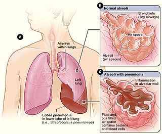
Lobar pneumonia is a form of pneumonia characterized by inflammatory exudate within the intra-alveolar space resulting in consolidation that affects a large and continuous area of the lobe of a lung.

<span class="mw-page-title-main">Alveolar lung disease</span> Medical condition

Alveolar lung diseases, are a group of diseases that mainly affect the alveoli of the lungs.

Restrictive lung diseases are a category of extrapulmonary, pleural, or parenchymal respiratory diseases that restrict lung expansion, resulting in a decreased lung volume, an increased work of breathing, and inadequate ventilation and/or oxygenation. Pulmonary function test demonstrates a decrease in the forced vital capacity.

<span class="mw-page-title-main">Mucociliary clearance</span>

Mucociliary clearance (MCC), mucociliary transport, or the mucociliary escalator describes the self-clearing mechanism of the airways in the respiratory system. It is one of the two protective processes for the lungs in removing inhaled particles including pathogens before they can reach the delicate tissue of the lungs. The other clearance mechanism is provided by the cough reflex. Mucociliary clearance has a major role in pulmonary hygiene.

<span class="mw-page-title-main">Chronic obstructive pulmonary disease</span> Lung disease involving long-term poor airflow

Chronic obstructive pulmonary disease (COPD) is a type of progressive lung disease characterized by long-term respiratory symptoms and airflow limitation. GOLD 2024 defined COPD as a heterogeneous lung condition characterized by chronic respiratory symptoms due to abnormalities of the airways and/or alveoli (emphysema) that cause persistent, often progressive, airflow obstruction.

<span class="mw-page-title-main">Pulmonary interstitial emphysema</span> Collection of air outside of the normal air space of the pulmonary alveoli

Pulmonary interstitial emphysema (PIE) is a collection of air outside of the normal air space of the pulmonary alveoli, found instead inside the connective tissue of the peribronchovascular sheaths, interlobular septa, and visceral pleura. This collection of air develops as a result of alveolar and terminal bronchiolar rupture. Pulmonary interstitial emphysema is more frequent in premature infants who require mechanical ventilation for severe lung disease. Infants with pulmonary interstitial emphysema are typically recommended for admission to a neonatal intensive care unit.

<span class="mw-page-title-main">Emphysema</span> Medical condition

Emphysema is any air-filled enlargement in the body's tissues. Most commonly emphysema refers to the enlargement of air spaces (alveoli) in the lungs, and is also known as pulmonary emphysema.

Bronchoscopic lung volume reduction(BLVR) is a procedure to reduce the volume of air within the lungs. BLVR was initially developed in the early 2000s as a minimally invasive treatment for severe COPD that is primarily caused by emphysema. BLVR evolved from earlier surgical approaches first developed in the 1950s to reduce lung volume by removing damaged portions of the lungs via pneumonectomy or wedge resection. Procedures include the use of valves, coils, or thermal vapour ablation.

<span class="mw-page-title-main">Ventilation–perfusion coupling</span> Relationship between respiratory and cardiovascular processes

Ventilation-perfusion coupling is the relationship between ventilation and perfusion processes, which take place in the respiratory system and the cardiovascular system. Ventilation is the movement of gas during breathing, and perfusion is the process of pulmonary blood circulation, which delivers oxygen to body tissues. Anatomically, the lung structure, alveolar organization, and alveolar capillaries contribute to the physiological mechanism of ventilation and perfusion. Ventilation-perfusion coupling maintains a constant ventilation/perfusion ratio near 0.8 on average, while the regional variation exists within the lungs due to gravity. When the ratio gets above or below 0.8, it is considered abnormal ventilation-perfusion coupling, also known as a ventilation–perfusion mismatch. Lung diseases, cardiac shunts, and smoking can cause a ventilation-perfusion mismatch that results in significant symptoms and diseases, which can be treated through treatments like bronchodilators and oxygen therapy.

References

  1. 1 2 3 4 5 6 7 8 Terry PB, Traystman RJ (December 2016). "The Clinical Significance of Collateral Ventilation". Annals of the American Thoracic Society. 13 (12): 2251–2257. doi:10.1513/AnnalsATS.201606-448FR. PMC   5466185 . PMID   27739872.
  2. 1 2 3 4 Eberlein M, Baldes N, Bölükbas S (May 2019). "A novel air leak test using surfactant: a step forward or a bubble that will burst?". Journal of Thoracic Disease. 11 (Suppl 9): S1119–S1122. doi: 10.21037/jtd.2019.05.06 . PMC   6560585 . PMID   31245059.
  3. 1 2 3 Mitzner, W. (1 January 2006). "Ventilation | Collateral". Ventilation | Collateral. pp. 434–438. doi:10.1016/B0-12-370879-6/00425-7. ISBN   9780123708793 . Retrieved 29 August 2021.
  4. Meier P, Schirmer SH, Lansky AJ, Timmis A, Pitt B, Seiler C (June 2013). "The collateral circulation of the heart". BMC Med. 11: 143. doi: 10.1186/1741-7015-11-143 . PMC   3689049 . PMID   23735225.
  5. 1 2 Gompelmann, D.; Eberhardt, R.; Herth, F.J.F. (2013). "Collateral Ventilation". Respiration. 85 (6): 515–520. doi: 10.1159/000348269 . ISSN   0025-7931. PMID   23485627.
  6. 1 2 3 4 Spencer's pathology of the lung (5th ed.). New York: McGraw-Hill. 1996. pp. 33–34. ISBN   0071054480.
  7. Higuchi, T.; Reed, A.; Oto, T. (1 May 2006). "Relation of interlobar collaterals to radiological heterogeneity in severe emphysema". Thorax. 61 (5): 409–413. doi:10.1136/thx.2005.051219. PMC   2111177 . PMID   16467071 . Retrieved 30 August 2021.
  8. Poggi C, Mantovani S, Pecoraro Y, Carillo C, Bassi M, D'Andrilli A, Anile M, Rendina EA, Venuta F, Diso D (November 2018). "Bronchoscopic treatment of emphysema: an update". Journal of Thoracic Disease. 10 (11): 6274–6284. doi: 10.21037/jtd.2018.10.43 . PMC   6297441 . PMID   30622803.
  9. Gordon M, Duffy S, Criner GJ (August 2018). "Lung volume reduction surgery or bronchoscopic lung volume reduction: is there an algorithm for allocation?". Journal of Thoracic Disease. 10 (Suppl 23): S2816–S2823. doi: 10.21037/jtd.2018.05.118 . PMC   6129811 . PMID   30210836.
  10. 1 2 3 Delaunois, L. (1 October 1989). "Anatomy and physiology of collateral respiratory pathways". European Respiratory Journal. 2 (9): 893–904. doi: 10.1183/09031936.93.02090893 . PMID   2680588. S2CID   7124561 . Retrieved 30 August 2021.
  11. Cetti, E. J.; Moore, A. J.; Geddes, D. M. (1 May 2006). "Collateral ventilation". Thorax. 61 (5): 371–373. doi:10.1136/thx.2006.060509. PMC   2111181 . PMID   16648350 . Retrieved 2 September 2021.