| Look up colliculus in Wiktionary, the free dictionary. |
Colliculus (Latin for "mound") can refer to:
| Look up colliculus in Wiktionary, the free dictionary. |
Colliculus (Latin for "mound") can refer to:

The inferior colliculus (IC) is the principal midbrain nucleus of the auditory pathway and receives input from several peripheral brainstem nuclei in the auditory pathway, as well as inputs from the auditory cortex. The inferior colliculus has three subdivisions: the central nucleus, a dorsal cortex by which it is surrounded, and an external cortex which is located laterally. Its bimodal neurons are implicated in auditory-somatosensory interaction, receiving projections from somatosensory nuclei. This multisensory integration may underlie a filtering of self-effected sounds from vocalization, chewing, or respiration activities.

The superior colliculus is a paired structure of the mammalian midbrain. In other vertebrates the homologous structure is known as the optic tectum or simply tectum. The adjective form tectal is commonly used for mammals as well as other vertebrates.
The anterior colliculus is the anterior portion of the medial malleolus of the distal tibia, forming part of the ankle mortise. It has an attachment of the anterior tibiotalar ligament.
The posterior colliculus is the posterior portion of the medial malleolus of the distal tibia which is smaller in size comparing to the anterior colliculus. It has an attachment of the posterior tibiotalar ligament which is a part of deltoid ligament on the medial side of the ankle.

The seminal colliculus, or verumontanum, of the prostatic urethra is a landmark near the entrance of the ejaculatory ducts. Verumontanum is translated from Latin to mean 'mountain ridge', a reference to the distinctive median elevation of urothelium that characterizes the landmark on magnified views. Embryologically, it is derived from the uterovaginal primordium. The landmark is important in classification of several urethral developmental disorders. The margins of seminal colliculus are the following:

The seminal vesicles, vesicular glands, or seminal glands, are a pair of simple tubular glands posteroinferior to the urinary bladder of some male mammals. Seminal vesicles are located within the pelvis. They secrete fluid that partly composes the semen.

The facial colliculus is an elevated area located on the pontine tegmentum in the floor of the fourth ventricle. It is formed by fibers from the facial motor nucleus of the facial nerve as they loop over the abducens nucleus. Thus a lesion to the facial colliculus would result in ipsilateral facial paralysis and ipsilateral unopposed eye medial deviation.
| This disambiguation page lists articles associated with the title Colliculus. If an internal link led you here, you may wish to change the link to point directly to the intended article. |

The brainstem is the posterior part of the brain, continuous with the spinal cord. In the human brain the brainstem includes the midbrain, and the pons and medulla oblongata of the hindbrain. Sometimes the diencephalon, the caudal part of the forebrain, is included.

The tibia, also known as the shinbone or shankbone, is the larger, stronger, and anterior (frontal) of the two bones in the leg below the knee in vertebrates, and it connects the knee with the ankle bones. The tibia is found on the medial side of the leg next to the fibula and closer to the median plane or centre-line. The tibia is connected to the fibula by the interosseous membrane of the leg, forming a type of fibrous joint called a syndesmosis with very little movement. The tibia is named for the flute tibia. It is the second largest bone in the human body next to the femur. The leg bones are the strongest long bones as they support the rest of the body.

The fibula or calf bone is a leg bone located on the lateral side of the tibia, with which it is connected above and below. It is the smaller of the two bones and in proportion to its length, the slenderest of all the long bones. Its upper extremity is small, placed toward the back of the head of the tibia, below the level of the knee joint, and excluded from the formation of this joint. Its lower extremity inclines a little forward, so as to be on a plane anterior to that of the upper end; it projects below the tibia, and forms the lateral part of the ankle-joint.
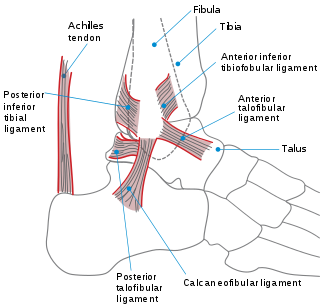
The ankle, or the talocrural region, is the region where the foot and the leg meet. The ankle includes three joints: the ankle joint proper or talocrural joint, the subtalar joint, and the inferior tibiofibular joint. The movements produced at this joint are dorsiflexion and plantarflexion of the foot. In common usage, the term ankle refers exclusively to the ankle region. In medical terminology, "ankle" can refer broadly to the region or specifically to the talocrural joint.

The midbrain or mesencephalon is a portion of the central nervous system associated with vision, hearing, motor control, sleep/wake, arousal (alertness), and temperature regulation.
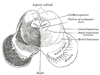
The medial longitudinal fasciculus (MLF) is one of a pair of crossed over tracts, on each side of the brainstem. These bundles of axons are situated near the midline of the brainstem and are made up of both ascending and descending fibers that arise from a number of sources and terminate in different areas. The MLF is the main central connection for the oculomotor nerve, trochlear nerve, and abducens nerve. The vertical gaze center is at the rostral interstitial nucleus (riMLF).
Pott's fracture, also known as Pott's syndrome I and Dupuytren fracture, is an archaic term loosely applied to a variety of bimalleolar ankle fractures. The injury is caused by a combined abduction external rotation from an eversion force. This action strains the sturdy medial (deltoid) ligament of the ankle, often tearing off the medial malleolus due to its strong attachment. The talus then moves laterally, shearing off the lateral malleolus or, more commonly, breaking the fibula superior to the tibiofibular syndesmosis. If the tibia is carried anteriorly, the posterior margin of the distal end of the tibia is also sheared off by the talus. A fractured fibula in addition to detaching the medial malleolus will tear the tibiofibular syndesmosis. The combined fracture of the medial malleolus, lateral malleolus, and the posterior margin of the distal end of the tibia is known as a "trimalleolar fracture".
The pretectal area, or pretectum, is a midbrain structure composed of seven nuclei and comprises part of the subcortical visual system. Through reciprocal bilateral projections from the retina, it is involved primarily in mediating behavioral responses to acute changes in ambient light such as the pupillary light reflex, the optokinetic reflex, and temporary changes to the circadian rhythm. In addition to the pretectum's role in the visual system, the anterior pretectal nucleus has been found to mediate somatosensory and nociceptive information.

In humans, the tectospinal tract is a nerve tract that coordinates head and eye movements. This tract is part of the extrapyramidal system. To be specific, the tectospinal tract connects the midbrain tectum and cervical regions of the spinal cord.

In the brain, the corpora quadrigemina are the four colliculi—two inferior, two superior—located on the tectum of the dorsal aspect of the midbrain. They are respectively named the inferior and superior colliculus.
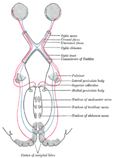
Parinaud's syndrome, also known as dorsal midbrain syndrome, vertical gaze palsy, and sunset sign, is an inability to move the eyes up and down. It is caused by compression of the vertical gaze center at the rostral interstitial nucleus of medial longitudinal fasciculus (riMLF). The eyes lose the ability to move upward and down.

The nucleus of the trochlear nerve is located in the midbrain, at an intercollicular level between the superior colliculus and inferior colliculus. It is a motor nucleus, and so is located near the midline, embedded within the medial longitudinal fasciculus. The oculomotor nerve and trochlear nerve are the only two cranial nerves with nuclei in the midbrain, other than the trigeminal nerve, which has a midbrain nucleus called the mesencephalic nucleus of trigeminal nerve, which functions in preserving dentition.
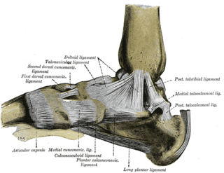
The deltoid ligament is a strong, flat, triangular band, attached, above, to the apex and anterior and posterior borders of the medial malleolus. The deltoid ligament is composed of: 1. Anterior tibiotalar ligament 2. Tibiocalcaneal ligament 3. Posterior tibiotalar ligament 4. Tibionavicular ligament. It consists of two sets of fibers, superficial and deep.

A malleolus is the bony prominence on each side of the human ankle.

The collicular artery or quadrigeminal artery arises from the posterior cerebral artery. This small artery supplies portions of the midbrain, especially the superior colliculus, inferior colliculus, and tectum.
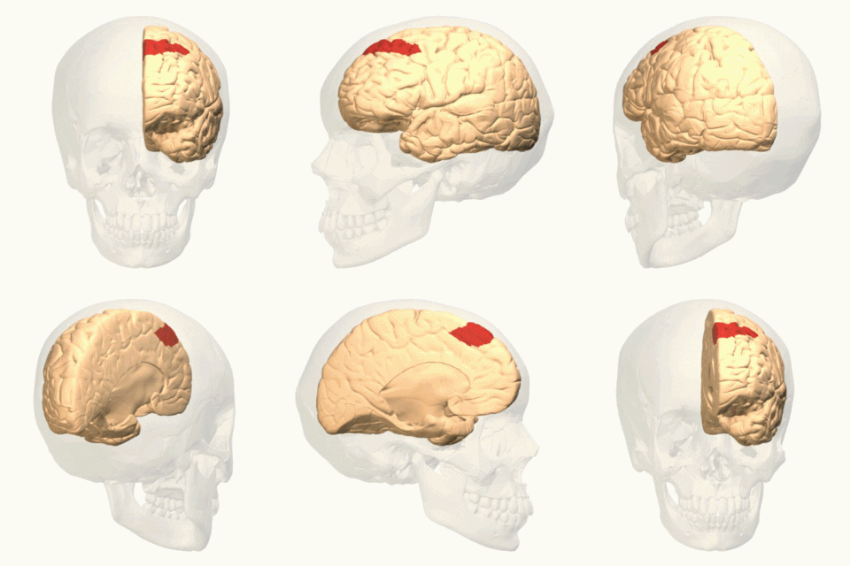
In neuroanatomy, corticomesencephalic tract is a descending nerve tract that originates in the frontal eye field and terminate in the midbrain. Its fibers mediate conjugate eye movement.