Colon cancer staging

Last updated

Colon cancer staging is an estimate of the amount of penetration of a particular cancer. It is performed for diagnostic and research purposes, and to determine the best method of treatment. The systems for staging colorectal cancers depend on the extent of local invasion, the degree of lymph node involvement and whether there is distant metastasis.

Contents

Definitive staging can only be done after surgery and histopathology of colorectal carcinoma. An exception to this principle would be after a colonoscopic polypectomy of a malignant pedunculated polyp with minimal invasion. Preoperative staging of rectal cancers may be done with endoscopic ultrasound. Adjunct staging of metastasis include abdominal ultrasound, MRI, CT, PET scanning, and other imaging studies.

TNM staging system

The most common staging system is the TNM (for tumors/nodes/metastases) system, from the American Joint Committee on Cancer. [1] This system assigns a number based on three categories. "T" denotes the degree of invasion of the intestinal wall, "N" the degree of lymphatic node involvement, and "M" the degree of metastasis. Possibly, the overall Joint Committee stage is a shorter format of the TNM stage, and is usually quoted as a number I, II, III, IV derived from the TNM value grouped by prognosis; a higher number indicates a more advanced cancer and likely a worse outcome. Following is the eighth edition from 2017:
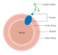
Tumor (T)

The T stages of bowel cancer Diagram showing T stages of bowel cancer CRUK 276.svg
The T stages of bowel cancer

Numbers 0 to 4, with subgroups, are used to describe deepest tumor depth: [2]

Node (N)

Numbers 0 to 2, and subgroups, are used to describe lymph node involvement: [2]

Metastasis (M)

Numbers 0 and 1, with subgroups, describe the metastasis status: [2]

Overall AJCC stage

AJCC stage [2] TNM stage [2] TNM stage criteria [2]
Stage 0Tis N0 M0Tis: Tumor confined to mucosa; cancer-in-situ
Stage IT1 N0 M0T1: Tumor invades submucosa
T2 N0 M0T2: Tumor invades muscularis propria
Stage II-AT3 N0 M0T3: Tumor invades subserosa or beyond (without other organs involved)
Stage II-BT4a N0 M0T4a: Tumor perforates the visceral peritoneum
Stage II-CT4b N0 M0T4b: Tumor invades adjacent organs
Stage III-A
  • T1-2 N1 M0 or
  • T1, N2a, M0
  • N1: Tumor cells in 1 to 3 regional lymph nodes. T1 or T2.
  • N2a: Tumor cells in 4 to 6 regional lymph nodes. T1
Stage III-B
  • T3-4a, N1 M0 or
  • T2-3, N2a, M0 or
  • T1-2 N2b M0
  • N1: Tumor cells in 1 to 3 regional lymph nodes. T3 or T4
  • N2a: Tumor cells in 4 to 6 regional lymph nodes. T2 or T3
  • N2b: Tumor cells in 7 or more regional lymph nodes. T1 or 2
Stage III-C
  • T4a N2a M0 or
  • T3-4a N2b M0 or
  • T4b N1-2, M0
  • N2a: Tumor cells in 4 to 6 regional lymph nodes. T4a
  • N2b: Tumor cells in 7 or more regional lymph nodes. T3-4a
  • N1-2: Tumor cells in at least one regional lymph node. T4b
Stage IVaany T, any N, M1aM1a: Metastasis to 1 other part of the body beyond the colon, rectum or regional lymph nodes. Any T, any N.
Stage IVbany T, any N, M1bM1b: Metastasis to more than 1 other part of the body beyond the colon, rectum or regional lymph nodes. Any T, any N.
Stage IVcany T, any N, M1cM1c: Metastasis to the peritoneal surface. Any T, any N.

Dukes classification

Micrograph of a colorectal adenocarcinoma metastasis to a lymph node. The cancerous cells are at the top center-left of the image, in glands (circular/ovoid structures) and eosinophilic (bright pink). H&E stain. Crc met to node1.jpg
Micrograph of a colo rectal adenocarcinoma metastasis to a lymph node. The cancerous cells are at the top center-left of the image, in glands (circular/ovoid structures) and eosinophilic (bright pink). H&E stain.

In 1932 the British pathologist Cuthbert Dukes (1890–1977) devised a classification system for colorectal cancer. [3] Several different forms of the Dukes classification were developed. [4] [5] However, this system has largely been replaced by the more detailed TNM staging system and is no longer recommended for use in clinical practice. [6]

Astler–Coller classification

An adaptation by the Americans Astler and Coller in 1954 further divided stages B and C [8] [9]

The stage gives valuable information for the prognosis and management of the particular cancer. [ citation needed ]

Full Dukes classification

Another modification of the original Dukes classification was made in 1935 by Gabriel, Dukes and Bussey. [10] This subdivided stage C. This staging system was noted to be prognostically relevant to rectal and colonic adenocarcinoma. [11] Stage D was added by Turnbull to denote the presence of liver and other distant metastases [12]

References

  1. "Cancer Staging". American Cancer Society. Retrieved 12 Sep 2020.
  2. 1 2 3 4 5 6 "Colorectal Cancer: Stages". Cancer.net (American Society of Clinical Oncology). Retrieved 2019-09-26. Approved by the Cancer.Net Editorial Board, 11/2018. In turn citing:
    Amin, Mahul B.; Greene, Frederick L.; Edge, Stephen B.; Compton, Carolyn C.; Gershenwald, Jeffrey E.; Brookland, Robert K.; Meyer, Laura; Gress, Donna M.; Byrd, David R.; Winchester, David P. (2017). "The Eighth Edition AJCC Cancer Staging Manual: Continuing to build a bridge from a population-based to a more "personalized" approach to cancer staging". CA: A Cancer Journal for Clinicians. 67 (2): 93–99. doi: 10.3322/caac.21388 . ISSN   0007-9235. PMID   28094848.
  3. Who Named It, showing correct grammatical usage
  4. Kyriakos M: The President cancer, the Dukes classification, and confusion, Arch Pathol Lab Med 109:1063, 1985
  5. Dukes CE. The classification of cancer of the rectum. Journal of Pathological Bacteriology 1932;35:323
  6. AJCC (American Joint Committee on Cancer) Cancer Staging Manual, 7th ed, Edge, SB, Byrd, DR, Compton, CC, et al (Eds), Springer, New York 2010. p 143.
  7. Single Best Answers in Surgery, Patten DK et al. Hodder Education 2009. p.107 ( ISBN   9780340972359)
  8. Astler VB, Coller FA: The prognostic significance of direct extension of carcinoma of the colon and rectum. Ann Surg 139:846, 1954
  9. Banias, Laura; Jung, Ioan; Chiciudean, Rebeca; Gurzu, Simona (August 2022). "From Dukes-MAC Staging System to Molecular Classification: Evolving Concepts in Colorectal Cancer". International Journal of Molecular Sciences. 23 (16): 9455. doi: 10.3390/ijms23169455 . PMC   9409470 . PMID   36012726.
  10. Gabriel WB, Dukes C, Busset HJR: Lymphatic spread in cancer of the rectum. Br J Surg 23:395–413, 1935
  11. Grinnell RS: The grading and prognosis of carcinoma of the colon and rectum. Ann Surg 109:500-33, 1939
  12. Turnbull RB Jr, Kyle K, Watson FR, et al: Cancer of the colon: the influence of the no touch isolation technique on survival rates. Ann Surg 166:420-7, 1967