Related Research Articles

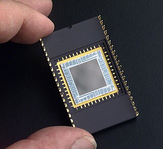
A charge-coupled device (CCD) is an integrated circuit containing an array of linked, or coupled, capacitors. Under the control of an external circuit, each capacitor can transfer its electric charge to a neighboring capacitor. CCD sensors are a major technology used in digital imaging.

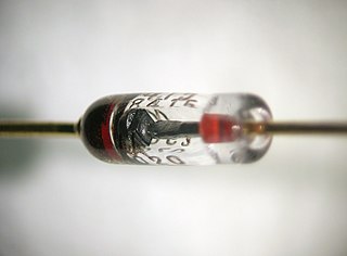
A diode is a two-terminal electronic component that conducts current primarily in one direction. It has low resistance in one direction and high resistance in the other.

A microscope is a laboratory instrument used to examine objects that are too small to be seen by the naked eye. Microscopy is the science of investigating small objects and structures using a microscope. Microscopic means being invisible to the eye unless aided by a microscope.

A scanning electron microscope (SEM) is a type of electron microscope that produces images of a sample by scanning the surface with a focused beam of electrons. The electrons interact with atoms in the sample, producing various signals that contain information about the surface topography and composition of the sample. The electron beam is scanned in a raster scan pattern, and the position of the beam is combined with the intensity of the detected signal to produce an image. In the most common SEM mode, secondary electrons emitted by atoms excited by the electron beam are detected using a secondary electron detector. The number of secondary electrons that can be detected, and thus the signal intensity, depends, among other things, on specimen topography. Some SEMs can achieve resolutions better than 1 nanometer.

An optical spectrometer is an instrument used to measure properties of light over a specific portion of the electromagnetic spectrum, typically used in spectroscopic analysis to identify materials. The variable measured is most often the irradiance of the light but could also, for instance, be the polarization state. The independent variable is usually the wavelength of the light or a closely derived physical quantity, such as the corresponding wavenumber or the photon energy, in units of measurement such as centimeters, reciprocal centimeters, or electron volts, respectively.

X-ray fluorescence (XRF) is the emission of characteristic "secondary" X-rays from a material that has been excited by being bombarded with high-energy X-rays or gamma rays. The phenomenon is widely used for elemental analysis and chemical analysis, particularly in the investigation of metals, glass, ceramics and building materials, and for research in geochemistry, forensic science, archaeology and art objects such as paintings.

Photomultiplier tubes (photomultipliers or PMTs for short) are extremely sensitive detectors of light in the ultraviolet, visible, and near-infrared ranges of the electromagnetic spectrum. They are members of the class of vacuum tubes, more specifically vacuum phototubes. These detectors multiply the current produced by incident light by as much as 100 million times or 108 (i.e., 160 dB), in multiple dynode stages, enabling (for example) individual photons to be detected when the incident flux of light is low.

A photocathode is a surface engineered to convert light (photons) into electrons using the photoelectric effect. Photocathodes are important in accelerator physics where they are utilised in a photoinjector to generate high brightness electron beams. Electron beams generated with photocathodes are commonly used for free electron lasers and for ultrafast electron diffraction. Photocathodes are also commonly used as the negatively charged electrode in a light detection device such as a photomultiplier, phototube and image intensifier.

Video camera tubes are devices based on the cathode-ray tube that were used in television cameras to capture television images, prior to the introduction of charge-coupled device (CCD) image sensors in the 1980s. Several different types of tubes were in use from the early 1930s, and as late as the 1990s.

A streak camera is an instrument for measuring the variation in a pulse of light's intensity with time. They are used to measure the pulse duration of some ultrafast laser systems and for applications such as time-resolved spectroscopy and LIDAR.

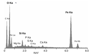
Energy-dispersive X-ray spectroscopy, sometimes called energy dispersive X-ray analysis or energy dispersive X-ray microanalysis (EDXMA), is an analytical technique used for the elemental analysis or chemical characterization of a sample. It relies on an interaction of some source of X-ray excitation and a sample. Its characterization capabilities are due in large part to the fundamental principle that each element has a unique atomic structure allowing a unique set of peaks on its electromagnetic emission spectrum. The peak positions are predicted by the Moseley's law with accuracy much better than experimental resolution of a typical EDX instrument.
A semiconductor detector in ionizing radiation detection physics is a device that uses a semiconductor to measure the effect of incident charged particles or photons.
Photoemission electron microscopy is a type of electron microscopy that utilizes local variations in electron emission to generate image contrast. The excitation is usually produced by ultraviolet light, synchrotron radiation or X-ray sources. PEEM measures the coefficient indirectly by collecting the emitted secondary electrons generated in the electron cascade that follows the creation of the primary core hole in the absorption process. PEEM is a surface sensitive technique because the emitted electrons originate from a shallow layer. In physics, this technique is referred to as PEEM, which goes together naturally with low-energy electron diffraction (LEED), and low-energy electron microscopy (LEEM). In biology, it is called photoelectron microscopy (PEM), which fits with photoelectron spectroscopy (PES), transmission electron microscopy (TEM), and scanning electron microscopy (SEM).

The Advanced Camera for Surveys (ACS) is a third-generation axial instrument aboard the Hubble Space Telescope (HST). The initial design and scientific capabilities of ACS were defined by a team based at Johns Hopkins University. ACS was assembled and tested extensively at Ball Aerospace & Technologies Corp. and the Goddard Space Flight Center and underwent a final flight-ready verification at the Kennedy Space Center before integration in the cargo bay of the Columbia orbiter. It was launched on March 1, 2002, as part of Servicing Mission 3B (STS-109) and installed in HST on March 7, replacing the Faint Object Camera (FOC), the last original instrument. ACS cost US$86 million at that time.

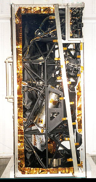
The Faint Object Camera (FOC) was a camera installed on the Hubble Space Telescope from launch in 1990 until 2002. It was replaced by the Advanced Camera for Surveys. In December 1993, Hubble's vision was corrected on STS-61 by installing COSTAR, which corrected the problem with Hubble's mirror before it reached an instrument like FOC. Later instruments had this correction built in, which is why it was possible to later remove COSTAR itself and replace it with a new science instrument.

The Faint Object Spectrograph (FOS) was a spectrograph installed on the Hubble Space Telescope. It was replaced by the Space Telescope Imaging Spectrograph in 1997, and is now on display in the National Air and Space Museum in Washington DC.

Photodetectors, also called photosensors, are sensors of light or other electromagnetic radiation. There are a wide variety of photodetectors which may be classified by mechanism of detection, such as photoelectric or photochemical effects, or by various performance metrics, such as spectral response. Semiconductor-based photodetectors typically use a p–n junction that converts photons into charge. The absorbed photons make electron–hole pairs in the depletion region. Photodiodes and photo transistors are a few examples of photo detectors. Solar cells convert some of the light energy absorbed into electrical energy.
An X-ray image intensifier (XRII) is an image intensifier that converts X-rays into visible light at higher intensity than the more traditional fluorescent screens can. Such intensifiers are used in X-ray imaging systems to allow low-intensity X-rays to be converted to a conveniently bright visible light output. The device contains a low absorbency/scatter input window, typically aluminum, input fluorescent screen, photocathode, electron optics, output fluorescent screen and output window. These parts are all mounted in a high vacuum environment within glass or, more recently, metal/ceramic. By its intensifying effect, It allows the viewer to more easily see the structure of the object being imaged than fluorescent screens alone, whose images are dim. The XRII requires lower absorbed doses due to more efficient conversion of X-ray quanta to visible light. This device was originally introduced in 1948.

X-ray detectors are devices used to measure the flux, spatial distribution, spectrum, and/or other properties of X-rays.

Dynamics Explorer 1 was a NASA high-altitude mission, launched on 3 August 1981, and terminated on 28 February 1991. It consisted of two satellites, DE-1 and DE-2, whose purpose was to investigate the interactions between plasmas in the magnetosphere and those in the ionosphere. The two satellites were launched together into polar coplanar orbits, which allowed them to simultaneously observe the upper and lower parts of the atmosphere.
References
- ↑ "The Digicon Detectors". www.stsci.edu.
- 1 2 Meaburn, J. (2012). Detection and Spectrometry of Faint Light. Dordrecht: Springer Science+Business Media, B.V. p. 36. ISBN 9789027711984.
- ↑ Morgan, B.L.; Airey, R.W.; McMullan, D. (1976). Advances in Electronics and Electron Physics. London: Academic Press. p. 761. ISBN 0120145545.
- ↑ Böhme, S.; Esser, U.; Hefele, H.; Heinrich, I.; Hofmann, W.; Krahn, D.; Matas, V. R.; Schmadel, L. D.; Zech, G. (2013). Astronomy and Astrophysics Abstracts: Volume 42 Literature 1986. Springer Science & Business Media. p. 186. ISBN 978-3-662-12382-9.
- ↑ Berger, Harold (1976). Practical Applications of Neutron Radiography and Gaging. Philadelphia, PA: ASTM International. p. 72. ISBN 0803105355.