
Within a nervous system, a neuron, neurone, or nerve cell is an electrically excitable cell that fires electric signals called action potentials across a neural network. Neurons communicate with other cells via synapses - specialized connections that commonly use minute amounts of chemical neurotransmitters to pass the electric signal from the presynaptic neuron to the target cell through the synaptic gap. The neuron is the main component of nervous tissue in all animals except sponges and placozoa. Non-animals like plants and fungi do not have nerve cells.

Glia, also called glial cells(gliocytes) or neuroglia, are non-neuronal cells in the central nervous system (brain and spinal cord) and the peripheral nervous system that do not produce electrical impulses. The neuroglia make up more than one half the volume of neural tissue in our body. They maintain homeostasis, form myelin in the peripheral nervous system, and provide support and protection for neurons. In the central nervous system, glial cells include oligodendrocytes, astrocytes, ependymal cells, and microglia, and in the peripheral nervous system they include Schwann cells and satellite cells.
Neurochemistry is the study of chemicals, including neurotransmitters and other molecules such as psychopharmaceuticals and neuropeptides, that control and influence the physiology of the nervous system. This particular field within neuroscience examines how neurochemicals influence the operation of neurons, synapses, and neural networks. Neurochemists analyze the biochemistry and molecular biology of organic compounds in the nervous system, and their roles in such neural processes including cortical plasticity, neurogenesis, and neural differentiation.
Synaptogenesis is the formation of synapses between neurons in the nervous system. Although it occurs throughout a healthy person's lifespan, an explosion of synapse formation occurs during early brain development, known as exuberant synaptogenesis. Synaptogenesis is particularly important during an individual's critical period, during which there is a certain degree of synaptic pruning due to competition for neural growth factors by neurons and synapses. Processes that are not used, or inhibited during their critical period will fail to develop normally later on in life.

Astrogliosis is an abnormal increase in the number of astrocytes due to the destruction of nearby neurons from central nervous system (CNS) trauma, infection, ischemia, stroke, autoimmune responses or neurodegenerative disease. In healthy neural tissue, astrocytes play critical roles in energy provision, regulation of blood flow, homeostasis of extracellular fluid, homeostasis of ions and transmitters, regulation of synapse function and synaptic remodeling. Astrogliosis changes the molecular expression and morphology of astrocytes, in response to infection for example, in severe cases causing glial scar formation that may inhibit axon regeneration.

Microglia are a type of neuroglia located throughout the brain and spinal cord. Microglia account for about 10-15% of cells found within the brain. As the resident macrophage cells, they act as the first and main form of active immune defense in the central nervous system (CNS). Microglia are distributed in large non-overlapping regions throughout the CNS. Microglia are key cells in overall brain maintenance—they are constantly scavenging the CNS for plaques, damaged or unnecessary neurons and synapses, and infectious agents. Since these processes must be efficient to prevent potentially fatal damage, microglia are extremely sensitive to even small pathological changes in the CNS. This sensitivity is achieved in part by the presence of unique potassium channels that respond to even small changes in extracellular potassium. Recent evidence shows that microglia are also key players in the sustainment of normal brain functions under healthy conditions. Microglia also constantly monitor neuronal functions through direct somatic contacts and exert neuroprotective effects when needed.

The neuroimmune system is a system of structures and processes involving the biochemical and electrophysiological interactions between the nervous system and immune system which protect neurons from pathogens. It serves to protect neurons against disease by maintaining selectively permeable barriers, mediating neuroinflammation and wound healing in damaged neurons, and mobilizing host defenses against pathogens.

Ben A. Barres was an American neurobiologist at Stanford University. His research focused on the interaction between neurons and glial cells in the nervous system. Beginning in 2008, he was chair of the Neurobiology Department at Stanford University School of Medicine. He transitioned to male in 1997, and became the first openly transgender scientist in the National Academy of Sciences in 2013.

Synaptic pruning, a phase in the development of the nervous system, is the process of synapse elimination that occurs between early childhood and the onset of puberty in many mammals, including humans. Pruning starts near the time of birth and continues into the late-20s. During pruning, both the axon and dendrite decay and die off. It was traditionally considered to be complete by the time of sexual maturation, but this was discounted by MRI studies.
Gliotransmitters are chemicals released from glial cells that facilitate neuronal communication between neurons and other glial cells. They are usually induced from Ca2+ signaling, although recent research has questioned the role of Ca2+ in gliotransmitters and may require a revision of the relevance of gliotransmitters in neuronal signalling in general.
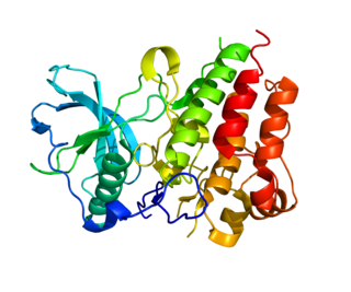
Colony stimulating factor 1 receptor (CSF1R), also known as macrophage colony-stimulating factor receptor (M-CSFR), and CD115, is a cell-surface protein encoded by the human CSF1R gene. CSF1R is a receptor that can be activated by two ligands: colony stimulating factor 1 (CSF-1) and interleukin-34 (IL-34). CSF1R is highly expressed in myeloid cells, and CSF1R signaling is necessary for the survival, proliferation, and differentiation of many myeloid cell types in vivo and in vitro. CSF1R signaling is involved in many diseases and is targeted in therapies for cancer, neurodegeneration, and inflammatory bone diseases.
Neuroinflammation is inflammation of the nervous tissue. It may be initiated in response to a variety of cues, including infection, traumatic brain injury, toxic metabolites, or autoimmunity. In the central nervous system (CNS), including the brain and spinal cord, microglia are the resident innate immune cells that are activated in response to these cues. The CNS is typically an immunologically privileged site because peripheral immune cells are generally blocked by the blood–brain barrier (BBB), a specialized structure composed of astrocytes and endothelial cells. However, circulating peripheral immune cells may surpass a compromised BBB and encounter neurons and glial cells expressing major histocompatibility complex molecules, perpetuating the immune response. Although the response is initiated to protect the central nervous system from the infectious agent, the effect may be toxic and widespread inflammation as well as further migration of leukocytes through the blood–brain barrier may occur.
Beth Stevens is an associate professor in the Department of Neurology at Harvard Medical School and the F. M. Kirby Neurobiology Center at Boston Children’s Hospital. She has helped to identify the role of microglia and complement proteins in the "pruning" or removal of synaptic cells during brain development, and has also determined that the impaired or abnormal microglial function could be responsible for diseases like autism, schizophrenia, and Alzheimer's.

Synaptic stabilization is crucial in the developing and adult nervous systems and is considered a result of the late phase of long-term potentiation (LTP). The mechanism involves strengthening and maintaining active synapses through increased expression of cytoskeletal and extracellular matrix elements and postsynaptic scaffold proteins, while pruning less active ones. For example, cell adhesion molecules (CAMs) play a large role in synaptic maintenance and stabilization. Gerald Edelman discovered CAMs and studied their function during development, which showed CAMs are required for cell migration and the formation of the entire nervous system. In the adult nervous system, CAMs play an integral role in synaptic plasticity relating to learning and memory.

Urtė Neniškytė is a Lithuanian neuroscientist. Her scientific interest and main area of work relates to the interaction of neurons and immune cells in the brain. She has studied the cellular mechanisms of Alzheimer's disease and is the co-author of the first articles about cell death in relation to phagocytosis.

Anna V. Molofsky is an American psychiatrist and glial biologist. She is an associate professor in the department of psychiatry at UC San Francisco. Her lab currently studies the communication between astrocytes, microglia, and neurons to understand how these signals regulate synaptic development in health and disease.
Camilla Bellone is an Italian neuroscientist and assistant professor in the Department of Basic Neuroscience at the University of Geneva, in Switzerland. Bellone's laboratory explores the molecular mechanisms and neural circuits underlying social behavior and probes how defects at the molecular and circuit level give rise to psychiatric disease states such as Autism Spectrum Disorders.
Cagla Eroglu is a Turkish neuroscientist and associate professor of cell biology and neurobiology at Duke University in Durham, North Carolina and an investigator with the Howard Hughes Medical Institute. Eroglu is also the director of graduate studies in cell and molecular biology at Duke University Medical Center. Eroglu is a leader in the field of glial biology, and her lab focuses on exploring the role of glial cells, specifically astrocytes, in synaptic development and connectivity.

Lindsay M. De Biase is an American neuroscientist and glial biologist as well as an assistant professor at the David Geffen School of Medicine at the University of California, Los Angeles. De Biase explores the diversity of microglia that exist within the basal ganglia circuitry to one day target regional or circuit-specific microglia in disease. De Biase's graduate work highlighted the existence and roles of neuron-OPC synapses in development and her postdoctoral work was critical in showing that microglia are not homogenous within the brain parenchyma.
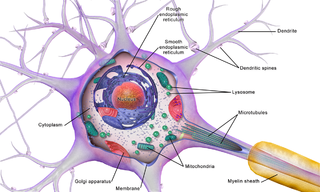
Brain cells make up the functional tissue of the brain. The rest of the brain tissue is structural or connective called the stroma which includes blood vessels. The two main types of cells in the brain are neurons, also known as nerve cells, and glial cells also known as neuroglia.