
Magnetoencephalography (MEG) is a functional neuroimaging technique for mapping brain activity by recording magnetic fields produced by electrical currents occurring naturally in the brain, using very sensitive magnetometers. Arrays of SQUIDs are currently the most common magnetometer, while the SERF magnetometer is being investigated for future machines. Applications of MEG include basic research into perceptual and cognitive brain processes, localizing regions affected by pathology before surgical removal, determining the function of various parts of the brain, and neurofeedback. This can be applied in a clinical setting to find locations of abnormalities as well as in an experimental setting to simply measure brain activity.

Functional magnetic resonance imaging or functional MRI (fMRI) measures brain activity by detecting changes associated with blood flow. This technique relies on the fact that cerebral blood flow and neuronal activation are coupled. When an area of the brain is in use, blood flow to that region also increases.

Functional neuroimaging is the use of neuroimaging technology to measure an aspect of brain function, often with a view to understanding the relationship between activity in certain brain areas and specific mental functions. It is primarily used as a research tool in cognitive neuroscience, cognitive psychology, neuropsychology, and social neuroscience.
Neurotechnology encompasses any method or device in which electronics interface with the nervous system to monitor or modulate neural activity.
Functional integration is the study of how brain regions work together to process information and effect responses. Though functional integration frequently relies on anatomic knowledge of the connections between brain areas, the emphasis is on how large clusters of neurons – numbering in the thousands or millions – fire together under various stimuli. The large datasets required for such a whole-scale picture of brain function have motivated the development of several novel and general methods for the statistical analysis of interdependence, such as dynamic causal modelling and statistical linear parametric mapping. These datasets are typically gathered in human subjects by non-invasive methods such as EEG/MEG, fMRI, or PET. The results can be of clinical value by helping to identify the regions responsible for psychiatric disorders, as well as to assess how different activities or lifestyles affect the functioning of the brain.

Neuroimaging is the use of quantitative (computational) techniques to study the structure and function of the central nervous system, developed as an objective way of scientifically studying the healthy human brain in a non-invasive manner. Increasingly it is also being used for quantitative studies of brain disease and psychiatric illness. Neuroimaging is a highly multidisciplinary research field and is not a medical specialty.
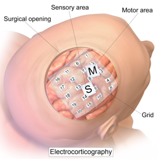
Electrocorticography (ECoG), or intracranial electroencephalography (iEEG), is a type of electrophysiological monitoring that uses electrodes placed directly on the exposed surface of the brain to record electrical activity from the cerebral cortex. In contrast, conventional electroencephalography (EEG) electrodes monitor this activity from outside the skull. ECoG may be performed either in the operating room during surgery or outside of surgery. Because a craniotomy is required to implant the electrode grid, ECoG is an invasive procedure.
Neurophysics is the branch of biophysics dealing with the development and use of physical methods to gain information about the nervous system. Neurophysics is an interdisciplinary science using physics and combining it with other neurosciences to better understand neural processes. The methods used include the techniques of experimental biophysics and other physical measurements such as EEG mostly to study electrical, mechanical or fluidic properties, as well as theoretical and computational approaches. The term "neurophysics" is a portmanteau of "neuron" and "physics".
Integrative neuroscience is the study of neuroscience that works to unify functional organization data to better understand complex structures and behaviors. The relationship between structure and function, and how the regions and functions connect to each other. Different parts of the brain carrying out different tasks, interconnecting to come together allowing complex behavior. Integrative neuroscience works to fill gaps in knowledge that can largely be accomplished with data sharing, to create understanding of systems, currently being applied to simulation neuroscience: Computer Modeling of the brain that integrates functional groups together.

Electroencephalography (EEG) is a method to record an electrogram of the electrical activity on the scalp that has been shown to represent the macroscopic activity of the surface layer of the brain underneath. It is typically non-invasive, with the electrodes placed along the scalp. Electrocorticography, involving invasive electrodes, is sometimes called "intracranial EEG".
Epilepsy is the most common childhood brain disorder in the United States. Nearly 3 million people have been diagnosed with this disease, while 450,000 of them are under the age of 17. Two thirds of the child population will overcome the side effects, including seizures, through treatment during adolescence.
Mark Steven Cohen is an American neuroscientist and early pioneer of functional brain imaging using magnetic resonance imaging. He currently is a Professor of Psychiatry, Neurology, Radiology, Psychology, Biomedical Physics and Biomedical Engineering at the Semel Institute for Neuroscience and Human Behavior and the Staglin Center for Cognitive Neuroscience. He is also a performing musician.
Clinical Electrophysiological Testing is based on techniques borrowed from electrophysiology used for the clinical diagnosis of patients. There are many processes that occur in the body which produce electrical signals that can be detected. Depending on the location and the source of these signals, distinct methods and techniques have been developed to properly target them.

Resting state fMRI is a method of functional magnetic resonance imaging (fMRI) that is used in brain mapping to evaluate regional interactions that occur in a resting or task-negative state, when an explicit task is not being performed. A number of resting-state brain networks have been identified, one of which is the default mode network. These brain networks are observed through changes in blood flow in the brain which creates what is referred to as a blood-oxygen-level dependent (BOLD) signal that can be measured using fMRI.
Functional magnetic resonance spectroscopy of the brain (fMRS) uses magnetic resonance imaging (MRI) to study brain metabolism during brain activation. The data generated by fMRS usually shows spectra of resonances, instead of a brain image, as with MRI. The area under peaks in the spectrum represents relative concentrations of metabolites.
Dynamic functional connectivity (DFC) refers to the observed phenomenon that functional connectivity changes over a short time. Dynamic functional connectivity is a recent expansion on traditional functional connectivity analysis which typically assumes that functional networks are static in time. DFC is related to a variety of different neurological disorders, and has been suggested to be a more accurate representation of functional brain networks. The primary tool for analyzing DFC is fMRI, but DFC has also been observed with several other mediums. DFC is a recent development within the field of functional neuroimaging whose discovery was motivated by the observation of temporal variability in the rising field of steady state connectivity research.
The Ludwig-Boltzmann-Institute for functional Brain Topography was a research institute for the investigation of the function of brain areas. It was founded in 1993 in Vienna, Austria by Lüder Deecke. With his retirement in 2006 the institute was closed.

Hal Blumenfeld is a Professor of Neurology, Neuroscience, and Neurosurgery at Yale University. He is an expert on brain mechanisms of consciousness and on altered consciousness in epilepsy. As director of the Yale Clinical Neuroscience Imaging Center (CNIC) he leads multi-disciplinary research and is also well known for his teaching contributions in neuroanatomy and clinical neuroscience.

Fabrice Bartolomei is a French neurologist and neurophysiologist, Hospital practitioner and University Professor at Aix-Marseille University (AMU), leading the Service de Neurophysiologie Clinique of the Timone Hospital at the Assistance Publique - Hôpitaux de Marseille, and he is the medical director of the ‘Centre Saint-Paul - Hopital Henri Gastaut’. He is the coordinator of the clinical network CINAPSE that is dedicated to the management of adult and pediatric cases of severe epilepsies and leader of the Federation Hospitalo-Universitaire Epinext. He is also member of the research unit Institut de Neurosciences des Systèmes](INS), UMR1106, Inserm - AMU.
Computational models in epilepsy mainly focus on describing an electrophysiological manifestation associated with epilepsy called seizures. For this purpose, computational neurosciences use differential equations to reproduce the temporal evolution of the signals recorded experimentally. A book published in 2008, Computational Neuroscience in Epilepsy,. summarizes different works done up to this time. The goals of using its models are diverse, from prediction to comprehension of underlying mechanisms.