Epiploic appendagitis

Last updated
Epiploic appendagitis
Appendicitis epiploica.jpg

Epiploic appendagitis (EA) is an uncommon, benign, self-limiting inflammatory process of the epiploic appendices. Other, older terms for the process include appendicitis epiploica and appendagitis, but these terms are used less now in order to avoid confusion with acute appendicitis.

Contents

Epiploic appendices are small, fat-filled sacs or finger-like projections along the surface of the upper and lower colon and rectum. They may become acutely inflamed as a result of torsion (twisting) or venous thrombosis. The inflammation causes pain, often described as sharp or stabbing, located on the left, right, or central regions of the abdomen. There is sometimes nausea and vomiting. The symptoms may mimic those of acute appendicitis, diverticulitis, or cholecystitis. The pain is characteristically intense during/after defecation or micturition (espec. in the sigmoid type) due to the effect of traction on the pedicle of the lesion caused by straining and emptying of the bowel and bladder. Initial lab studies are usually normal. EA is usually diagnosed incidentally on CT scan which is performed to exclude more serious conditions.

Although it is self-limiting, epiploic appendagitis can cause severe pain and discomfort. It is usually thought to be best treated with an anti-inflammatory and a moderate to severe pain medication (depending on the case) as needed. Surgery is not recommended in nearly all cases. Sand and colleagues, [1] however, recommend laparoscopic surgery to excise the inflamed appendage in most cases in order to prevent recurrence.

Signs and symptoms

Abdominal CT scan, Epiploic Appendagitis (circle) Epiploic Appendagitis .jpg
Abdominal CT scan, Epiploic Appendagitis (circle)

The condition commonly occurs in patients in their 40s and 50s predominantly in men. Epiploic appendagitis is normally misdiagnosed in most patients. [2] Epiploic appendagitis presents with an acute onset of pain, commonly in the left lower quadrant the symptoms often lead to a misdiagnosis for diverticulitis. Diverticulitis manifests with evenly distributed lower abdominal pain accompanied with nausea, fever, and leukocytosis. Patients with acute epiploic appendagitis do not normally report a change in bowel habits, while a small number may have constipation or diarrhea. [2]

Peritoneal loose body

Gross pathology of a peritoneal loose body, caused torsion and autoamputation of an epiploic appendage, which eventually becomes embedded in a fibrous capsule. Gross pathology of a peritoneal loose body.jpg
Gross pathology of a peritoneal loose body, caused torsion and autoamputation of an epiploic appendage, which eventually becomes embedded in a fibrous capsule.

It is rare however possible for epiploic appendagitis to result in a peritoneal loose body. Peritoneal loose body is a free floating mass of dead fibrous tissue surrounded by several layers of calcification (deposit of calcium salts). The loose body is the result of torsed, infarcted or detached epiploic appendages that eventually become fibrotic (inflammation and scarring) masses. If the loose body becomes large enough it can cause urinary retention (inability to empty bladder) or bowel obstructions. [3]

Background

Epiploic appendages are also called appendices epiploicae. The appendages themselves are 50–100 appendages that are oriented in two rows anterior and posterior. The appendages are parallel to the superficial section of the taenia coli. Furthermore, the appendages are between 0.5 and 5 cm long, each appendage is attached with one or two arterioles and a venule within vascular stalks attached to the colon. Torsion (twisting or wrenching motion) of the appendages can cause ischemia which can cause painful symptoms that mimic other conditions such as diverticulitis, and appendicitis; however, it is rare. The pain associated with the inflamed appendages is located in the left and sometimes in the right lower abdominal quadrant. Diagnosis of epiploic appendagitis can be challenging due to its infrequency. [4]

Diagnosis

Epiploic appendagitis is more common in patients older than 40 years of age; however, it can occur at any age. "The reported ages range from 12 to 82 years. Men are slightly more affected than women." [2] Patients with epiploic appendagitis describe having a localized, strong, non-migratory sharp pain after eating. Patients generally have tender abdomens as a symptom. Symptoms do not include fever, vomiting, or leukocytosis. The pain is typically located in the right or left lower abdominal quadrant. When there is pain in the right lower quadrant, it can mimic appendicitis; however, it more commonly mimics diverticulitis, with pain present on the left side. [2]

Differential diagnosis

There are several conditions that mimic the symptoms of epiploic appendicitis.

Omental infarction: Omental infarction is uncommon reason for acute abdomen. It is similar to acute appendicitis. The pain is of a few days duration centering in the right lower or upper quadrant. Imaging is required to obtain an accurate diagnosis due to the common misdiagnosis of omental infarction as appendicitis or cholecystitis. Omental infarction occurs commonly in pediatric patients approximately 15 percent of cases. The most frequent cause of non- torsion related omental infarction is due to trauma as well as thrombosis or the omental veins. The predisposition for omental infarction includes obesity, strenuous activity, congestive heart failure, digitalis administration, recent abdominal surgery and trauma. [2] "The typical CT findings are a solitary large non-enhancing omental mass with heterogeneous attenuation, which is most often located in the right lower quadrant, deep to the rectus abdominis muscle and either anterior to the transverse colon or anteromedial to the ascending colon". [2] Omental Infarction can be difficult to differentiate from diverticulitis however omental infarction is not normally attributed with bowel wall thickening. It is rare that the colonic wall will be thickened due to spread of the inflammation from the omentum (a fold of peritoneum connecting or supporting abdominal structures) to the tenia omentalis of the colon. [2]

Diverticulitis: Diverticulitis normally happens in older patients than in epiploic appendagitis. The two inflammatory conditions are quite indistinguishable based on physical manifestations. Patients with diverticulitis will present with nausea, vomiting, fever, elevated leukocyte count, rebound tenderness, and will have more extensive lower abdominal pain than patients with epiploic appendagitis. Additionally inflammation from diverticulitis may spread to the epiploic appendages making it difficult to diagnose, for inflammation of the appendices epiploicae may be resultant to other inflammatory conditions in the colonic wall and surrounding mesocolon. [2]

Radiologic evaluation

Ultrasound and CT scans are the normal means of positive diagnosis of epiploic appendagitis. Ultrasound scans show "an oval, non-compressible hyperechoic mass with a subtle hypoechoic rim directly under the site of maximum tenderness". [4] Normally, epiploic appendages cannot be seen on CT scan. [4] After cross-sectional imaging and the increased use of abdominal CT for evaluating lower abdominal pain, EA is increasingly diagnosed. Pathognomonic CT scan data represent EA as 2–4 cm, oval shaped, fat density lesions, surrounded by inflammation. Contrasting with diverticulitis findings, the colonic wall is mostly unchanged.[ citation needed ]

Management

Epiploic appendagitis is self-limiting and can be managed conservatively with NSAIDs.[ citation needed ]

Epidemiology

Acute epiploic appendagitis is usually associated with obesity, hernia and unaccustomed exercise. The inflammation of the epiploic appendages normally resolves on its own for most patients. It is possible however uncommon for acute epiploic appendagitis to result in adhesion, bowel obstruction, intussusception, intraperitoneal loose body, peritonitis, and/or abscess formation. [2] Treatment consists of reassurance of the patient and analgesics. Under non invasive treatment, symptoms resolve in two weeks. Hospitalization is not necessary. [5]

Related Research Articles

<span class="mw-page-title-main">Appendicitis</span> Inflammation of the appendix

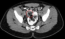
Appendicitis is inflammation of the appendix. Symptoms commonly include right lower abdominal pain, nausea, vomiting, and decreased appetite. However, approximately 40% of people do not have these typical symptoms. Severe complications of a ruptured appendix include widespread, painful inflammation of the inner lining of the abdominal wall and sepsis.

<span class="mw-page-title-main">Peritonitis</span> Medical condition

Peritonitis is inflammation of the localized or generalized peritoneum, the lining of the inner wall of the abdomen and cover of the abdominal organs. Symptoms may include severe pain, swelling of the abdomen, fever, or weight loss. One part or the entire abdomen may be tender. Complications may include shock and acute respiratory distress syndrome.

<span class="mw-page-title-main">Bowel obstruction</span> Medical condition

Bowel obstruction, also known as intestinal obstruction, is a mechanical or functional obstruction of the intestines which prevents the normal movement of the products of digestion. Either the small bowel or large bowel may be affected. Signs and symptoms include abdominal pain, vomiting, bloating and not passing gas. Mechanical obstruction is the cause of about 5 to 15% of cases of severe abdominal pain of sudden onset requiring admission to hospital.

<span class="mw-page-title-main">Abdominal pain</span> Stomach aches

Abdominal pain, also known as a stomach ache, is a symptom associated with both non-serious and serious medical issues. Since the abdomen contains most of the body's vital organs, it can be an indicator of a wide variety of diseases. Given that, approaching the examination of a person and planning of a differential diagnosis is extremely important.

<span class="mw-page-title-main">Diverticulitis</span> Digestive disease of the large intestine

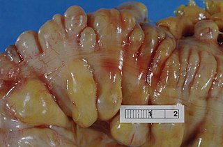
Diverticulitis, also called colonic diverticulitis, is a gastrointestinal disease characterized by inflammation of abnormal pouches—diverticula—that can develop in the wall of the large intestine. Symptoms typically include lower abdominal pain of sudden onset, but the onset may also occur over a few days. There may also be nausea, diarrhea or constipation. Fever or blood in the stool suggests a complication. People may experience a single attack, repeated attacks, or ongoing "smouldering" diverticulitis.

<span class="mw-page-title-main">Diverticulosis</span> Condition of the wall of the intestine

Diverticulosis is the condition of having multiple pouches (diverticula) in the colon that are not inflamed. These are outpockets of the colonic mucosa and submucosa through weaknesses of muscle layers in the colon wall. Diverticula do not cause symptoms in most people. Diverticular disease occurs when diverticula become clinically inflamed, a condition known as diverticulitis.

<span class="mw-page-title-main">Fitz-Hugh–Curtis syndrome</span> Medical condition

Fitz-Hugh–Curtis syndrome is a rare complication of pelvic inflammatory disease (PID) involving liver capsule inflammation leading to the formation of adhesions presenting with the clinical syndrome of right upper quadrant (RUQ) pain.

<span class="mw-page-title-main">Meckel's diverticulum</span> Medical condition

A Meckel's diverticulum, a true congenital diverticulum, is a slight bulge in the small intestine present at birth and a vestigial remnant of the vitelline duct. It is the most common malformation of the gastrointestinal tract and is present in approximately 2% of the population, with males more frequently experiencing symptoms.

<span class="mw-page-title-main">Gastrointestinal disease</span> Medical condition

Gastrointestinal diseases refer to diseases involving the gastrointestinal tract, namely the esophagus, stomach, small intestine, large intestine and rectum, and the accessory organs of digestion, the liver, gallbladder, and pancreas.

<span class="mw-page-title-main">Volvulus</span> Twisting of part of the intestine, causing a bowel obstruction

A volvulus is when a loop of intestine twists around itself and the mesentery that supports it, resulting in a bowel obstruction. Symptoms include abdominal pain, abdominal bloating, vomiting, constipation, and bloody stool. Onset of symptoms may be rapid or more gradual. The mesentery may become so tightly twisted that blood flow to part of the intestine is cut off, resulting in ischemic bowel. In this situation there may be fever or significant pain when the abdomen is touched.

<span class="mw-page-title-main">Diverticular disease</span> Medical condition

Diverticular disease is when problems occur due to diverticulosis, a benign condition defined by the formation of pouches (diverticula) from the weak spots in the wall of the large intestine. This disease spectrum includes diverticulitis, symptomatic uncomplicated diverticular disease (SUDD), and segmental colitis associated with diverticulosis (SCAD). The most common symptoms across the disease spectrum are abdominal pain and bowel habit changes such as diarrhea or constipation. Otherwise, diverticulitis presents with systemic symptoms such as fever and elevated white blood cell count whereas SUDD and SCAD don’t. Treatment ranges from conservative bowel rest to medications such as antibiotics, antispasmodics, acetaminophen, mesalamine, rifaximin, and corticosteroids depending on the specific conditions.

<span class="mw-page-title-main">Gastrointestinal perforation</span> Medical condition

Gastrointestinal perforation, also known as gastrointestinal rupture, is a hole in the wall of the gastrointestinal tract. The gastrointestinal tract is composed of hollow digestive organs leading from the mouth to the anus. Symptoms of gastrointestinal perforation commonly include severe abdominal pain, nausea, and vomiting. Complications include a painful inflammation of the inner lining of the abdominal wall and sepsis.

Valentino's syndrome is pain presenting in the right lower quadrant of the abdomen caused by a duodenal ulcer with perforation through the retroperitoneum.

<span class="mw-page-title-main">Ischemic colitis</span> Medical condition

Ischemic colitis is a medical condition in which inflammation and injury of the large intestine result from inadequate blood supply. Although uncommon in the general population, ischemic colitis occurs with greater frequency in the elderly, and is the most common form of bowel ischemia. Causes of the reduced blood flow can include changes in the systemic circulation or local factors such as constriction of blood vessels or a blood clot. In most cases, no specific cause can be identified.

An acute abdomen refers to a sudden, severe abdominal pain. It is in many cases a medical emergency, requiring urgent and specific diagnosis. Several causes need immediate surgical treatment.

<span class="mw-page-title-main">Pylephlebitis</span> Medical condition

Pylephlebitis is an uncommon thrombophlebitis of the portal vein or any of its branches that is caused by infection. It is usually a complication of intra-abdominal sepsis, most often following diverticulitis, perforated appendicitis, or peritonitis. Considered uniformly lethal in the pre-antibiotic era, it still carries a mortality of 10-30%.

<span class="mw-page-title-main">Omental cake</span> Medical condition

Omental cake is a radiologic sign indicative of an abnormally thickened greater omentum. It refers to infiltration of the normal omental structure by other types of soft-tissue or chronic inflammation resulting in a thickened, or cake-like appearance.

Abdominal guarding is the tensing of the abdominal wall muscles to guard inflamed organs within the abdomen from the pain of pressure upon them. The tensing is detected when the abdominal wall is pressed. Abdominal guarding is also known as 'défense musculaire'.

<span class="mw-page-title-main">Omental infarction</span> Medical condition

Omental infarction, or omental torsion, is an acute vascular disorder which compromises tissue of the greater omentum—the largest peritoneal fold in the abdomen.

Pelvic abscess is a collection of pus in the pelvis, typically occurring following lower abdominal surgical procedures, or as a complication of pelvic inflammatory disease (PID), appendicitis, or lower genital tract infections. Signs and symptoms include a high fever, pelvic mass, vaginal bleeding or discharge, and lower abdominal pain. It can lead to sepsis and death.

References

  1. Sand, M., et al. (2007). Epiploic appendagitis – clinical characteristics of an uncommon surgical diagnosis. BMC Surgery 7:11.
  2. 1 2 3 4 5 6 7 8 9 Singh, Ajay K.; Gervais, Debra A.; Hahn, Peter F.; Sagar, Pallavi; Mueller, Peter R.; Novelline, Robert A. (2005-11-01). "Acute Epiploic Appendagitis and Its Mimics". RadioGraphics. 25 (6): 1521–1534. doi:10.1148/rg.256055030. ISSN   0271-5333. PMID   16284132.
  3. Sussman, Rachael; Murdock, Jonah (2015-04-02). "Peritoneal Loose Body". New England Journal of Medicine. 372 (14): 1359. doi:10.1056/NEJMicm1316094. ISSN   0028-4793. PMID   25830426.
  4. 1 2 3 Sand, Michael; Gelos, Marcos; Bechara, Falk G.; Sand, Daniel; Wiese, Till H.; Steinstraesser, Lars; Mann, Benno (2007-01-01). "Epiploic appendagitis – clinical characteristics of an uncommon surgical diagnosis". BMC Surgery. 7: 11. doi: 10.1186/1471-2482-7-11 . ISSN   1471-2482. PMC   1925058 . PMID   17603914.
  5. van Breda Vriesman, A. C. (2004). Infaction and idiopathic inflammation of intraperitoneal fat. Implications of diagnostic imaging of the acute abdomen