This article needs additional citations for verification .(December 2008) |
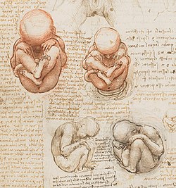
Fetal position (British English: also foetal) is the positioning of the body of a prenatal fetus as it develops. In this position, the back is curved, the head is bowed, and the limbs are bent and drawn up to the torso. A compact position is typical for fetuses. Many newborn mammals, especially rodents, remain in a fetal position well after birth.
This type of compact position is used in the medical profession to minimize injury to the neck and chest.
Some people assume a fetal position when sleeping, especially when the body becomes cold. [1] In some cultures bodies have been buried in fetal position. [2]
Sometimes, when a person has suffered extreme physical or psychological trauma (including massive stress), they will assume a similar compact position in which the back is curved forward, the legs are brought up as tightly against the abdomen as possible, the head is bowed as close to the abdomen as possible, and the arms are wrapped around the head to prevent further trauma.[ citation needed ]
This type of position has been observed in drug addicts, who enter the position when experiencing withdrawal. Sufferers of anxiety are also known to assume the same type of position during panic attacks.
Assuming this type of position and playing dead is often recommended as a strategy to end a bear attack. [3]
Amniocentesis is a medical procedure used primarily in the prenatal diagnosis of genetic conditions. It has other uses such as in the assessment of infection and fetal lung maturity. Prenatal diagnostic testing, which includes amniocentesis, is necessary to conclusively diagnose the majority of genetic disorders, with amniocentesis being the gold-standard procedure after 15 weeks' gestation.
Rh disease is a type of hemolytic disease of the fetus and newborn (HDFN). HDFN due to anti-D antibodies is the proper and currently used name for this disease as the Rh blood group system actually has more than 50 antigens and not only the D-antigen. The term "Rh Disease" is commonly used to refer to HDFN due to anti-D antibodies, and prior to the discovery of anti-Rho(D) immune globulin, it was the most common type of HDFN. The disease ranges from mild to severe, and occurs in the second or subsequent pregnancies of Rh-D negative women when the biologic father is Rh-D positive.
Prenatal and perinatal psychology explores the psychological and psychophysiological effects and implications of the earliest experiences of the individual, before birth, prenatal, as well as during and immediately after childbirth, perinatal. Prenatal and perinatal psychology can be seen as a part of developmental psychology, although historically it was developed in the heterogenous field of psychoanalysis. Prenatal and perinatal psychology are often discussed together to group the period during pregnancy, childbirth, and through the early stages of infancy. The role of prenatal and perinatal psychology is to explain the experience and behavior of the individual before birth, postnatal consequences, and the lasting effects on development that occur during this time period.
A breech birth is when a baby is born bottom first instead of head first, as is normal. Around 3–5% of pregnant women at term have a breech baby. Due to their higher than average rate of possible complications for the baby, breech births are generally considered higher risk. Breech births also occur in many other mammals such as dogs and horses, see veterinary obstetrics.
Kouros is the modern term given to free-standing Ancient Greek sculptures that depict nude male youths. They first appear in the Archaic period in Greece and are prominent in Attica and Boeotia, with a less frequent presence in many other Ancient Greek territories such as Sicily. Such statues are found across the Greek-speaking world; the preponderance of these were found in sanctuaries of Apollo with more than one hundred from the sanctuary of Apollo Ptoion, Boeotia, alone. These free-standing sculptures were typically marble, but the form is also rendered in limestone, wood, bronze, ivory and terracotta. They are typically life-sized, though early colossal examples are up to 3 meters tall.
Obstetric ultrasonography, or prenatal ultrasound, is the use of medical ultrasonography in pregnancy, in which sound waves are used to create real-time visual images of the developing embryo or fetus in the uterus (womb). The procedure is a standard part of prenatal care in many countries, as it can provide a variety of information about the health of the mother, the timing and progress of the pregnancy, and the health and development of the embryo or fetus.
Coffin birth, also known as postmortem fetal extrusion, is the expulsion of a nonviable fetus through the vaginal opening of the decomposing body of a deceased pregnant woman due to increasing pressure from intra-abdominal gases. This kind of postmortem delivery occurs very rarely during the decomposition of a body. The practice of chemical preservation, whereby chemical preservatives and disinfectant solutions are pumped into a body to replace natural body fluids, have made the occurrence of "coffin birth" so rare that the topic is rarely mentioned in international medical discourse.
The Kleihauer–Betke ("KB") test, Kleihauer–Betke ("KB") stain, Kleihauer test or acid elution test is a blood test used to measure the amount of fetal hemoglobin transferred from a fetus to a mother's bloodstream. It is usually performed on Rh-negative mothers to determine the required dose of Rho(D) immune globulin (RhIg) to inhibit formation of Rh antibodies in the mother and prevent Rh disease in future Rh-positive children. It is named after Enno Kleihauer and Klaus Betke who described it in 1957.
Sacrococcygeal teratoma (SCT) is a type of tumor known as a teratoma that develops at the base of the coccyx (tailbone) and is thought to be primarily derived from remnants of the primitive streak. Sacrococcygeal teratomas are benign 75% of the time, malignant 12% of the time, and the remainder are considered "immature teratomas" that share benign and malignant features. Benign sacrococcygeal teratomas are more likely to develop in younger children who are less than 5 months old, and older children are more likely to develop malignant sacrococcygeal teratomas.
In obstetrics, Leopold maneuvers are a common and systematic way to determine the position of a fetus inside the woman's uterus. They are named after the gynecologist Christian Gerhard Leopold. They are also used to estimate term fetal weight.
Obstetrical forceps are a medical instrument used in childbirth. Their use can serve as an alternative to the ventouse method.
Fetal movement refers to motion of a fetus caused by its own muscle activity. Locomotor activity begins during the late embryological stage and changes in nature throughout development. Muscles begin to move as soon as they are innervated. These first movements are not reflexive, but arise from self-generated nerve impulses originating in the spinal cord. As the nervous system matures, muscles can move in response to stimuli.
Percutaneous umbilical cord blood sampling (PUBS), also called cordocentesis, fetal blood sampling, or umbilical vein sampling is a diagnostic genetic test that examines blood from the fetal umbilical cord to detect fetal abnormalities. Fetal and maternal blood supply are typically connected in utero with one vein and two arteries to the fetus. The umbilical vein is responsible for delivering oxygen rich blood to the fetus from the mother; the umbilical arteries are responsible for removing oxygen poor blood from the fetus. This allows for the fetus’ tissues to properly perfuse. PUBS provides a means of rapid chromosome analysis and is useful when information cannot be obtained through amniocentesis, chorionic villus sampling, or ultrasound ; this test carries a significant risk of complication and is typically reserved for pregnancies determined to be at high risk for genetic defect. It has been used with mothers with immune thrombocytopenic purpura.
In obstetrics, asynclitic birth, or asynclitism, refers to the malposition of the fetal head in the uterus relative to the birth canal. Many babies enter the pelvis in an asynclitic presentation, but in most cases, the issue is corrected during labor. Asynclitic presentation is not the same as shoulder presentation, where the shoulder enters first.
In obstetrics, position is the orientation of the fetus in the womb, identified by the location of the presenting part of the fetus relative to the pelvis of the mother. Conventionally, it is the position assumed by the fetus before the process of birth, as the fetus assumes various positions and postures during the course of childbirth.
A fetus or foetus is the unborn mammalian offspring that develops from an embryo. Following the embryonic stage, the fetal stage of development takes place. Prenatal development is a continuum, with no clear defining feature distinguishing an embryo from a fetus. However, in general a fetus is characterized by the presence of all the major body organs, though they will not yet be fully developed and functional, and some may not yet be situated in their final anatomical location.
In obstetrics, a cephalic presentation or head presentation or head-first presentation is a situation at childbirth where the fetus is in a longitudinal lie and the head enters the pelvis first; the most common form of cephalic presentation is the vertex presentation, where the occiput is the leading part. All other presentations are abnormal (malpresentations) and are either more difficult to deliver or not deliverable by natural means.
Fetal abduction refers to the rare crime of child abduction by kidnapping of an at term pregnant woman and extraction of her fetus through a crude cesarean section. Dr. Michael H. Stone and Dr. Gary Brucato have alternatively referred to this crime as "fetus-snatching" or "fetus abduction." Homicide expert Vernon J. Geberth has used the term "fetal kidnapping." In the small number of reported cases, a few pregnant victims and about half of their fetuses survived the assault and non-medically performed cesarean.
Fetal electroencephalography, also known as prenatal EEG includes any recording of electrical fluctuations arising from the brain of a fetus. Doctors and scientists use EEGs to detect and characterize brain activity, such as sleep states, potential seizures, or levels of a coma. EEG captures the electrical activity in the vicinity of the recording electrodes. The majority of the neural electrical activity arises from the flow of current from the cell bodies of pyramidal neurons to their apical dendrites, which become depolarized by excitatory inputs from other neurons. To record the most accurate signals, scientists try to minimize the distance between the recording electrode and the neural activity that they want to detect. Given the difficulty of attaching electrodes to a fetus inside a uterus, doctors and scientists use a variety of techniques to record fetal brain activity.